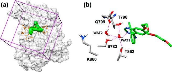
Figure 4.
(a) DOCK setup for HER2 showing protein in a gray surface, energy grid bounding box in purple, binding pocket spheres in yellow, and erlotinib in a green surface. (b) Close-up view of erlotinib, showing the water-mediated network. Atoms within H-bonding distance shown as dashed magenta lines.

