Abstract
Targeting proteins to regions where they are required is essential for proper development of organisms. For achievement of this, subcellular mRNA localization is one of the critical mechanisms. Subcellular mRNA localization is an evolutionarily conserved phenomenon from E. coli to human and contributes to limiting the regions at which its products function and efficiently supplies substrates for protein translation. During early Drosophila embryogenesis, while 71% of the 3370 mRNAs analyzed have shown prominent subcellular localization, the underlying molecular mechanisms have not been elucidated. Here, we reveal that anillin mRNA, one of the localized mRNAs in early Drosophila embryo, localizes to the tip of the pseudo-cleavage furrow in the Drosophila syncytial blastoderm using in situ hybridization combined with immunohistochemistry. Localization analyses with transgenic fly lines carrying a series of deletion mRNAs indicate that this localization is dependent on its own nascent polypeptides including the actin binding domain (ABD). In addition to the mRNA localization, it is revealed that the pleckstrin homology (PH) domain of Anillin protein is also required for its proper localization. Thus, we indicate that the precise localization of Anillin protein is tightly regulated by the ABD on the nascent polypeptide and PH domain in the Drosophila syncytial blastoderm.
Subject terms: Cell division, Embryogenesis
Introduction
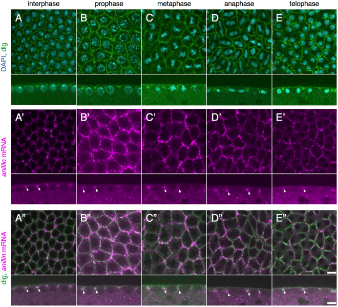
In early Drosophila embryogenesis, the first thirteen nuclear divisions occur without accompanying cytokinesis1. In the embryo between nuclear division cycle 10 and 14, which is called the syncytial blastoderm, thousands of nuclei at the embryo surface rapidly and synchronously divide four times. In order to prevent the collision of neighboring mitotic spindles, a transient membrane invagination called the pseudo-cleavage furrow (PCF) or metaphase furrow occurs between nuclei2,3. This furrow progressively ingresses from prophase to metaphase, and then starts to retract at anaphase (Fig. 1A–E)4. Most of the components in the PCF are shared with those of the cleavage furrow during cytokinesis5–10. The Anillin protein is one of the main components in the cleavage furrow and functions as a scaffold protein to maintain the contractile ring during cytokinesis and also localizes to the PCF in the syncytial blastoderm with strong accumulation at the tip of the PCF7. However, the mechanisms by which the Anillin protein is localized to the location at which it functions remain unclear.
Figure 1.
Endogenous anillin mRNA localizes to the tip of the PCF through the cell cycle in the syncytial blastoderm. (A–E) The plasma membrane of the syncytial blastoderm at interphase, prophase, metaphase, anaphase, and telophase was marked with anti-dlg IgG and DNA was stained with DAPI. (A’–E’) FISH images with anillin mRNA probes. anillin mRNA was detected in the syncytial blastoderm by in situ hybridization with the DIG-labeled RNA probe against anillin mRNA (arrowheads in the lower panels of A’–E’). (A”–E”) Merged images of anillin mRNA signals and dlg signals. anillin mRNA localized to the tip of the PCF, an invagination of the plasma membrane, throughout the cell cycle (arrowheads in the lower panels of A”–E”). The localization of anillin mRNA at the tip of the PCF in at least three different embryos was observed. There was no significant difference among these three embryos. Each upper and lower panel shows horizontal and vertical sections, respectively. Each panel is shown at the same magnification. The scale bar indicates 50 µm.
Subcellular mRNA localization provides the appropriate amount of a protein at the region at which it is required. Since subcellularly localized mRNA was initially visualized in Ascidian embryos in 198311, a large number of localized mRNAs have been reported in various species and types of cells (Holt and Bullock, 2009). The Drosophila embryo is an attractive model in analyses of subcellular mRNA localization, and has been utilized for more than three decades. A number of developmentally important genes have been identified and characterized in the Drosophila embryo, and, thus, the subcellular localization of their mRNAs may be directly linked to their functions, as is the case for bicoid and nanos mRNAs (Ephrussi et al., 1991; Gavis and Lehmann, 1992). Furthermore, a large-scale analysis of mRNA localization in the Drosophila early embryo by fluorescence in situ hybridization has been conducted and several types of prominent mRNA localization patterns have been reported, such as accumulation in the posterior yolk or pole plasm, apical localization above nuclei of the embryo surface, and nuclear division-related localization12.
In the present study, we focus on anillin mRNA, which shows a hexagonal pattern that is one of the distinguishing patterns in the syncytial blastoderm, and this category includes several mRNAs such as canoe, Diaphanous, Polychaetoid, and CG43427 12. In order to investigate the molecular mechanisms underlying the subcellular localization of anillin mRNA in the syncytial blastoderm, we established transgenic fly lines expressing anillin mRNAs carrying a set of deletions and analyzed their localization using in situ hybridization analysis. In the present study, we reveal that anillin mRNA localized to the tip of the PCF in the syncytial blastoderm in its own nascent polypeptide-dependent manner. In addition to the mRNA localization, the pleckstrin homology domain of Anillin protein is required for its proper localization at the tip of the PCF in the syncytial blastoderm.
Results
anillin mRNA localizes to the tip of the PCF throughout the cell cycle in the syncytial blastoderm
The diagnostic localization of the anillin mRNA like honeycomb-like pattern in the Drosophila syncytial blastoderm12 closely resembles the pattern of the PCF, which is an incomplete and temporal membrane invagination2,3,13. In addition, anillin mRNA partially colocalizes with the Anillin protein in the plasma membrane, particularly at the tip of the PCF in the syncytial blastoderm (Supplementary Fig. 1, arrowheads in lower panels)7,12. These findings strongly suggest that anillin mRNA localizes to the PCF. In order to investigate more precisely whether anillin mRNA localizes to the PCF, we performed in situ hybridization combined with immunostaining with anti-discs large (dlg) IgG, a plasma membrane and PCF marker (Lee et al., 2003; Cao et al.10. In the syncytial blastoderm, the cell cycle consists of interphase and a mitotic phase without gap phases and progresses quickly (Zalokar and Erk, 1976; Foe and Alberts, 1983). The results of double staining showed that anillin mRNA partially colocalized to the front edge of the dlg signal throughout the cell cycle in the syncytial blastoderm (Fig. 1, arrowheads in the lower panels of A”–E”).
The coding region of a splicing variant of anillin mRNA, anillin SV-A is necessary for its localization at the tip of the PCF
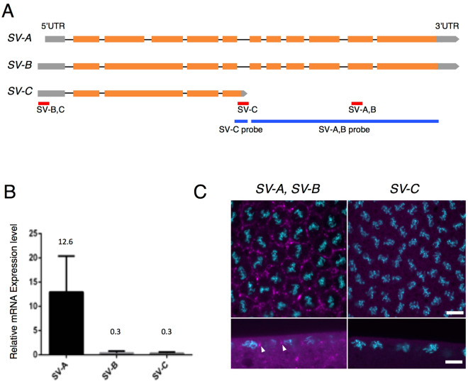
According to the database (Flybase: http://flybase.org/reports/FBgn0261385.html), three different splice variants (SV-A, B and C) of anillin mRNA have been predicted to exist (Fig. 2A). In order to identify the PCF localization element in anillin mRNA, we initially investigated which SV of anillin mRNA was expressed and localized to the PCF in the syncytial blastoderm. We measured the expression levels of three SVs using RT-qPCR. Since it was not possible to detect SV-A or B independently (Fig. 2A), we calculated the expression level of SV-A or B based on differences between the expression levels of SV-A, B or SV-B, C and SV-C, respectively. RT-qPCR revealed that the expression level of SV-A in the syncytial blastoderm was 42-fold higher than those of SV-B and C, which were close to the limit of detection by this method (Fig. 2B). In addition to the quantitative analysis, we examined the localization of each SV by in situ hybridization. We were unable to detect any signals of SV-C (Fig. 2C). Similar to the quantitative analysis, we were unable to find a probe with the ability to detect SV-A and B separately (Fig. 2A). However, the signals observed appeared to represent SV-A signals because SV-A was dominantly expressed in the syncytial blastoderm (Fig. 2B). SV-C was expressed as weakly as SV-B and, thus, a signal for SV-C was not detected (Fig. 2C). These results indicate that most of the anillin mRNA signals at the tip of the PCF are derived from SV-A signals (Fig. 2C and Supplementary Fig. 2A).
Figure 2.
SV-A is dominantly expressed and localized to the tip of the PCF. (A) Diagram of three different predicted SVs of anillin mRNA. Gray box: 5′UTR, Orange box: exon, Gray pentagon: 3′UTR, Black bar: intron, Red bar: the amplified region for detecting each SV, Blue bar: the region at which each probe hybridizes for FISH. (B) The expression level of each SV was detected by RT-qPCR. Only SV-A was expressed at a high level in the syncytial blastoderm. Data are means ± SD (n = 3). (C) The localization patterns of the SVs that hybridized with the SV-A, B and SV-C probes. The signals of SVs are shown in magenta and DNA stained with DAPI is shown in cyan. The localization pattern of each SV in at least three different embryos was observed. There was no significant difference among the examined embryos. Upper and lower panels show horizontal and vertical sections, respectively. Each panel is shown at the same magnification. The scale bar indicates 50 µm.
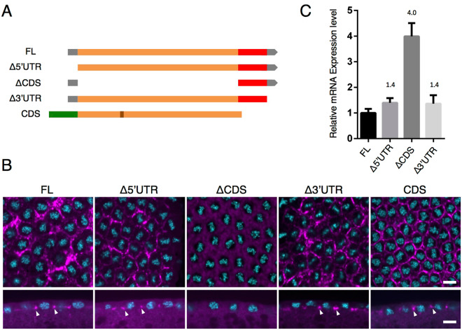
In order to identify the region in SV-A that is necessary for localization to the tip of the PCF, we established transgenic fly lines expressing a series of deletion derivatives of SV-A fused to DsRed-monomer (Fig. 3A). We performed in situ hybridization with the probe for DsRed-monomer to identify exogenous RNAs without detecting endogenous anillin mRNA. While 5′ or 3′ untranslated region (UTR)-deleted mRNAs (Δ5′UTR and Δ3′UTR) localized to the tip of the invaginating furrow, as observed with FL (Fig. 3B, arrowheads and Supplementary Fig. 2B), coding sequence (CDS)-deleted RNA (ΔCDS) did not and was evenly distributed throughout the embryo (Fig. 3B, ΔCDS and Supplementary Fig. 2B). RT-qPCR analyses revealed that ΔCDS RNA was sufficiently expressed to be detectable even if it was not specifically localized because the expression level of ΔCDS RNA was found to be 4.0-fold higher than that of the PCF-localized FL (Fig. 3C). These results suggest that CDS is crucial for the furrow tip localization of anillin mRNA in the syncytial blastoderm. In order to further demonstrate the importance of the CDS region of anillin mRNA for localization to the tip of the PCF, we investigated the localization of GFP-fused anillin CDS 14 using in situ hybridization with the probe for GFP. We used the transgenic strain carrying GFP-fused SV-B (CDS), which is available from a stock center. CDS is derived from SV-B, which includes all SV-A CDS and additional 81-bp sequences (Fig. 2A), and the SV-B protein has been reported to properly localize at the PCF in the syncytial blastoderm14. The results obtained showed that CDS mRNA localized to the tip of the PCF (Fig. 3B). Collectively, these results suggest that the CDS region of anillin mRNA is necessary and sufficient for localization to the tip of the PCF in the syncytial blastoderm (Fig. 3C).
Figure 3.
The CDS of SV-A is necessary and sufficient for the proper localization of SV-A. (A) Diagrams of deletion constructs of anillin fused with the DsRed-monomer or GFP coding sequence. FL, Δ5′UTR, ΔCDS and Δ3′UTR were derived from SV-A whereas CDS was derived from SV-B carrying additional 81 bp comparing with SV-A. Gray box: 5′UTR, Orange box: exon, Gray pentagon: 3′UTR, Red box: DsRed-monomer-coding sequence, Green box: GFP-coding sequence, Brawn box: additional 81 bp. (B) The localization pattern of each deletion derivative of mRNA detected with an anti-sense RNA probe against the DsRed-monomer-coding sequence. Each deletion mRNA is shown in magenta and DNA stained with DAPI is shown in cyan. The localization pattern of each mRNA in at least three different embryos was observed. There was no significant difference among the examined embryos. Upper and lower panels show horizontal and vertical sections. Each panel is shown at the same magnification. The scale bar indicates 50 µm. (C) The expression level of each deletion construct was detected by RT-qPCR. ΔCDS was more strongly expressed than FL and other deletion derivatives. Data are means ± SD (n = 3).
In order to further narrow down the region required for the furrow tip localization of anillin CDS, we divided SV-A CDS into three parts (Supplementary Fig. 3A) and generated transgenic fly lines expressing each divided CDS. None of the SV-A carrying each of the three parts of CDS mRNAs localized to the PCF (Supplementary Figs 3C–E and 2C), despite all of them being sufficiently expressed for detection by RT-qPCR (Supplementary Fig. 3F). These results indicate that none of the segments for CDS mRNA were sufficient for localization to the furrow tip.
However, the proteins encoded by each divided CDS segment showed different localization patterns, such as on the plasma membrane, in the nucleus, or colocalization with Peanut (Supplementary Fig. 3C”–E”). These localization patterns appeared to depend on the domain feature carried in each CDS segment. There are well-known domains in each CDS segment of Anillin, such as the myosin binding domain (MBD) and actin binding domain (ABD) in CDS 1, nuclear localization signal (NLS) in CDS 2 and NLS, the pleckstrin homology (PH) domain, which binds to Peanut, and amino acids in the PH domain that are critical for PI(4,5)P2 binding in CDS 37,14–16. The localization pattern of each CDS segment was consistent with those predicted from the function of each domain included in each CDS segment (Supplementary Fig. 3C’–E’).
anillin mRNA localizes to the tip of the PCF in a manner that is dependent on its nascent polypeptides
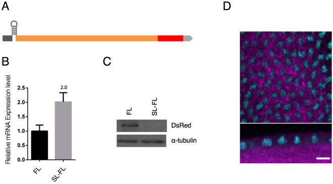
In divided CDS segment localization analyses, we were unable to identify any obvious RNA localization element within anillin CDS. Previous studies reported that the mRNAs encoding transmembrane or secreted proteins localized to the endoplasmic reticulum (ER) in a manner that was mediated by their own nascent polypeptides17. Furthermore, as is the case with these mRNAs, some of the mRNAs encoding cytoplasmic or nuclear proteins, such as X-box binding protein 1, Diaphanous 1, and ABP140 mRNAs, also localized subcellularly depending on their nascent polypeptides18–20. In order to investigate whether the localization of anillin mRNA depends on its own nascent polypeptides, we established transgenic fly lines harboring full-length anillin mRNA in which a stable stem-loop structure (∆G = −42.10 kcal/mol) was inserted between 5′UTR and CDS (SL-FL) to inhibit protein translation (Fig. 4A). This stem-loop structure is commonly used to prevent translation by impairing ribosome loading onto CDS21,22. We found that while the expression levels of SL-FL were 2-fold higher level than those of FL mRNA (Fig. 4B), no predicted protein was detected in the SL-FL fly early embryo (Fig. 4C). Under these conditions, SL-FL did not localize to the PCF (Fig. 4D). These results indicate that the nascent polypeptides translated from anillin mRNA are important for its mRNA localization. However, since CDS 1, 2, and 3 are translated into a part of the Anillin protein, translation itself may not have been sufficient for the proper localization of anillin mRNA (Supplementary Figs 2C and 3).
Figure 4.
Translation of anillin mRNA is necessary for its localization to the tip of the PCF. (A) Diagram of a construct in which a stable stem-loop was inserted between 5′UTR and CDS of FL (SL-FL). (B) FL mRNA and SL-FL mRNA levels were quantified by RT-qPCR. SL-FL mRNA expression levels were 2-fold higher than those of FL mRNA. Data are means ± SD (n = 3). (C) The SL-FL protein was not detected in a Western blot analysis. (D) SL-FL mRNA did not localize to the tip of the PCF. mRNA is shown in magenta and DNA stained with DAPI is shown in cyan. Upper and lower panels show horizontal and vertical sections. The scale bar indicates 50 µm.
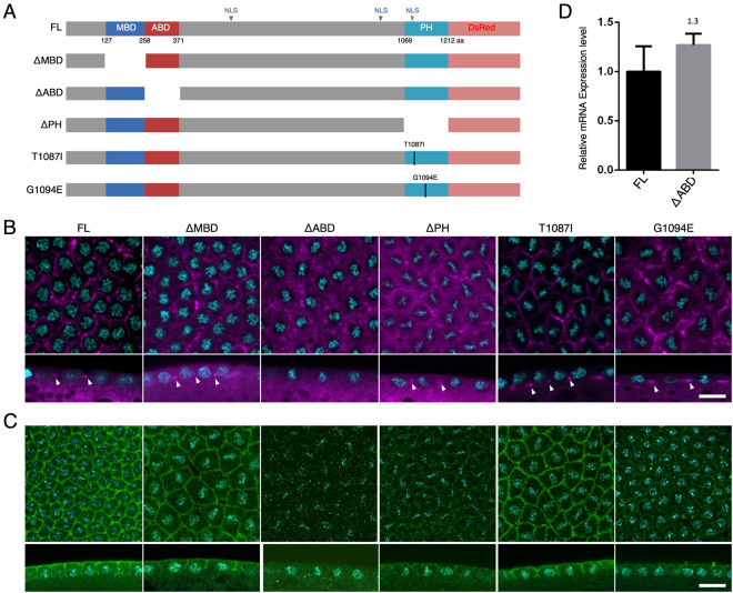
Therefore, we examined whether the specific domain of the Anillin protein is required for the localization of anillin mRNA to the PCF. Anillin contains three known domains: the MBD, ABD, and PH domain (Fig. 5A)7,14,23. In order to investigate the role of each domain region of the Anillin protein in its mRNA localization, we generated transgenic fly lines expressing each domain-deleted Anillin, namely, ΔMBD, ΔABD, and ΔPH (Fig. 5A). In addition to these deletions, we investigated the effects of base substitutions corresponding to each amino acid in the PH domain (T1087I, G1094E) that are suggested to be responsible for PI(4,5)P2 binding16 (Fig. 5A). PI(4,5)P2 has been predicted to function in cell cleavage during cytokinesis16. Domain-deleted or base-substituted mRNAs, except for ΔABD, localized to the tip of the PCF (Fig. 5B, arrowheads and Supplementary Fig. 2D). Based on RT-qPCR data, the expression level of ΔABD appeared to be sufficient for detection even if its mRNA was not specifically localized to the PCF (Fig. 5D). These results demonstrate that ABD is necessary for the proper localization of anillin mRNA.
Figure 5.
ABD is necessary for the localization of anillin mRNA to the tip of the PCF. (A) Diagram of deletion or amino acid-substitution variants. (B) The localization pattern of each deletion or mutated mRNA was detected with an anti-sense RNA probe against the DsRed-monomer-coding sequence. The localization pattern of each mRNA in at least three different embryos was observed. There was no significant difference among the examined embryos. Each mRNA signal and DNA stained with DAPI is shown in magenta and cyan, respectively. (C) The localization pattern of proteins encoded by each deletion or mutated mRNA was detected with anti-RFP IgG. The localization pattern of each fusion protein in at least three different embryos was observed. There was no significant difference among the examined embryos. Each protein signal and DNA stained with DAPI are shown in green and cyan, respectively. Upper and lower panels show horizontal and vertical sections. Each panel is shown at the same magnification. The scale bar indicates 50 µm. (D) The expression level of ΔABD mRNA was quantified by RT-qPCR and compared to that of FL. Data are means ± SD (n = 3).
In ΔMBD or T1087I base-substituted transgene-expressing embryos, those mRNAs and proteins were precisely localized (Fig. 5B,C). However, while ΔPH and G1094E base-substituted mRNAs localized to the PCF (Fig. 5B), the localization of their proteins to the PCF was very faint (Fig. 5C). The results of a Western blot analysis and its quantified data revealed that the protein level of ΔABD, ΔPH, or G1094E was similar to that of ΔMBD or T1087I (Supplementary Fig. 4). These results suggest that not only ABD, but also the PH domain is required for the proper localization of the Anillin protein in the syncytial blastoderm, as reported for the localization of the human Anillin protein to the cleavage furrow of mammalian cultured cells23.
Discussion
It is well known that subcellular mRNA localization is one of the most important processes to express proper protein function in organisms. However, in contrast to the rapidly increasing number of studies describing subcellularly localized mRNAs, our knowledge on the molecular mechanisms underlying mRNA localization is still limited. In an attempt to reduce this gap, we herein focused on anillin mRNA, which shows a characteristic hexagonal localization pattern in the Drosophila syncytial blastoderm12, and revealed that anillin mRNA localizes to the tip of the PCF and interior embryo throughout the cell cycle in the syncytial blastoderm (Lower panels of Fig. 1A’–E”).
The localization patterns of a series of deficient mRNAs indicated that the CDS of anillin mRNA is necessary and sufficient for its PCF localization (Fig. 3 and Supplementary Fig. 2B). The in situ hybridization analysis of translationally inactive mRNA-expressing embryos revealed that the PCF localization of anillin mRNA was achieved in its own translation-dependent manner (Fig. 4). Translation-dependent mRNA localization is widely observed in several different types of cells and organisms. In most cases of co-translational mRNA subcellular localization, the translation of these mRNAs is transiently paused18,20,24,25. Regarding anillin mRNA, it is important to note that ribosome footprint data with Drosophila early embryos (RPFDB: http://sysbio.gzzoc.com/rpfdb/index.html)26 match the predicted translational pause or delay occurring downstream of the sequences encoding ABD. Consistent with this finding, 7 successive prolines are downstream of the ABD. Since the polyproline stretch over 6 prolines in the nascent polypeptide was shown to strongly repress its own translation under eIF5A-absent conditions27, the translation of anillin mRNA may be paused at the polyproline stretch in anillin mRNA.
We revealed that nascent polypeptides including ABD of SV-A are necessary for the localization of its mRNA to the tip of the PCF, and SV-B mRNA was localized to the furrow tip even when an additional 27 amino acids were inserted into its ABD (CDS in Fig. 3B). A previous study reported that some actin-binding proteins expressed in the syncytial blastoderm were not localized to the PCF27. These findings suggest that actin-binding activity may not be sufficient to make its mRNA localize to the tip of the PCF. However, CDS 1 mRNA does not localize to the furrow tip even though it encodes ABD (Supplementary Fig. 3C). In S. cerevisiae, ABP140 mRNA localizes to the distal pole of the mother cell through its own nascent polypeptide, which mainly includes ABD27. However, not only ABD, but also additional polypeptides without sequence specificities are needed for the proper localization of its mRNA. As is the case for ABP140 mRNA, additional polypeptides of the Anillin protein besides ABD may be necessary for the localization of anillin mRNA to the tip of the PCF. ΔPH mRNA, which is truncated but carries a longer additional sequence than CDS 1 mRNA, localized to the furrow tip, whereas CDS 1 mRNA did not (Fig. 5B).
The localization patterns of CDS 1 and 3 revealed that the CDS 1 protein including ABD colocalized with actin at the apical region of the PCF (Supplementary Fig. 3C’–C”’), while the CDS 3 protein may recruit Peanut and narrowly localized to the tip of the PCF, whereas CDS 3 mRNA did not (Supplementary Fig. 3E–E”’). However, the localization of both of these proteins was not same as that of FL or CDS (Fig. 5C and Supplementary Fig. 3B’,E’). The PCF may form in embryos expressing CDS 1 or CDS 3, and endogenous Anillin protein was also expressed and localized normally (data not shown). Nevertheless, actin and Peanut exhibited abnormal localization patterns from those in wild-type embryos (Supplementary Fig. 3C”,E”)15,28. These results suggest that the expression of CDS 1 or CDS 3 exerts a dominant negative effect on the function of endogenous Anillin protein. Furthermore, these results indicate that the precise localization of the Anillin protein in the syncytial blastoderm requires multiple domains, similar to mammalian Anillin (Oegema et al.23). The localization patterns of ΔABD and ΔPH protein are also consistent with the requirement of multiple domains for the precise Anillin protein localization in the syncytial blastoderm (Fig. 5C). We also revealed that G1094E base-substituted protein shows impaired ability to localize to the tip of the PCF, while T1087I base-substitution does not affect on the Anillin protein localization (Fig. 5C). Although previous study reported that both the glycine at position 1094 and threonine at position 1087 are required for the binding to phosphatidylinositol phosphate lipids including PI(4,5)P2 or liposome16, the glycine at position 1094 might therefore have another function other than the binding to PIPs that may be important for the Anillin protein localization.
In order to localize the Anillin protein precisely in the syncytial blastoderm, we revealed that it is necessary to localize its mRNA to the tip of the PCF in its nascent polypeptide-dependent manner. However, while anillin mRNA in the cellular blastoderm following the syncytial blastoderm does not localize subcellularly to the tip of the invagination, Anillin protein can localize to the furrow tip (Field and Alberts, 1995) (data not shown). These results suggest that other trans-factor(s) is (are) necessary for the localization of anillin protein to the furrow tip in the later cellular blastoderm.
Materials and Methods
Drosophila stocks
Fly stocks were cultured at 25 °C on standard food containing 0.65% agar, 10% glucose, 5% corn flour, and 3% rice bran. Canton S was used as the wild type. The nanos-GAL4 (4937) and the UASp-GFP-scra (51348) (CDS) flies used in the present study were obtained from the Bloomington Drosophila Stock Center. Transgenic flies carrying UASp-Full length-DsRed-monomer (FL), UASp-∆5′UTR-DsRed-monomer (Δ5′UTR), UASp-ani-DsRed-monomer-∆CDS (ΔCDS), UASp-∆3′UTR-DsRed-monomer (Δ3′UTR), UASp-stem loop-Full length-DsRed-monomer (SL-FL), UASp-CDS 1-400aa -DsRed-monomer (CDS1), UASp-CDS 401-800aa -DsRed-monomer (CDS2), or UASp-CDS 801-1212aa -DsRed-monomer (CDS3) were generated using standard methods15,28.
DNA constructs
In order to construct pUASP-FL-DsRed-monomer, the ani SV-A sequence was amplified with LD23793 (BDGP). FL, Δ5′UTR, ΔCDS, Δ3′UTR, CDS1, CDS2, and CDS3 were cloned into pUASP (Rørth, 1998) in order to generate C-terminal DsRed-monomer fusion proteins. SL-FL was cloned into pUASP, and the stem loop sequence was inserted between 5′UTR and CDS of FL.
Fixation of Drosophila embryos
Embryos were collected for 2.5 hours. They were washed with H2O and dechorionated with 10% sodium hypochlorite solution. After washing with H2O, embryos were fixed in 1:1 4% paraformaldehyde in phosphate-buffered saline (PBS): heptane with shaking. After washing three times with methanol, embryos were stored at −30 °C and then used for in situ hybridization or immunohistochemical staining within one month.
RNA probe production
PCR was performed to prepare the template for transcription. The PCR products obtained were purified with the High Pure PCR Product Purification Kit (Sigma-Aldrich; 11 732 676 001). Anti-sense RNA probes labeled with digoxigenin (DIG) were synthesized according to the protocol of Lécuyer, E. et al.12.
In situ hybridization and immunohistochemistry
The conditions and methods for fluorescence in situ hybridization and fluorescence in situ hybridization combined with immunostaining are described in detail by Lécuyer, E. et al.12,15,28. The following antibodies were used for fluorescence in situ hybridization: Biotin-SP (long spacer) IgG fraction monoclonal mouse anti-Digoxin (1:400; Jackson ImmunoResearch Laboratories Inc.; 200-062-156), Peroxidase IgG fraction monoclonal mouse anti-Biotin (1:100; Jackson ImmunoResearch Laboratories Inc.; 200-032-211), TSA Fluorescein (1:50; PerkinElmer; FP1013), and TSA Reagent Alexa Fluor 594 Tyramide (1:50; Molecular Probes; T20950). The following antibodies were used for immunostaining: rabbit polyclonal anti-anillin IgG (1:500; Provided by Dr. Maria G. Giansanti) (Giansati et al., 2015; Sechi et al., 2017), mouse anti-dlg IgG (1:100; Developmental Studies Hybridoma Bank (DSHB); 4F3), rabbit polyclonal anti-RFP IgG (1:100; MBL; PM005), mouse monoclonal anti-Lamin IgG (1:100; DSHB; ADL67.10) and mouse monoclonal anti-Peanut IgG (1:100; DSHB; 4C9H4). Actin was stained with Alexa FluorTM 488 Phalloidin (1:100; Thermo Fisher Scientific; A12379). Samples were inspected with a confocal laser-scanning microscope (Olympus FLUOVIEW FV10i).
RT-qPCR
RNAs were isolated using TRIzol reagent (Thermo Fisher Scientific). cDNA was synthesized using the PrimeScript RT reagent kit (TaKaRa). PCR was performed with SYBR Premix Ex Taq II in the CFX96 TouchTM Real-Time PCR Detection System (BIO-RAD), and data were analyzed with a standard curve-based method calculated with CFX ManagerTM software. The specificities of primers were tested with melt curves created by CFX ManagerTM software and agarose gel electrophoresis of the amplified fragments. The following primers were used for RT-qPCR: DsRed-monomer (fw 5′-ACGGCACCTTCATCTACAAG-3′/rev 5′-CGGCAGTCTTCTTCTGCATTA-3′), GAPDH (fw 5′-GGAGCCACCAGACGAAAC-3′/rev 5′-CGAACACAGACGAAGGG-3′).
Western blotting
Flies laid embryos for 2 hours on embryo collection plates. Embryos dechorionated with 10% sodium hypochlorite were homogenized in a double volume of 2 × Laemmli SDS sample buffer (4% SDS, 20% glycerol, 0.004% BPB 0.125 M Tris-HCl [pH6.8], and 10% 2-mercaptoethanol) and then centrifuged at 14,000 × g at 4 °C for 20 min after boiling at 95 °C for 5 min. The supernatants were separated on SDS-polyacrylamide gels containing 6% acrylamide. Western blots were performed using mouse monoclonal anti-α-tubulin IgG (1:8000; Sigma; T5168) or rabbit polyclonal anti-RFP IgG (1:1000; MBL; PM005). Horseradish peroxidase-conjugated secondary antibodies were from Thermo Fisher Scientific (#31430 and #31460). Regarding detection using anti-RFP IgG, primary and secondary antibodies were diluted with Can Get Signal Solution-1 and 2 (TOYOBO), respectively.
Electronic supplementary material
Acknowledgements
We are very grateful to Drs M. G. Giansanti, J. Brill, and C. Field for providing reagents. We also thank the Bloomington Drosophila Stock Center for the fly lines. This study was partially supported by grants to H.Y. (JSPS KAKENHI Grant Number 17K07385) and M. Y. (JSPS KAKENHI Grant Number 16K07346) and grants from the JSPS Core-to-Core Program, Asia-Africa Science Platforms and Program for Advancing Strategic International Networks to Accelerate the Circulation of Talented Researchers (Grant No. S2802) and grant to T. H. from The Japan Science Society.
Author Contributions
T.H. and H.Y. designed the project. T.H. and R.T. performed experiments. T.H. analyzed the results. H.Y., M.Y. and T.H. wrote the main manuscript text and T.H. and H.Y. prepared all figures. All authors reviewed the manuscript.
Competing Interests
The authors declare no competing interests.
Footnotes
Publisher's note: Springer Nature remains neutral with regard to jurisdictional claims in published maps and institutional affiliations.
Change history
11/18/2021
A Correction to this paper has been published: 10.1038/s41598-021-01791-5
Electronic supplementary material
Supplementary information accompanies this paper at 10.1038/s41598-018-31106-0.
References
- 1.Foe VE, Alberts BM. Studies of nuclear and cytoplasmic behaviour during the five mitotic cycles that precede gastrulation in Drosophila embryogenesis. J. Cell Sci. 1983;61:31–70. doi: 10.1242/jcs.61.1.31. [DOI] [PubMed] [Google Scholar]
- 2.Stafstrom JP, Staehelin LA. Dynamics of the nuclear envelope and of nuclear pore complexes during mitosis in the Drosophila embryo. Eur. J. Cell Biol. 1984;34:179–189. [PubMed] [Google Scholar]
- 3.Sullivan W, Theurkauf WE. The cytoskelton and morphogenesis of the early Drosophila embryo. Curr. Opin. Cell Biol. 1995;7:18–22. doi: 10.1016/0955-0674(95)80040-9. [DOI] [PubMed] [Google Scholar]
- 4.Sherlekar A, Rikhy R. Syndapin promotes pseudocleavage furrow formation by actin organization in the syncytial Drosophila embryo. Mol. Biol. Cell. 2016;27:2064–2079. doi: 10.1091/mbc.E15-09-0656. [DOI] [PMC free article] [PubMed] [Google Scholar]
- 5.Mazumdar A, Mazumdar M. How one becomes many: Blastoderm cellularization in Drosophila melanogaster. BioEssays. 2002;24:1012–1022. doi: 10.1002/bies.10184. [DOI] [PubMed] [Google Scholar]
- 6.Fares H, Peifer M, Pringle JR. Localization and possible functions of Drosophila septins. Mol. Biol. Cell. 1995;6:1843–1859. doi: 10.1091/mbc.6.12.1843. [DOI] [PMC free article] [PubMed] [Google Scholar]
- 7.Field CM, Alberts BM. Anillin, a contractile ring protein that cycles from the nucleus to the cell cortex. J. Cell Biol. 1995;131:165–178. doi: 10.1083/jcb.131.1.165. [DOI] [PMC free article] [PubMed] [Google Scholar]
- 8.Afshar K, Stuart B, Wasserman S. A. Functional analysis of the Drosophila diaphanous FH protein in early embryonic development. Development. 2000;127:1887–1897. doi: 10.1242/dev.127.9.1887. [DOI] [PubMed] [Google Scholar]
- 9.Foe VE, et al. Microtubules and mitotic cycle phase modulate spatiotemporal distributions of F-actin and myosin II in Drosophila syncytial blastoderm embryos. Development. 2000;127:1767–87. doi: 10.1242/dev.127.9.1767. [DOI] [PubMed] [Google Scholar]
- 10.Cao J, Albertson R, Riggs B, Field CM, Sullivan W. Nuf, a Rab11 effector, maintains cytokinetic furrow integrity by promoting local actin polymerization. J. Cell Biol. 2008;182:301–313. doi: 10.1083/jcb.200712036. [DOI] [PMC free article] [PubMed] [Google Scholar]
- 11.Jeffery WR, Tomlinson CR, Brodeur D. Localization of actin messenger RNA during early ascidian development. Dev. Biol. 1983;7:408–417. doi: 10.1016/0012-1606(83)90290-7. [DOI] [PubMed] [Google Scholar]
- 12.Lécuyer E, et al. Global analysis of mRNA localization reveals a prominent role in organizing cellular architecture and function. Cell. 2007;131:174–187. doi: 10.1016/j.cell.2007.08.003. [DOI] [PubMed] [Google Scholar]
- 13.Schejter ED, Wieschaus E. Functional elements of the cytoskeleton in the early Drosophila embryo. Annu. Rev. Cell Biol. 1993;9:67–99. doi: 10.1146/annurev.cb.09.110193.000435. [DOI] [PubMed] [Google Scholar]
- 14.Silverman-gavrila RV, Hales KG, Wilde A. Anillin-mediated targeting of peanut to pseudocleavage furrows is regulated by the GTPase Ran. Mol. Biol. Cell. 2008;19:3735–3744. doi: 10.1091/mbc.e08-01-0049. [DOI] [PMC free article] [PubMed] [Google Scholar]
- 15.Field CM, Coughlin M, Doberstein S, Marty T, Sullivan W. Characterization of anillin mutants reveals essential roles in septin localization and plasma membrane integrity. Development. 2005;132:2849–60. doi: 10.1242/dev.01843. [DOI] [PubMed] [Google Scholar]
- 16.Liu J, Fairn GD, Ceccarelli DF, Sicheri F, Wilde A. Cleavage furrow organization requires PIP2-mediated recruitment of anillin. Curr. Biol. 2012;22:64–69. doi: 10.1016/j.cub.2011.11.040. [DOI] [PubMed] [Google Scholar]
- 17.Walter P, Blobel G. Translocation of proteins across the endoplasmic reticulum III. Signal recognition protein (SRP) causes signal sequence-dependent and site-specific arrest of chain elongation that is released by microsomal membranes. J. Cell Biol. 1981;91:551–556. doi: 10.1083/jcb.91.2.551. [DOI] [PMC free article] [PubMed] [Google Scholar]
- 18.Yanagitani K, et al. Article Cotranslational targeting of XBP1 protein to the membrane promotes cytoplasmic splicing of its own mRNA. Mol. Cell. 2009;34:191–200. doi: 10.1016/j.molcel.2009.02.033. [DOI] [PubMed] [Google Scholar]
- 19.Liao G, Ma X, Liu G. An RNA-zipcode-independent mechanism that localizes Dia1 mRNA to the perinuclear ER through interactions between Dia1 nascent peptide and Rho-GTP. J. Cell Sci. 2011;124:589–599. doi: 10.1242/jcs.072421. [DOI] [PMC free article] [PubMed] [Google Scholar]
- 20.Kilchert C, Spang A. Cotranslational transport of ABP140 mRNA to the distal pole of S. cerevisiae. EMBO J. 2011;30:3567–3580. doi: 10.1038/emboj.2011.247. [DOI] [PMC free article] [PubMed] [Google Scholar]
- 21.Pelletier J, Sonenberg N. Insertion mutagenesis to increase secondary structure within the 5′ noncoding region of a eukaryotic mRNA reduces translational efficiency. Cell. 1985;40:515–526. doi: 10.1016/0092-8674(85)90200-4. [DOI] [PubMed] [Google Scholar]
- 22.Pyhtila B, Zheng T, Lager PJ, Keene JD. Signal sequence-and translation-independent mRNA localization to the endoplasmic reticulum. RNA. 2008;14:445–453. doi: 10.1261/rna.721108. [DOI] [PMC free article] [PubMed] [Google Scholar]
- 23.Oegema K, Savoian MS, Mitchison TJ, Field CM. Functional analysis of a human homologue of the Drosophila actin binding protein anillin suggests a role in cytokinesis. J. Cell Biol. 2000;150:539–552. doi: 10.1083/jcb.150.3.539. [DOI] [PMC free article] [PubMed] [Google Scholar]
- 24.Ahmed AU, Fisher PR. Import of nuclear-encoded mitochondrial proteins: A cotranslational perspective. International Review of Cell and Molecular Biology. 2009;273:49–68. doi: 10.1016/S1937-6448(08)01802-9. [DOI] [PubMed] [Google Scholar]
- 25.Dudek SM, et al. Abl tyrosine kinase phosphorylates nonmuscle Myosin light chain kinase to regulate endothelial barrier function. Mol. Biol. Cell. 2010;21:4042–4056. doi: 10.1091/mbc.e09-10-0876. [DOI] [PMC free article] [PubMed] [Google Scholar]
- 26.Dunn, J. G., Foo, C. K., Belletier, N. G., Gavis, E. R. & Weissman, J. S. Ribosome profiling reveals pervasive and regulated stop codon readthrough in Drosophila melanogaster. 3, 1–32 (2013). [DOI] [PMC free article] [PubMed]
- 27.Gutierrez E, et al. Article eIF5A promotes translation of polyproline motifs. Mol. Cell. 2013;51:35–45. doi: 10.1016/j.molcel.2013.04.021. [DOI] [PMC free article] [PubMed] [Google Scholar]
- 28.Miller KG, Field CM, Alberts BM. Actin-binding proteins from Drosophila embryos - a complex network of interacting proteins detected by F-actin affinity-chromatography. J. Cell Biol. 1989;109:2963–2975. doi: 10.1083/jcb.109.6.2963. [DOI] [PMC free article] [PubMed] [Google Scholar]
Associated Data
This section collects any data citations, data availability statements, or supplementary materials included in this article.


