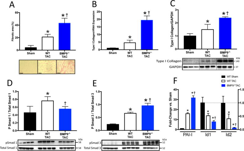
Figure 3. BMP9 Deficiency Increases Fibrosis and Promotes Smad3 Signaling Following 2 weeks of TAC.

A) Representative histological staining for collagen and quantitation of LV fibrotic area in WT and BMP9−/− mice after TAC. B–C) LV mRNA and protein expression of type I collagen normalized to GAPDH expression. D–E) Representative immunoblots and quantitation of phosphorylated Smad 1 (pSmad1), phosphorylated Smad 3 (pSmad3), and GAPDH. F) LV mRNA expression of inhibitor of differentiation 1 and 2 (Id1, Id2), and plasminogen activator inhibitor-1 (PAI-I). Data are expressed as fold change vs. Sham. p<0.05: *, vs. Sham; †, vs. WT TAC.
