Abstract
The sensitivity of many ion channels to phosphatidylinositol-4,5-bisphosphate (PIP2) levels in the cell membrane suggests that PIP2 fluctuations are important and general signals modulating neuronal excitability. Yet the PIP2 dynamics of central neurons in their native environment remained largely unexplored. Here, we examined the behavior of PIP2 concentrations in response to activation of Gq-coupled neurotransmitter receptors in rat CA1 hippocampal neurons in situ in acute brain slices. Confocal microscopy of the PIP2-selective molecular sensors tubbyCT-GFP and PLCδ1-PH-GFP showed that pharmacological activation of muscarinic acetylcholine (mAChR) or group I metabotropic glutamate (mGluRI) receptors induces transient depletion of PIP2 in the soma as well as in the dendritic tree. The observed PIP2 dynamics were receptor-specific, with mAChR activation inducing stronger PIP2 depletion than mGluRI, whereas agonists of other Gαq-coupled receptors expressed in CA1 neurons did not induce measureable PIP2 depletion. Furthermore, the data show for the first time neuronal receptor-induced oscillations of membrane PIP2 concentrations. Oscillatory behavior indicated that neurons can rapidly restore PIP2 levels during persistent activation of Gq and PLC. Electrophysiological responses to receptor activation resembled PIP2 dynamics in terms of time course and receptor specificity. Our findings support a physiological function of PIP2 in regulating electrical activity.
Introduction
Phosphatidylinositol-4,5-bisphosphate (PIP2) directly controls many cellular functions, including membrane and cytoskeletal dynamics and the activity of membrane proteins1–4. These regulatory effects of PIP2 are mediated by modulation of activity or of membrane association of PIP2-interacting proteins. In particular, many ion channels are highly sensitive to manipulation of PIP2 levels5,6.
In cell culture models activation of Gαq-coupled receptors can deplete the PIP2 content of the plasma membrane by activating phospholipase Cβ (PLCβ)1–3,7. Such PIP2 concentration changes were also observed in primary cultures of purkinje8 and hippocampal9–12 neurons. A series of thorough analyses of PIP2 signaling in response to muscarinic receptor activity in isolated sympathetic ganglion neurons (SGC)13–18, established that in these neurons activation of some Gq-coupled receptors leads to transient depletion of PIP2, which in turn inhibits Kv7.2/3-mediated M-currents. Hence, PIP2 depletion downstream of receptor-mediated pathways may be a ubiquitous principle controlling neuronal activity by modulating ion channels19,20. Thus it seems likely that the well-known increase of excitability by modulatory neurotransmitters, e.g. in hippocampal pyramidal neurons21,22, is mediated by deactivation of PIP2 -dependent channels23. There is also some evidence for PIP2-dependent regulation of Kir and K2P channels in striatal and thalamic neurons, respectively24,25. However, channel modulation may as well be mediated by other cellular signals downstream of Gq activity. Thus Kv7 channels are also inhibited by intracellular Ca2+ elevation26 and inhibition of Gq-sensitive TASK channels is mediated by DAG27. Both, Ca2+ and DAG signals may occur without a substantial drop in PIP2 downstream of PLCβ28–30.
Another issue is the method used to assess PIP2 dynamics. The standard approach is live-cell fluorescence microscopy using genetically encoded sensors built upon PIP2-binding domains fused to fluorescent proteins3. The most popular sensor domain also used by the studies cited above is the pleckstrin homology domain from PLCδ1 (PLCδ1-PH)31,32. However, interpretation of the observations is complicated by the IP3 affinity of PLCδ1-PH, compromising its suitability as a sensor of PIP2 following PLC activation33,34. More recently, the PIP2-specific tubbyCT domain enabled unequivocal measurement of Gαq-induced PIP2 depletion in cultured sympathetic and hippocampal neurons15,33. While these findings derived from isolated neurons are consistent with substantial PIP2 concentration changes, they might still not reflect physiological conditions. Studies in cardiac myocytes showed that PIP2 content can considerably differ between isolated cells and cells in situ35. Besides differences in the extent of PIP2 depletion, differences in time course may also have relevance for signaling via this pathway. Thus, knowledge of PIP2 dynamics in native neurons is required for understanding their physiological significance.
To address these issues, we characterized the PIP2 concentration behavior induced by activation of Gαq/PLC-coupled transmitter receptors in hippocampal CA1 pyramidal neurons in situ in acute brain slices. These neurons receive modulatory cholinergic input from the septohippocampal pathway36, which is mediated postsynaptically by Gq-coupled M1/M3 receptors and results in transient changes of excitability37–39. We find that mAChR and mGluRI receptors induce robust PIP2 depletion in soma and main apical dendrites. Strikingly, both receptors induced PIP2 oscillations. Moreover, PIP2 depletion was receptor and neuron type-specific. Correlation with changes in electrophysiological activity supports an instructive signaling role of these neuronal PIP2 dynamics.
Results
Muscarinic receptors mediate PIP2 dynamics in CA1 neurons in situ
We began our investigation of PIP2 dynamics in acute brain slices by examining the response of hippocampal CA1 pyramidal neurons to activation of their muscarinic ACh receptors. In order to measure Gαq induced PIP2 dynamics in situ, we expressed genetically encoded PIP2 sensors by stereotaxic injection of lentiviral expression vectors into the hippocampi of juvenile (P21) rats (see Methods). Two different GFP-fused sensor domains were used, tubbyCT-GFP15,40,41 and PLCd1-PH-GFP31,32. Both sensors work as ‘translocation sensors’, i.e. their degree of membrane association is a direct measure for PIP2 concentration and its temporal dynamics3.
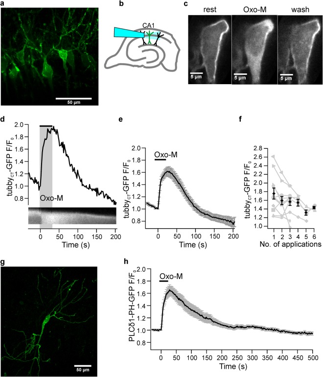
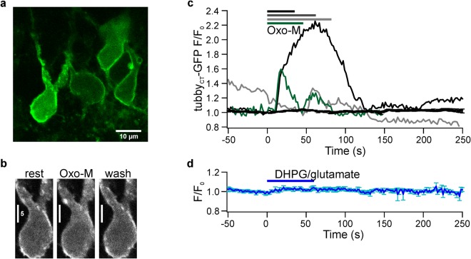
Fluorescence of neurons in acute slices from rats (P26-32) infected with the vector encoding tubbyCT-GFP indicated successful expression in CA1 pyramidal neurons. As shown in Fig. 1a GFP fluorescence was primarily localized to the plasma membrane of the soma and the dendritic tree.
Figure 1.
mAChR-mediated PIP2 depletion in acute brain slices. (a) Representative confocal image of CA1 pyramidal neurons expressing GFP-tagged tubbyCT PIP2 probes one week after injection of viral vector. (b) Schematic diagram of hippocampal slice and positioning of the application capillary in relation to pyramidal neurons in the CA1 area. (c) tubbyCT-GFP translocation during mAChR activation. Under resting conditions probes are associated with PIP2 at the membrane. Application of Oxo-M (10 µM) induced probe translocation to the cytosol indicating PIP2 depletion, followed by reassociation to the membrane indicating PIP2 recovery. (d) Representative somatic response to Oxo-M. Shown are cytoplasmatic fluorescence relative to fluorescence before stimulation (F/F0, upper panel) and a kymograph of probe translocation between membrane and cytosol (lower panel). (e) Average response from 28 individual neurons (26 slices; 16 rats). Application bar is set according to mean response latency. Reduced baseline fluorescence level at the end of the experiments in (d,e) results from photobleaching of GFP. (f) Translocation amplitudes upon repeated Oxo-M application (n = 11, 11, 11, 5, 4, and 3 for successive applications). Responses of individual neurons are shown in grey. (g) Confocal image (maximum intensity projection) of a representative CA1 pyramidal neuron expressing PLCδ1-PH-GFP. (h) Average translocation of PLCδ1-PH-GFP upon Oxo-M application (n = 16 neurons, 11 slices, 7 rats). Scale bars in (a,f): 50 µm. Contrast enhancement of 0.4% and 3% was applied to images shown in (c,d), respectively.
Activation of mAChR receptors by application of the specific agonist, oxotremorine-M (Oxo-M), induced massive and reversible translocation of the tubbyCT probes from the membrane to the cytoplasm in >95% of CA1 pyramidal cell somata examined, indicating strong PIP2 depletion (Fig. 1c). To quantify extent and time-course of probe translocation and hence PIP2 dynamics we measured fluorescence intensity changes in cytosolic ROIs, as cytosolic signals turned out to be less sensitive to tissue movement than when measuring from the small membrane compartment. Consequently an increase of fluorescence signal corresponds to probe dissociation from the membrane, indicating a loss of PIP2. Figure 1d shows a representative response to application of Oxo-M for 30 s. The mean tubbyCT translocation reached a peak cytoplasmic amplitude (F/F0) of 1.73 ± 0.06 (mean ± SEM; n = 28; 26 slices; 16 rats; Fig. 1e). Mean response latency was 8.8 ± 2.5 s and 90% of the peak response (t90) was reached within 19.9 ± 2.4 s. Upon washout of the agonist PIP2 levels recovered within about 100 s as indicated by re-association of the probe to the membrane. Cytoplasmic fluorescence returned to 10% of peak amplitude (t10, i.e. 90% recovery) in 65.1 ± 7.6 s. To explore the variability of muscarinic PIP2 dynamics, Oxo-M was applied repetitively with subsequent stimulations separated by a time interval of >10 minutes (Fig. 1f). On average, the degree of PIP2 depletion exhibited a slight but consistent decline in the course of repetitive stimulation. This is consistent with desensitization of muscarinic signaling previously observed in primary hippocampal neurons11.
Previous experiments with cultured neurons have used another sensor domain, PLCδ1-PH, to examine PIP2 dynamics9,17,18. In contrast to the tubbyCT sensor, however, PLCδ1-PH has a significant affinity for IP3, which is produced whenever PIP2 is cleaved by PLCβ33,34,40,42–44. Therefore the reliability of PLCδ1-PH as an indicator of PIP2 dynamics during GqPCR/PLC signaling has remained an unresolved issue33,34, which provided the rationale for choosing tubbyCT in the present study. Indeed, previous studies showed considerable differences in the behavior of tubbyCT-GFP and PLCδ1-PH-GFP sensors in terms of translocation following PLC activation15,34,40. Thus, we were interested in comparing both sensors in acute brain slices. When PLCδ1-PH-GFP was expressed in CA1 neurons (Fig. 1g), stimulation of mAChRs resulted in robust translocation of fluorescence in all neurons examined (Fig. 1h), similar to the results obtained with tubbyCT-GFP (PLCδ1-PH-GFP: latency 4.9 ± 1.2 s; t90 22.3 ± 1.9 s; F/F0 = 1.72 ± 0.07; n = 16, 11 slices, 7 rats). However, the recovery was significantly slower compared to tubbyCT-GFP (t10 = 132.7 ± 20.8 s; t-test p = 0.008), suggesting that responses of the PH sensor are co-determined by IP3 production.
Receptor-specific PIP2 depletion in CA1 neurons
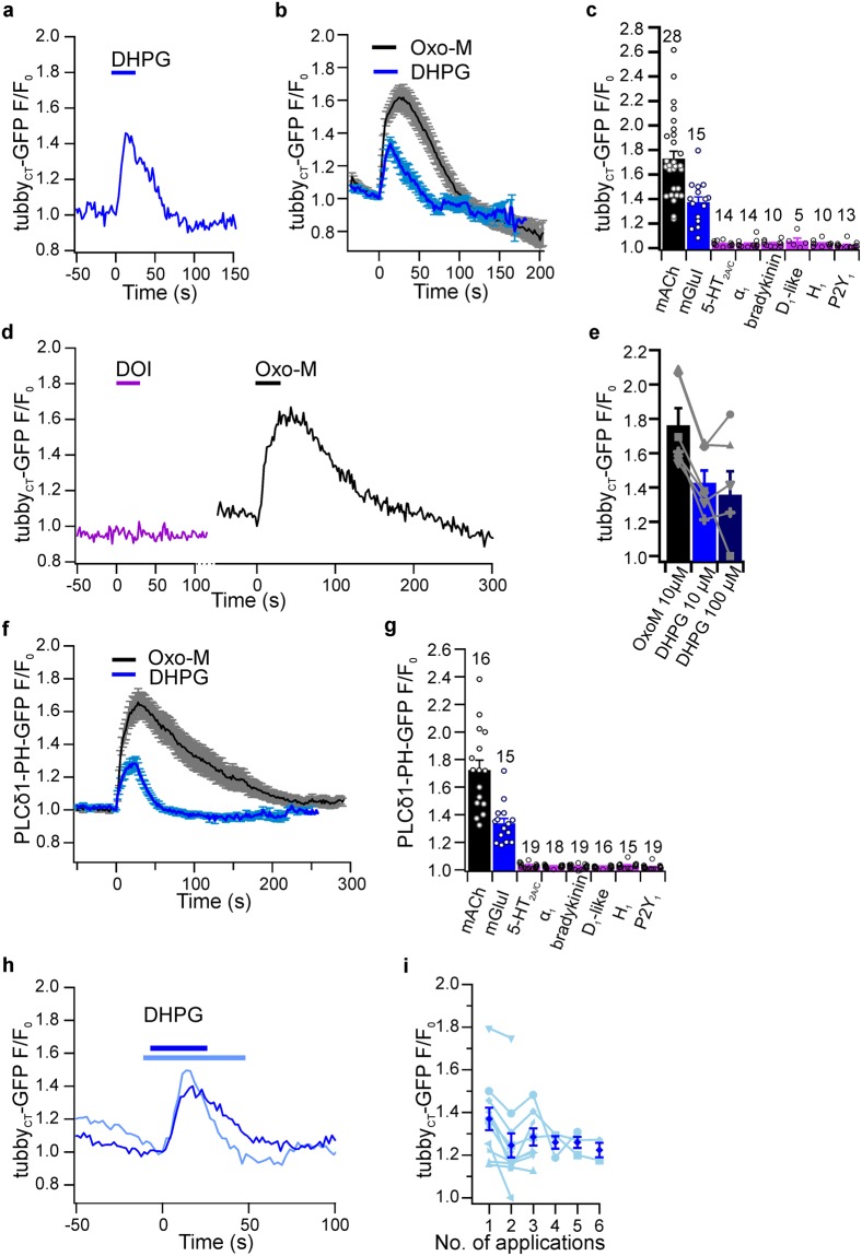
Activation of PLCβ and subsequent hydrolysis of PIP2 to IP3 and DAG is the main signaling pathway of Gαq-coupled receptors. Yet it is not known if PIP2 depletion is generally associated with the activation of Gαq coupled receptors other than M1/M3 receptors in central neurons. For example, in sympathetic ganglion neurons activity of muscarinic and purinergic receptors results in a depletion of PIP2, whereas bradykinin receptors generate IP3-dependent Ca2+ signals without substantial changes of the PIP2 concentration17,18,29,45,46. CA1 pyramidal neurons express various Gαq-coupled receptors which could potentially induce PIP2 depletion, including group I metabotropic glutamate receptor47, α1A-adrenoreceptor48, bradykinin B2 receptor49, Gq-coupled dopamineD1-like50–55, histamine H1 receptor56,57, P2Y1 receptor58,59, and 5-HT2A/2C receptors60–62. We applied specific agonists of group I mGluRs, 5-HT2A/2C receptors α1A adrenoreceptors, bradykinin receptors, Gq-coupled dopamineD1-like receptors, H1 histamine receptor, or P2Y1 receptor and monitored PIP2 concentrations with tubbyCT-GFP. Of these receptors, only mGluRs induced detectable probe translocation indicative for depletion of PIP2 (Fig. 2a). mGluRI-induced PIP2 depletion in neuronal somata was consistent across the population of neurons examined (F/F0 = 1.37 ± 0.05; n = 15; 14 slices from 11 rats; Fig. 2b).
Figure 2.
Somatic PIP2 dynamics are receptor specific. (a) Representative experiment showing PIP2 depletion in a CA1 soma in response to application of mGluRI agonist DHPG (10 µM) as determined by tubbyCT-GFP translocation. (b) Average PIP2 concentration changes obtained from 15 cells as in (a) (blue). Response to activation of mAChRs (grey) is redrawn from Fig. 1e for comparison (n = 28). (c) Summary of peak PIP2 depletion (translocation of tubbyCT-GFP) upon application of specific agonists for the Gq-coupled receptors indicated. Agonists applied were Oxo-M (10 µM, n = 28 neurons/26 slices/16 rats), DHPG (10 µM, n = 15/14/11), DOI, 10–20 µM, n = 14/14/6), methoxamine (10–20 µM; n = 14/14/6), bradykinin (10–20 µM, n = 10/10/4), SKF 83959 (10–20 µM, n = 5/5/3), 2-pyridylethylamin (10–50 µM, n = 10/10/4), ADPβS (10 µM; application for 30 to 60 s, n = 13/13/4). Numbers of experiments also indicated above bars. (d) Representative sensor responses measured from the same CA1 neuron during successive application of 5-HT2A/2C agonist DOI and mAChR agonist Oxo-M. (e) Response to higher concentration (100 µM) of DHPG did not increase tubbyCT-GFP translocation (application for 5 min each; n = 6, 6 slices, 3 rats). Successive responses of individual neurons shown in grey. (f) Average response of PLCδ1-PH-GFP sensor to mGluRI activation (blue, DHPG) and mAChR activation (black; Oxo-M; replotted from Fig. 1h). (g) Peak translocation of PLCδ1-PH-GFP sensor during application of agonists as in (c); Oxo-M (n = 16 neurons, 11 slices, 7 rats), DHPG (n = 15/11/7), DOI (n = 19/13/7), methoxamine (n = 18/12/7), bradykinin (n = 19/13/7), SKF 83959 (n = 16/11/6), 2-pyridylethylamin (n = 15/11/6), ADPβS (n = 19/13/7). (h) Example responses from two different neurons to application of DHPG (10 µM). Note that one response (dark blue) closely matches the average response while the other (light blue) shows pronounced recovery in the presence of the agonist. (i) Responses to repeated DHPG application (n = 11, 11, 8, 3, 3, and 2 for successive applications, respectively; delay between applications ≥ 10 min; individual responses indicated in light blue).
As summarized in Fig. 2c, PIP2 levels in CA1 neurons were insensitive to activation of any of the other receptors. Importantly, subsequent control application of Oxo-M triggered robust translocation of tubbyCT-GFP in each neuron, indicating proper responsiveness of the cell and appropriate sensitivity of the detection approach (Fig. 2d). Further, the prolonged application of each of the agonists (except muscarinic and glutamatergic) for up to 120 seconds or application of the endogenous ligands serotonin and dopamine did not evoke detectable responses (not shown). Equivalent results were obtained with neurons expressing the alternative PIP2 sensor domain, PLCδ1-PH-GFP. As shown in Fig. 2f and g, the stimulation of mAChR and mGluRI but none of the other receptors examined induced translocation of PLCδ1-PH-GFP. In summary, results obtained with both sensor domains indicate that α1A-adrenoreceptor, bradykinin, dopaminD1-like, histamin-H1, P2Y1, and 5-HT2A/2C do not induce significant PIP2 depletion in the soma of CA1 pyramidal neurons. Thus, PIP2 depletion is specific to mAChR and mGluRI, at least in the context of standard experimental conditions. However, response magnitude of mGluR activation was significantly lower compared to muscarinic PIP2 depletion in a population of cells challenged by both agonists (n = 12, 11 slices, 8 rats; paired t-test p = 0.032). While Oxo-M and DHPG have similar binding affinities for their cognate receptors63, EC50 values for downstream effects such as Ca2+ responses are often higher for DHPG than for Oxo-M, raising the possibility that 10 µM of DHPG might not be sufficient to evoke a saturating PIP2 response. However, increasing the concentration to 100 µM or the duration of agonist application of the glutamatergic agonist (DHPG) did not further increase the response mediated by mGluRs (Fig. 2e) and these responses were significantly smaller than muscarinic responses in the same neurons (p < 0.05, one way ANOVA followed by Tukey post-hoc test, n = 6, 6 slices, 3 rats).
In addition to the smaller responses, mGluRI-induced depletion of PIP2 also differed in its time course compared to muscarinic stimulation (Fig. 2a,b,f). As measured with tubbyCT-GFP, response latencies (6.81 ± 0.89 s) and rise time (t90 = 12.68 ± 1.15 s) where comparable, but time course of recovery was faster compared to mAChR activation (t10 = 28.38 ± 3.57 s; t-test p = 0.0001) Remarkably, in 6 out of 15 recordings, PIP2 levels recovered in the continued presence of the agonist DHPG, as illustrated by individual recordings shown in Fig. 2h. Similarly, PIP2 dynamics as measured with the PLCd1-PH-GFP probe showed a much faster recovery after activation of mGluRI (t10 = 30.44 ± 2.05 s, n = 15, 14 slices, 7 rats) when compared to mAChRs (t10 = 132.74 ± 20.77 s; t-test p = 0.0003; Fig. 2f). As noted with mAChR activation, PIP2 dynamics induced by mGluRIs showed slight desensitization in response to repeated application of the agonist (Fig. 2i).
Dendritic PIP2 dynamics
Next, we were interested in the spatial pattern of PIP2 depletion, in particular with respect to dendritic compartments. Because we probed PIP2 dynamics with translocation sensors that require microscopic resolution of membrane versus cytoplasm the measurements were confined to dendrites with a diameter of more than 1 µm that were localized close to the slice surface allowing for good optical access. Thus we achieved recordings from the main apical dendrite and its major branches up to 300 µm and basal dendrites to 20 µm distal to the soma. The distance between stratum pyramidale to fissura hippocampi defining the total length of apical dendrites was about 400 µm for our slices.
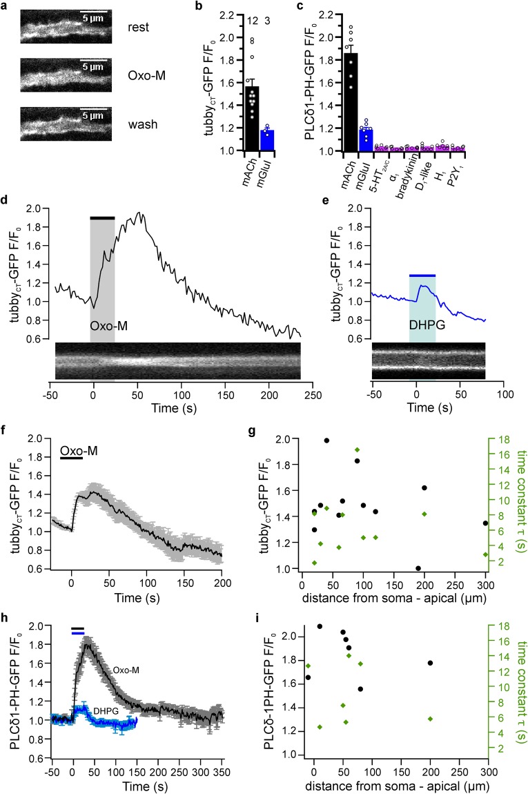
We found robust but receptor-specific PIP2 depletion in all dendritic compartments examined. Figure 3a shows a representative example illustrating membrane localization of tubbyCT-GFP in a dendrite and its transient redistribution into the dendritic cytosol during pharmacological activation of mAChRs, indicating reversible depletion of PIP2. With the exception of one apical dendrite (190 µm distance from soma), all dendrites examined (n = 12) responded with the translocation of tubbyCT-GFP. The time course of representative mAChR-induced dendritic PIP2 dynamics is further shown in Fig. 3d as a kymograph and quantitatively as the change of cytosolic fluorescence intensity. The average peak amplitude derived from dendrites of 12 neurons was F/F0 = 1.57 ± 0.06 (12 slices, 10 rats). The average latency of the responses was 14.29 ± 5.26 s. We observed a rise time t90 of 17.52 ± 2.67 s and 90% recovery time (t10) was 41.27 ± 7.25 s after the end of the application. As shown in Fig. 3a,e, activation of mGluRI also resulted in translocation of tubbyCT-GFP. However, the degree of PIP2 depletion was significantly weaker compared to activation of mAChRs (F/F0 = 1.18 ± 0.02; paired t-test, p = 0.041, n = 3 dendrites in 3 slices from 2 rats. Similar observations were made with PLCδ1-PH-GFP as the PIP2 probe (Fig. 3c,h). Thus, activation of muscarinic receptors by Oxo-M induced strong translocation (F/F0 = 1.86 ± 0.07; t90 = 21.86 ± 1.90 s; t10 = 142 ± 30.53 s; n = 7 dendrites, 7 slices, 3 rats), whereas activation of mGluRI receptors by DHPG induced small yet reproducible translocation of PLCdδ1-PH-GFP (F/F0 = 1.19 ± 0.02; n = 7). Stimulation of various other Gq-coupled receptors did not induce detectable translocation of PLCδ1-PH-GFP (Fig. 3c).
Figure 3.
Dendritic PIP2 dynamics. (a) Translocation of tubbyCT-GFP in response to Oxo-M application (10 µM) in an apical dendrite 60 µm from the soma. (b) Translocation of tubbyCT-GFP from dendritic membranes induced by mAChR (Oxo-M, 10 µM, n = 12 neurons/12 slices/10 rats) and mGluRI (DHPG, 10 µM, n = 3/3/2) activation, quantified as changes of axial (cytoplasmic) fluorescence changes relative to pre-stimulus fluorescence. (c) Translocation of PLCδ1-PH-GFP from dendritic membranes in response to activation of various Gq-coupled receptors. Agonists used as in Fig. 2 (n = 7 neurons, 7 slices, 3 rats for each condition). (d) Time course of PIP2 depletion from a dendritic recording 220 µm from the soma, shown as the relative increase of axial fluorescence (upper panel) and corresponding kymograph (lower panel). (e) Representative fluorescence change and kymograph of dendritic tubbyCT-GFP translocation in response to application of DHPG (10 µM; apical dendrite; 20 µm from soma). (f) Average time course of mAChR-induced dendritic PIP2 dynamics. (g) Amplitudes (black) and time constants (green) of tubbyCT-GFP translocation in response to application of Oxo-M plotted as a function of the distance of the recording location from the neuronal soma. Data from 13 individual neurons. (h) Average time course of dendritic PLCδ1-PH-GFP sensor translocation in response to application of the agonists indicated (n = 7 each). (i) Properties of translocation of PLCδ1-PH-GFP in response to Oxo-M plotted as a function of dendritic recording site relative to soma. Contrast enhancement of 10%, 1% and 3% was applied to images shown in a, d and e, respectively.
We wondered about spatial, basal-to-apical signaling gradients along the dendrites. Figure 3g,i display amplitudes and time constants of PIP2 depletion as a function of the distance from the soma. We find that neither parameter shows an evident trend along the dendritic distance, suggesting that PIP2 depletion is mostly homogenous throughout the larger dendritic compartments examined here (Pearson correlation coefficients: tubbyCT-GFP amplitudes, −0.071; tubbyCT-GFP time constants, 0.168;, PLCδ1-PH-GFP amplitudes, −0.209;, PLCδ1-PH-GFP time constants, −0.233).
Taken together, these results indicate that receptor-induced PIP2 dynamics in the larger dendritic compartments amenable to examination with translocation sensors is similar to the somatic dynamics. Specifically, activation of muscarinic receptors resulted in strong depletion of PIP2, glutamatergic receptors were considerably less effective, and PIP2 levels recovered rapidly.
Prolonged receptor activation revealed complexity of PIP2 dynamics
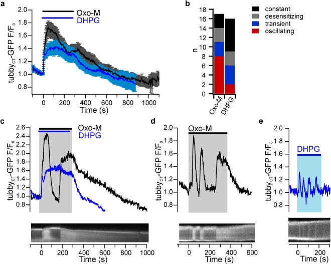
The occasionally observed early recovery of PIP2 level during activation of mGluRI prompted us to examine the time course of PIP2 dynamics during sustained stimulation. Figure 4 shows the resulting PIP2 dynamics measured with the tubbyCT sensor in neuronal somata during continuous application of the receptor agonists for 5 min. Notably, PIP2 depletion generally showed a phasic-tonic time course with an initial strong PIP2 depletion followed by partial recovery in the sustained presence of the agonist as apparent from the average from a larger number of neurons (n = 17 and 16 for mAChR and mGluRI activation, respectively; Fig. 4a). The initial rate of PIP2 decrease was similar for both receptors (t90 = 39.3 ± 5.3 s and 43.3 ± 8.8 s, respectively), but as previously seen with brief receptor stimulation (Fig. 2), the muscarinic responses had a higher average amplitude compared to glutamatergic responses (F/F0 = 1.84 ± 0.08 and 1.55 ± 0.07, respectively; paired t-test p = 0.0002, n = 16, 14 slices; 5 rats). PIP2 recovery after muscarinic receptor activation was more pronounced than for mGluR stimulation such that PIP2 levels tended towards similar values at the end of receptor stimulation period. For both receptors, recovery of PIP2 levels after removal of the receptor agonist was slower than observed with brief receptor stimulation (mAChR: t10 243.2 ± 43.9 s; mGluRI: t10 167.8 ± 41.5 s; cf. Fig. 2b) and thus depended on the duration of receptor activation.
Figure 4.
Prolonged receptor activation reveals PIP2 oscillations. (a) Average time course of somatic PIP2 dynamics during extended application of mAChR and mGluRI agonists Oxo-M (n = 17 neurons/15 slices/5 rats) and DHPG (n = 16/14/5) as measured by translocation of tubbyCT-GFP. (b) Distribution of distinct temporal patterns of tubbyCT-GFP sensor translocation within the population of neurons challenged by sustained application (5 min) of agonists. Responses were categorized as constant, desensitizing (partial recovery), transient (full recovery) and oscillating (≥2 peaks). (c) Distinct temporal behavior of PIP2 dynamics of an individual pyramidal neuron to successive applications of Oxo-M and DHPG. Lower panel shows a kymograph illustrating the response to Oxo-M. (d) Oscillatory PIP2 dynamics in a CA1 neuron in response to prolonged activation of mACh receptors. Note the pronounced repetitive phases of rapid PIP2 depletion and replenishment during stimulation. (e) Oscillatory PIP2 dynamics of a neuron in response to continuous activation of mGluRs. Contrast enhancement of 0.4%, 1% and 5% was applied to kymographic images shown in c, d and e, respectively.
While demonstrating partial desensitization of PIP2 responses as a common pattern, the prolonged stimulation also revealed considerable variability and complexity in the time course of PIP2 dynamics (Fig. 4b). Most strikingly, PIP2 depletion was often multiphasic or oscillatory. Examples for such complex PIP2 dynamics are shown in Fig. 4c–e. In these cells, PIP2 levels returned to baseline despite sustained presence of the agonist, and moreover, multiple depletion events occurred in rapid succession (Fig. 4d,e). Altogether, 8 out of 17 neurons showed oscillatory PIP2 concentration dynamics with Oxo-M and two out of 16 during application of DHPG. These observations suggest that PIP2 dynamics may be subject to complex temporal regulation and indicate potent PIP2 resynthesis capability of CA1 neurons during receptor activation.
Modulation of electrical behavior by receptor stimulation
The well-described effects of muscarinic activity on the electrical properties of CA1 neurons – including inhibition of M-currents – may (at least partially) be mediated by PIP2 concentration dynamics. We thus were interested in differential effects on excitability of the various Gq/PLC-coupled receptors examined for their coupling to PIP2 dynamics.
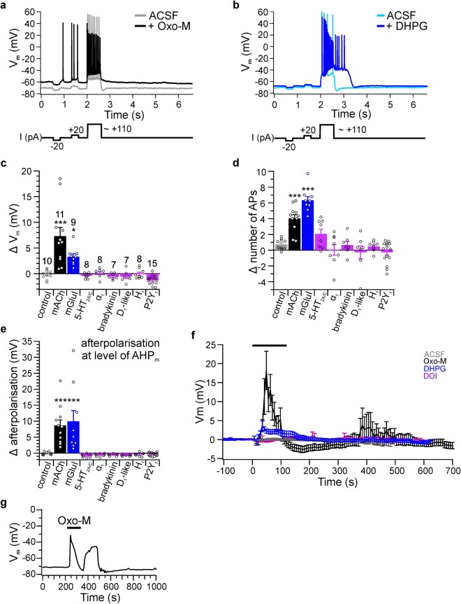
To this end, we performed patch clamp experiments in current clamp mode in acute brain slices prepared from rats at P14 to P21. Current step protocols were used to assess membrane potential, input resistance, spiking behavior, and afterpolarisation (Fig. 5a,b). Experiments were performed in the presence of inhibitors of GABAA/B and ionotropic glutamate receptors (see Methods) to exclude effects resulting from network activity.
Figure 5.
Electrophysiological responses to activation of Gq-coupled receptors. (a,b) Current clamp protocol (lower panel) and representative recordings of corresponding membrane potential (Vm) responses of CA1 pyramidal neurons before (light traces) and during (dark traces) activation of mAChRs or mGluRIs by 10 µM Oxo-M or DHPG, respectively. (c) Changes of resting membrane potential (ΔVm) displayed as the difference before and upon application of various GqPCR activators. Agonists applied were Oxo-M (10 µM; n = 10 neurons/10 slices from 7 rats; data points with plateau potentials were excluded for this analysis) for AChRs, DHPG (10 µM; n = 9/8/5) for mGluRIs, DOI (20 µM; n = 8/8/7) for 5-HT2A/C, methoxamine (20 µM; n = 8/8/7) for α1-adrenergic receptors, bradykinin (20 µM; n = 7/7/6), SKF 83959 (20 µM; n = 7/7/6) for D1-like dopamine receptors, 2-pyridylethylamin (20 µM; n = 8/8/7) for H1 histamine receptors, ADPβS (10 µM; n = 15/15/11) for P2Y1-R. Asterisks indicate significance of difference to control application of ACSF (n = 10/10/7) with p < 0.05 (*), 0.01 (**) and 0.001 (***; one-way ANOVA followed by Dunnett multiple comparison test). (d) Difference in number of action potentials (AP) triggered during 600 ms depolarizing current step. Numbers of experiments as in (c). (e) Changes in afterpolarisation where a negative Δ indicates an AHPm increase and positive Δ indicates an AHP reduction or afterdepolarisation. Numbers of experiments as in (c). (f) Mean time courses of Vm modulation during application of ACSF as a control (grey), Oxo-M (black), DHPG (blue), and DOI (purple). Vm was measured at the first 500 ms of each trace in the absence of spiking; see (a). (g) Example membrane voltage oscillation during mAChR activation in a P27 slice.
Overall, we found pronounced changes in electrical behavior following the activation of muscarinic mAChR receptors and mGluRI but little effects of other Gq-coupled receptors. Consistent with previous findings64–67 agonists of both mAChR (n = 11, 11 slices, 10 rats) and mGluRI (DHPG 10 µM, n = 9, 8 slices, 5 rats) induced depolarization of the resting membrane potential and an increase in firing frequency during depolarization (number of action potentials, NAP; Fig. 5a–d,f). In CA1 cells, a train of action potentials is usually followed by an afterhyperpolarization (AHP)37,68,69. Application of either Oxo-M or DHPG resulted in the disappearance of the AHP and the appearance of an afterdepolarisation (ADP; Fig. 5a,b,e). All of these receptor-induced changes are consistent with the deactivation of potassium conductances such as M currents37,68,70,71. In some neurons, activation of mAChR receptors induced sustained depolarization (plateau potentials) subsequent to the 600 ms current step, as also described previously72.
As shown in Fig. 5c–f, stimulation of other Gq-coupled receptors, including 5-HT2A/C receptors, α1 adrenergic receptors, bradykinin receptors, D1-like dopamine receptors, H1 histamine receptors, and purinergic P2Y1 receptors had no significant effects on either of these electrophysiological characteristics. Noteworthy, the mAChR and mGluRI induced depolarization did not always persist during the agonist application, as occasionally initial transient hyperpolarisation and oscillations of the membrane potential was observed during mAChR activation. During muscarinic stimulation, 9 of 11 neurons showed substantial recovery from depolarization in the presence of Oxo-M; of those, 6 neurons showed complete repolarization or even hyperpolarisation before washout. In the presence of DHPG, 3 neurons showed a substantial recovery from initial depolarization. Oscillatory behavior was observed in neurons from younger animals (P14-21), but also in slices age-matched to the PIP2 imaging experiments, as shown in an exemplary recording (Fig. 5g) obtained from a neuron in a P27 slice.
In summary, we find that pronounced changes in membrane potential and firing rates paralleled neuronal PIP2 depletion in terms of effect size, time course and receptor specificity. Thus, depolarization, increased spike rates and PIP2 dynamics were largely restricted to the activation of mAChR and mGluRI receptors.
Neuron type-specific PIP2 dynamics: dentate gyrus granule cells
To extend our observations on PIP2 dynamics to additional neuronal cell types, we measured PIP2 dynamics following activation of the same receptors (mAChR and mGluRI) in dentate gyrus granule neurons (Fig. 6a). Granule neurons express both mAChR and mGluRI receptors at the soma47,73,74. However, detectable depletion of PIP2 upon stimulation with Oxo-M was observed in only three independent experiments (n = 24; Fig. 6b,c). None of the six neurons stimulated with DHPG (10 µM, n = 2) or Glutamate (100 µM, n = 4) showed any detectable sensor translocation (Fig. 6d). Thus, the induction of PIP2 dynamics may be highly specific between different types of neurons and this specificity seems to be dependent on mechanisms other than the expression of Gq/PLC-coupled transmitter receptors.
Figure 6.
Dentate gyrus granule neurons largely lack receptor-induced PIP2 depletion. (a) Confocal image of gyrus dentatus granule neurons in an acute hippocampal slice, expressing tubbyCT-GFP after sterotactic injection of lentiviral vector. (b) Confocal images demonstrating a minor degree of probe translocation (1% contrast enhanced; first neuron from left in (a)). (c) Only 3 out of n = 24 neurons showed mAChR-induced PIP2 depletion, as indicated by sensor translocation (3 individual neurons from separate animals). Black trace shows mean fluorescence time course of 21 non-responsive neurons. Time 0 indicates start of the application shown by bars. (d) Application of the mGluRI agonist DHPG (n = 2 neurons, both of which were responsive to Oxo-M) or glutamate (n = 4) did not induce PIP2 depletion.
Discussion
Direct observation of PIP2 dynamics in central neurons in situ
While there is good evidence for PIP2 depletion in response to activation of Gq-coupled receptors for some types of neurons studied in the cell culture dish, surprisingly little is known about the prevalence and spatiotemporal properties of PIP2 dynamics in central neurons under physiological conditions. PIP2 levels and their dynamic regulation may be largely different in vivo, as embedding in the native environment and full differentiation of neurons may impact on relevant factors such as expression and spatial subcellular organization of receptors and downstream components of the signaling cascade and the enzymes that resynthesize PIP2. Therefore, information on PIP2 concentration behavior in intact tissue preparations such as brain slices is required. Previous studies with organotypic slices were consistent with PIP2 depletion in-situ triggered by synaptic release of glutamate onto cerebellar Purkinje neurons75 or by muscarinic agonist application in cortical pyramidal cells76. However, both studies were not fully conclusive with respect to PIP2 signaling because PLCδ1-PH was used as a sensor domain, which has a similar affinity for PIP2 and IP3 and may report the production of IP3 rather than depletion of PIP244,77. In fact, Okubo et al.75 interpreted probe translocation in terms of IP3 production rather than depletion of PIP2.
Here, by using tubbyCT, as an alternative PIP2 sensor insensitive to IP333,34,40, our present data now show unequivocally that muscarinic and metabotropic glutamate receptors indeed trigger PIP2 dynamics in a prototypic central neuron in situ. Of note, in cultured cell lines tubbyCT previously failed to respond or only weakly translocated upon PLC-mediated PIP2 depletion34,40, which initially was attributed to a higher affinity for PIP2 compared to PLCδ1-PH. However, quantitative titration of PIP2 in living cells showed that its affinity to PIP2 is actually lower, which should make it a useful PIP2 sensor41. In fact, our current results demonstrate that tubbyCT readily translocates in a neuronal cellular environment. The difference in behavior between experimental conditions is not yet understood, but may suggest cell type-specific segregation of PIP2 into distinct pools selectively accessible to the different PIP2-binding domains78. In any case, our findings show that in the native neuronal system tubbyCT is a much better reporter of PIP2 dynamics than might have been anticipated20. Thus, using tubbyCT-GFP allowed us to systematically assay PIP2 dynamics without confounding effects of IP3 dynamics.
Spatiotemporal properties of PIP2 dynamics
We found that in the larger compartments accessible to measuring sensor translocation, receptor-induced PIP2 depletion appeared largely homogenous without evidence for substantial subcellular differences. This suggested that induced PLC activity is similar in somatic and dendritic compartments. This finding was not entirely expected, because while M1 receptors show high densities throughout soma and dendrites73, the most prominent mGluRI receptor of CA1 neurons, mGluR5, has a relatively low density in soma compared to dendrites47. Since glutamatergic PIP2 depletion in dendrites was not stronger than in the somatic compartment, the PIP2 depletion pattern does not appear to correlate closely with receptor distribution. It is worth noting that the moderate degree of PIP2 sensor translocation in dendrites did not result from the distinct dendritic geometry, because the smaller volume-to-membrane area ratio in the dendrites should rather result in a stronger relative increase of sensor fluorescence when sensors dissociate from the membrane. Also, muscarinic stimulation elicited larger dendritic responses than glutamatergic stimulation, showing that PIP2 sensor response was not saturated by mGluR stimulation. Of note, a similar observation was made by Nakamura et al.79 for Ca2+ dynamics in CA1 pyramidal cells: activation of mGluRI, mAChR and 5-HT2R elicited comparable Ca2+ waves despite different receptor distribution. Thus, these neurons may possess mechanisms to globalize Gq signaling including PIP2 depletion.
To our knowledge our results for the first time demonstrate oscillations of the PIP2 concentration in a neuron. While IP3, DAG and Ca2+ are known to undergo oscillatory concentration dynamics in neurons80, previous observations on PIP2 dynamics in primary dissociated neurons16,18,33 seemed to indicate that PIP2 concentrations essentially remained depleted during prolonged receptor activity. Oscillatory translocation of the PLCδ1-PH sensor domain observed occasionally has been understood as dynamics of the IP3 signal picked up by the PH domain23,44,81. More recently, careful observations also including the specific tubbyCT sensor showed bona-fide oscillations of PIP2 in mast cells82,83. Our observations suggest that such dynamics may be a more general phenomenon with implications for neuronal biology.
The mechanisms underlying PIP2 oscillations may include both positive and negative feedback regulation of PIP2 cleavage by PLC. Such mechanisms have previously been shown to be involved in Ca2+ and IP3 oscillations and include Ca2+-dependent activation of PLC80,81 as a positive feedback. In mast cells, PIP2 oscillations are probably driven by Ca2+ oscillations82. Inhibition of Gq signaling by, e.g., PKC, receptor kinases, or RGS molecules81,84 may contribute to a negative feedback loop controlling PIP2 degradation. Moreover, our observations reveal an impressive capability of PIP2 replenishment, as indicated by the rapid and complete recovery of PIP2 levels in presence of agonists. PIP2 resynthesis may be increased during GqPCR activation85, providing negative feedback to PIP2 depletion and possibly contributing to observed oscillations. Specifically, PIP2 replenishment may involve Ca2+ and phosphatidic acid-dependent phospholipid exchange at plasma membrane-endoplasmic reticulum (PM-ER) junctions86.
Whatever the mechanism underlying the oscillations is, our findings indicate that PIP2 dynamics may provide neurons with another dimension of effector modulation beyond a simple on/off switch for downstream effectors. Although the consequences of PIP2 oscillations for electrical neuronal activity remain to be explored, we note that indeed, neurons showed fluctuations of membrane potential and firing frequency during agonist application. It is worth mentioning that mAChR and mGluRI agonists can induce and shift gamma and theta oscillations87. In the light of the present data, it is tempting to speculate that PIP2 oscillations might participate in such frequency modulation.
Neuronal ion channels as effectors of PIP2 dynamics
Given the known high sensitivity of some ion channels to even a moderate drop in the PIP2 concentration5,78, a main potential target of PIP2 depletion are ion channels and thus electric excitability. Based on studies on isolated neurons, inhibition of Kv7 channels in sympathetic neurons as the direct consequence of PIP2 depletion is well established13,17,29. Our data permit the correlation of PIP2 dynamics and electrophysiology in situ. Activation of mAChR and mGluRI, but not other PLC-coupled receptors known to be present and functional in CA1 neurons induced robust PIP2 depletion. The same pattern of receptor specificity was observed for modulation membrane potential, firing frequency and afterhyperpolarization, providing at least circumstantial evidence for the causation of channel regulation by PIP2. Simultaneous recordings of electrical activity and PIP2 dynamics from the same neuron should be performed in the future to provide more direct evidence.
In conclusion, our data support and generalize the as yet largely hypothetical mechanism of PIP2 dynamics as a major cellular signal in the control of neuronal activity through regulation of PIP2-sensitive ion channels such as Kv7. Future studies need to address this issue rigorously by manipulating PIP2 levels in-situ20. Along those lines a recent study aimed at PIP2 depletion in hippocampal slice cultures by chemically induced recruitment of a PIP2 phosphatase88. While this approach did not reveal any effects on electrical properties of the neurons, the results appear inconclusive since changes in PIP2 concentration were not verified.
One of the most intriguing unknowns are the spatiotemporal properties of PIP2 dynamics during entirely physiological neuronal activity, i.e, during synaptic activity of the modulatory (e.g. cholinergic) and principal (i.e. glutamatergic) inputs into the hippocampal neurons and of the PIP2 dynamics associated with intrinsic neural (network) activity. Another question is the PIP2 signaling in the distal smaller dendritic compartments not amenable to analysis by the translocation probes used in this study. In particular, in the immediate postsynaptic compartment, i.e. spines, PIP2 may have a role in controlling synaptic plasticity89–91.
Materials and Methods
Virus production and constructs
Lentiviral plasmids pCMVΔR8.9, pVSVG and FUGW were kindly provided by Pavel Osten (MPI for medical research Heidelberg, Germany). The PLCδ1-PH and tubbyCT constructs were provided by Tamás Balla (NIH, Bethesda, USA) and Lawrence Shapiro (Columbia University, USA), respectively. Lentiviral particles were derived by triple transfection of HEK293FT cells with Lipofectamin 2000 (Invitrogen, Darmstadt, Germany). Virus purification from supernatant was achieved by 15 minute centrifugation at 3000 rpm, filtration through a Millex ® HV 0.45 µm filter (Millipore, Darmstadt, Germany) and two successive ultracentrifugation steps (25000 rpm, 1 h 30 min, 4 °C). Pellets were resuspended in TBS-5 buffer (50 mM Tris-HCl, 130 mM NaCl, 10 mM KCl, 5 mM KCl2) and subjected to a final 30 s centrifugation at 5000 rpm. Aliquots were stored at −80 °C and thawed up to two times.
Animals, stereotactic injection and slice preparation
Wistar rats were obtained from the animal facility of the Philipps University of Marburg (Marburg, Germany) or Charles River (Cologne, Germany) and kept and handled according to German law and institutional guidelines at the Philipps University. All procedures were approved by the Regierungspräsidium Giessen, Germany. Animals were housed with access to ad libitum water and food on a 12-h light/dark cycle. At weaning (postnatal day 21) male and female rats were anesthetized by intraperitoneal injection of a mixture of ketamine (Bela-Pharm, Vechta, Germany) and xylazine (Rompun®, Bayer AG, Leverkusen, Germany) at a dose of 100 and 10 mg per kilogram body weight. Additionally, the mixture included 0.05 mg/kg Atropine (B. Braun, Melsungen, Germany) and 0.1 ml/10 g body weight of a 0.9% NaCl solution for injection (Diaco, Triest, Italy). Under stereotactic control, lentivirus was injected bilaterally using Paxinos and Watson92 as a reference. Coordinates were optimized for targeting in juvenile rats by setting the adult references to x = +/−6.125, y = −6.15 and z = −6.2 mm and multiplying by the ratio of the juvenile to atlas (8.7 mm) distance of bregma to lambda. Up to 2.5 μl virus per hemisphere were injected in 500 nl portions going from ventral to dorsal in 0.3–0.35 mm steps during 5–10 minutes. For imaging and electrophysiological experiments, rats were anesthetized with Isoflurane (Baxter, Unterschleißheim, Germany) or Sevoflurane (Sevorane®, Abbott, Wiesbaden, Germany) and sacrificed by decapitation at the ages indicated in results. The head was placed in ice cold sucrose-ACSF (sucrose-artificial cerebrospinal fluid, in mM: 87 NaCl, 25 NaHCO3, 25 D-glucose, 75 sucrose, 2.5 KCl, 0.5 CaCl2 and 7 MgCl2, oxygenated with 95% O2/5% CO2) and the hippocampi rapidly removed. 300 μm transversal slices were cut with a vibratome (VT1200, Leica Biosystems, Wetzlar, Germany) and placed into a chamber with 4 °C sucrose-ACSF. After a 35 min recovery period at 35 °C slices were kept at room temperature. For recordings slices were transferred to a submerged chamber and perfused with ACSF (in mM: 125 NaCl, 25 NaHCO3, 25 D-glucose, 2.5 KCl, 2 CaCl2 and 1 MgCl2, oxygenated with 95% O2/5% CO2) for at least 20 minutes.
Imaging, electrophysiological recording and data analysis
Confocal imaging was performed with a Zeiss LSM710 (Zeiss, Oberkochen, Germany). The sampling rate for time series experiments was 1.75 s with a pixel size of 0.13 µm. In some cases (especially dendrite measurements) the sampling rate was increased to 1 s. In all cases where the sampling rate slightly deviated the data were resampled to allow averaging across experiments. Overlay with the original was performed to ensure preservation of time scale. Average cytoplasmic fluorescence intensities were determined from regions of interest (ROI) excluding both the plasma membrane (defined as the local intensity max at the cell’s border in the resting cell) and the nucleus. Distance of ROIs to the plasma membrane was >0.5 µm even when slight shifts of the cell’s position occurred during the experiment. ROIs were defined post-hoc using the microscope software ZEN (Versions 2008 and 2009) and obtained average intensities were exported to Igor Pro (Version 6.03 A, Wave Metrics, Portland, OR USA). Traces were background subtracted and normalized to the last time point before beginning of a response (F/F0 normalized to t0). Measurements without evident response were corrected for photobleaching according to a biexponential fit to the decaying fluorescence signal and normalized to signal at the onset of agonist application. We found that probe translocation generally prevented the reliable determination of the time course of photobleaching. Therefore most data were not corrected for bleaching which results in apparently lower signals following transient depletion of PIP2, with bleaching generally being more pronounced for tubbyCT-GFP than for PLCδ1-PH-GFP. Confocal images were further analyzed with ImageJ (National Institutes of Health, USA) to isolate individual images of a time series, create kymographs and set scale bars. Electrophysiological data were recorded with a HEKA EPC10USB amplifier and Patch Master software (Version 2.43 HEKA, Lambrecht, Germany) in current clamp mode. Data were low pass filtered with a 2.9 kHz Bessel filter and digitized at 20 kHz. Borosilicate recording pipettes had a resistance of 3–4 MΩ and were filled with intracellular solution containing (in mM): K-gluconate 135, KCl 20, MgCl2 2, Na2-ATP 2, Na2-GTP 0.3, HEPES 10 and EGTA 0.1 (adjusted to pH 7.2 with KOH). Series resistance was monitored in voltage clamp mode before and after each current clamp recording, but not corrected for. Measurements with a change in series resistance >40% during the course of the experiment were discarded. Input resistance was assessed by injection of small positive and negative currents steps, followed by a depolarizing current step above action potential threshold to quantify spiking behavior and afterpolarisation (see Fig. 5a,b). Sweep length was 7 seconds. In applications of the P2Y1 agonist ADPβS a shorter protocol without the positive 20 pA step was used. Medium afterhyperpolarisation (AHPm) was obtained as the difference of resting Vm and mean Vm at 70 to 120 ms after the depolarizing current step. Changes in AHP value resulting from application of receptor agonists are given as Δafterpolarization such that positive values indicate reduction of AHP or eventually the emergence of an afterdepolarisation. Amplitudes were calculated from averaging at least 10 baseline data points and a minimum of 3 peak points, with avoidance of plateau potentials.
Statistical analysis
Statistical significance was tested in Igor Pro. Randomness, equal variances and normal distribution of the data was tested with Igor’s Runs, Kolmogorow-Smirnow and Jarque-Bera test. In cases where validity of a parametric test was compromised, a Wilcoxon-Mann-Whitney test was performed. Where applicable, groups of two were compared with paired and unpaired Student’s t. Two tailed one-way ANOVA was followed by a Dunett test for comparing multiple groups to a single control or a Tukey test to compare all groups to each other. Unless noted otherwise all values are given ± standard error of the mean.
Chemicals and perfusion system
Oxotremorine-M, DHPG, Bradykinin, SKF83959, DOI, Serotonin and Dopamine were purchased from Tocris and Methoxamine and 2-Pyridylehylamin from Sigma. All other chemicals were from Sigma/Fluka or Merck (Germany). For application of test substances a capillary of 200 to 250 µM inner diameter (TSP200350, BGB Analytik AG, Boeckten, Germany or MicroFil MF28G-5, World Precision Instruments, Berlin, Germany) was placed directly next to the hippocampal recording region. Solution exchange at the tip occurred within 1–2 s. Unless noted otherwise recordings represent first applications of each test substance. To block fast glutamatergic and GABAA/B signaling in electrophysiological recordings, receptor antagonists (4 μM NBQX, 50 μM D-AP5, 50 μM Picrotoxin and 1 μM CGP 55845, all from Tocris) were added both to the bath and local perfusion.
Acknowledgements
We like to thank Olga Ebers for excellent technical assistance. This work was supported by grants from the Deutsche Forschungsgemeinschaft to D.O. (OL 240/1–1/2 and SFB 593 TP12).
Author Contributions
D.O. and S.H. designed the study. S.H. performed the experiments and analyzed the data. S.H. and D.O. wrote the manuscript. All authors approved of the manuscript.
Data availability
Most data generated or analysed during this study are included in this published article. Additional datasets generated and analysed during the current study are available from the corresponding authors on reasonable request.
Competing Interests
The authors declare no competing interests.
Footnotes
Publisher's note: Springer Nature remains neutral with regard to jurisdictional claims in published maps and institutional affiliations.
References
- 1.Falkenburger BH, Jensen JB, Hille B. Kinetics of PIP2 metabolism and KCNQ2/3 channel regulation studied with a voltage-sensitive phosphatase in living cells. J. Gen. Physiol. 2010;135:99–114. doi: 10.1085/jgp.200910345. [DOI] [PMC free article] [PubMed] [Google Scholar]
- 2.Di Paolo G, De Camilli P. Phosphoinositides in cell regulation and membrane dynamics. Nature. 2006;443:651–7. doi: 10.1038/nature05185. [DOI] [PubMed] [Google Scholar]
- 3.Balla T, Szentpetery Z, Kim YJ. Phosphoinositide signaling: new tools and insights. Physiology (Bethesda). 2009;24:231–44. doi: 10.1152/physiol.00014.2009. [DOI] [PMC free article] [PubMed] [Google Scholar]
- 4.Balla T. Phosphoinositides: Tiny Lipids With Giant Impact on Cell Regulation. Physiol. Rev. 2013;93:1019–1137. doi: 10.1152/physrev.00028.2012. [DOI] [PMC free article] [PubMed] [Google Scholar]
- 5.Suh B-C, Hille B. PIP2 Is a Necessary Cofactor for Ion Channel Function: How and Why? Annu. Rev. Biophys. 2008;37:175–195. doi: 10.1146/annurev.biophys.37.032807.125859. [DOI] [PMC free article] [PubMed] [Google Scholar]
- 6.Logothetis DE, Petrou VI, Adney SK, Mahajan R. Channelopathies linked to plasma membrane phosphoinositides. Pflugers Arch. 2010;460:321–41. doi: 10.1007/s00424-010-0828-y. [DOI] [PMC free article] [PubMed] [Google Scholar]
- 7.Horowitz LF, et al. Phospholipase C in living cells: activation, inhibition, Ca2+ requirement, and regulation of M current. J. Gen. Physiol. 2005;126:243–62. doi: 10.1085/jgp.200509309. [DOI] [PMC free article] [PubMed] [Google Scholar]
- 8.Okubo Y, Kakizawa S, Hirose K, Iino M. Visualization of IP(3) dynamics reveals a novel AMPA receptor-triggered IP(3) production pathway mediated by voltage-dependent Ca(2+) influx in Purkinje cells. Neuron. 2001;32:113–22. doi: 10.1016/S0896-6273(01)00464-0. [DOI] [PubMed] [Google Scholar]
- 9.Micheva KD. Regulation of presynaptic phosphatidylinositol 4,5-biphosphate by neuronal activity. J. Cell Biol. 2001;154:355–368. doi: 10.1083/jcb.200102098. [DOI] [PMC free article] [PubMed] [Google Scholar]
- 10.Nahorski SR, Young KW, John Challiss RA, Nash MS. Visualizing phosphoinositide signalling in single neurons gets a green light. Trends Neurosci. 2003;26:444–52. doi: 10.1016/S0166-2236(03)00178-4. [DOI] [PubMed] [Google Scholar]
- 11.Willets JM, Nash MS, Challiss RAJ, Nahorski SR. Imaging of muscarinic acetylcholine receptor signaling in hippocampal neurons: evidence for phosphorylation-dependent and -independent regulation by G-protein-coupled receptor kinases. J. Neurosci. 2004;24:4157–62. doi: 10.1523/JNEUROSCI.5506-03.2004. [DOI] [PMC free article] [PubMed] [Google Scholar]
- 12.Nash MS, Willets JM, Billups B, John Challiss RA, Nahorski SR. Synaptic Activity Augments Muscarinic Acetylcholine Receptor-stimulated Inositol 1,4,5-Trisphosphate Production to Facilitate Ca2+ Release in Hippocampal Neurons. J. Biol. Chem. 2004;279:49036–49044. doi: 10.1074/jbc.M407277200. [DOI] [PubMed] [Google Scholar]
- 13.Suh B-C, Hille B. Recovery from Muscarinic Modulation of M Current Channels Requires Phosphatidylinositol 4,5-Bisphosphate Synthesis. Neuron. 2002;35:507–520. doi: 10.1016/S0896-6273(02)00790-0. [DOI] [PubMed] [Google Scholar]
- 14.Zaika O, et al. Angiotensin II regulates neuronal excitability via phosphatidylinositol 4,5-bisphosphate-dependent modulation of Kv7 (M-type) K+ channels. J. Physiol. 2006;575:49–67. doi: 10.1113/jphysiol.2006.114074. [DOI] [PMC free article] [PubMed] [Google Scholar]
- 15.Hughes S, Marsh SJ, Tinker A, Brown DA. PIP(2)-dependent inhibition of M-type (Kv7.2/7.3) potassium channels: direct on-line assessment of PIP(2) depletion by Gq-coupled receptors in single living neurons. Pflugers Arch. 2007;455:115–24. doi: 10.1007/s00424-007-0259-6. [DOI] [PubMed] [Google Scholar]
- 16.Kruse M, Vivas O, Traynor-Kaplan A, Hille B. Dynamics of Phosphoinositide-Dependent Signaling in Sympathetic Neurons. J. Neurosci. 2016;36:1386–1400. doi: 10.1523/JNEUROSCI.3535-15.2016. [DOI] [PMC free article] [PubMed] [Google Scholar]
- 17.Gamper N, Reznikov V, Yamada Y, Yang J, Shapiro MS. Phosphotidylinositol 4,5-Bisphosphate Signals Underlie Receptor-Specific Gq/11-Mediated Modulation of N-Type Ca2+ Channels. J. Neurosci. 2004;24:10980–10992. doi: 10.1523/JNEUROSCI.3869-04.2004. [DOI] [PMC free article] [PubMed] [Google Scholar]
- 18.Winks JS, et al. Relationship between Membrane Phosphatidylinositol-4,5-Bisphosphate and Receptor-Mediated Inhibition of Native Neuronal M Channels. J. Neurosci. 2005;25:3400–3413. doi: 10.1523/JNEUROSCI.3231-04.2005. [DOI] [PMC free article] [PubMed] [Google Scholar]
- 19.Gamper N, Shapiro MS. Regulation of ion transport proteins by membrane phosphoinositides. Nat. Rev. Neurosci. 2007;8:921–34. doi: 10.1038/nrn2257. [DOI] [PubMed] [Google Scholar]
- 20.Leitner MG, Halaszovich CR, Ivanova O, Oliver D. Phosphoinositide dynamics in the postsynaptic membrane compartment: Mechanisms and experimental approach. Eur. J. Cell Biol. 2015;94:401–414. doi: 10.1016/j.ejcb.2015.06.003. [DOI] [PubMed] [Google Scholar]
- 21.Halliwell JV, Adams PR. Voltage-clamp analysis of muscarinic excitation in hippocampal neurons. Brain Res. 1982;250:71–92. doi: 10.1016/0006-8993(82)90954-4. [DOI] [PubMed] [Google Scholar]
- 22.Dutar P, Nicoll RA. Classification of muscarinic responses in hippocampus in terms of receptor subtypes and second-messenger systems: electrophysiological studies in vitro. J. Neurosci. 1988;8:4214–4224. doi: 10.1523/JNEUROSCI.08-11-04214.1988. [DOI] [PMC free article] [PubMed] [Google Scholar]
- 23.Young KW, et al. Muscarinic acetylcholine receptor activation enhances hippocampal neuron excitability and potentiates synaptically evoked Ca(2+) signals via phosphatidylinositol 4,5-bisphosphate depletion. Mol. Cell. Neurosci. 2005;30:48–57. doi: 10.1016/j.mcn.2005.05.006. [DOI] [PubMed] [Google Scholar]
- 24.Shen W, et al. Cholinergic modulation of Kir2 channels selectively elevates dendritic excitability in striatopallidal neurons. Nat. Neurosci. 2007;10:1458–1466. doi: 10.1038/nn1972. [DOI] [PubMed] [Google Scholar]
- 25.Bista P, et al. Differential phospholipase C-dependent modulation of TASK and TREK two-pore domain K+ channels in rat thalamocortical relay neurons. J. Physiol. 2015;593:127–144. doi: 10.1113/jphysiol.2014.276527. [DOI] [PMC free article] [PubMed] [Google Scholar]
- 26.Gamper N, Shapiro MS. Calmodulin Mediates Ca2+ -dependent Modulation of M-type K+ Channels. J. Gen. Physiol. 2003;122:17–31. doi: 10.1085/jgp.200208783. [DOI] [PMC free article] [PubMed] [Google Scholar]
- 27.Wilke, B. U. et al. Diacylglycerol mediates regulation of TASK potassium channels by Gq-coupled receptors. Nat Commun5 (2014). [DOI] [PubMed]
- 28.Delmas P, Wanaverbecq N, Abogadie FC, Mistry M, Brown DA. Signaling Microdomains Define the Specificity of Receptor-Mediated InsP3 Pathways in Neurons. Neuron. 2002;34:209–220. doi: 10.1016/S0896-6273(02)00641-4. [DOI] [PubMed] [Google Scholar]
- 29.Brown DA, Hughes SA, Marsh SJ, Tinker A. Regulation of M(Kv7.2/7.3) channels in neurons by PIP(2) and products of PIP(2) hydrolysis: significance for receptor-mediated inhibition. J. Physiol. 2007;582:917–25. doi: 10.1113/jphysiol.2007.132498. [DOI] [PMC free article] [PubMed] [Google Scholar]
- 30.Falkenburger BH, Dickson EJ, Hille B. Quantitative properties and receptor reserve of the DAG and PKC branch of Gq-coupled receptor signaling. J. Gen. Physiol. 2013;141:537–555. doi: 10.1085/jgp.201210887. [DOI] [PMC free article] [PubMed] [Google Scholar]
- 31.Stauffer TP, Ahn S, Meyer T. Receptor-induced transient reduction in plasma membrane PtdIns(4,5)P2 concentration monitored in living cells. Curr. Biol. 1998;8:343–6. doi: 10.1016/S0960-9822(98)70135-6. [DOI] [PubMed] [Google Scholar]
- 32.Várnai P, Balla T. Visualization of phosphoinositides that bind pleckstrin homology domains: calcium- and agonist-induced dynamic changes and relationship to myo-[3H]inositol-labeled phosphoinositide pools. J. Cell Biol. 1998;143:501–10. doi: 10.1083/jcb.143.2.501. [DOI] [PMC free article] [PubMed] [Google Scholar]
- 33.Nelson CP, Nahorski SR, Challiss RAJ. Temporal profiling of changes in phosphatidylinositol 4,5-bisphosphate, inositol 1,4,5-trisphosphate and diacylglycerol allows comprehensive analysis of phospholipase C-initiated signalling in single neurons. J. Neurochem. 2008;107:602–15. doi: 10.1111/j.1471-4159.2008.05587.x. [DOI] [PMC free article] [PubMed] [Google Scholar]
- 34.Szentpetery Z, Balla A, Kim YJ, Lemmon MA, Balla T. Live cell imaging with protein domains capable of recognizing phosphatidylinositol 4,5-bisphosphate; a comparative study. BMC Cell Biol. 2009;10:67. doi: 10.1186/1471-2121-10-67. [DOI] [PMC free article] [PubMed] [Google Scholar]
- 35.Hilgemann DW, Feng S, Nasuhoglu C. The Complex and Intriguing Lives of PIP2 with Ion Channels and Transporters. Sci. Signal. 2001;2001:re19. doi: 10.1126/stke.2001.111.re19. [DOI] [PubMed] [Google Scholar]
- 36.Dutar P, Bassant MH, Senut MC, Lamour Y. The septohippocampal pathway: structure and function of a central cholinergic system. Physiol. Rev. 1995;75:393–427. doi: 10.1152/physrev.1995.75.2.393. [DOI] [PubMed] [Google Scholar]
- 37.Cole AE, Nicoll RA. The pharmacology of cholinergic excitatory responses in hippocampal pyramidal cells. Brain Res. 1984;305:283–90. doi: 10.1016/0006-8993(84)90434-7. [DOI] [PubMed] [Google Scholar]
- 38.Rouse ST, Hamilton SE, Potter LT, Nathanson NM, Conn PJ. Muscarinic-induced modulation of potassium conductances is unchanged in mouse hippocampal pyramidal cells that lack functional M1 receptors. Neurosci. Lett. 2000;278:61–4. doi: 10.1016/S0304-3940(99)00914-3. [DOI] [PubMed] [Google Scholar]
- 39.Dasari S, Gulledge AT. M1 and M4 receptors modulate hippocampal pyramidal neurons. J. Neurophysiol. 2011;105:779–92. doi: 10.1152/jn.00686.2010. [DOI] [PMC free article] [PubMed] [Google Scholar]
- 40.Quinn KV, Behe P, Tinker A. Monitoring changes in membrane phosphatidylinositol 4,5-bisphosphate in living cells using a domain from the transcription factor tubby. J. Physiol. 2008;586:2855–71. doi: 10.1113/jphysiol.2008.153791. [DOI] [PMC free article] [PubMed] [Google Scholar]
- 41.Halaszovich CR, Schreiber DN, Oliver D. Ci-VSP is a depolarization-activated phosphatidylinositol-4, 5-bisphosphate and phosphatidylinositol-3, 4, 5-trisphosphate 5′-phosphatase. J. Biol. Chem. 2009;284:2106–13. doi: 10.1074/jbc.M803543200. [DOI] [PubMed] [Google Scholar]
- 42.Rebecchi M, Peterson A, McLaughlin S. Phosphoinositide-specific phospholipase C-.delta.1 binds with high affinity to phospholipid vesicles containing phosphatidylinositol 4,5-bisphosphate. Biochemistry. 1992;31:12742–12747. doi: 10.1021/bi00166a005. [DOI] [PubMed] [Google Scholar]
- 43.Lemmon MA, Ferguson KM, O’Brien R, Sigler PB, Schlessinger J. Specific and high-affinity binding of inositol phosphates to an isolated pleckstrin homology domain. Proc. Natl. Acad. Sci. USA. 1995;92:10472–6. doi: 10.1073/pnas.92.23.10472. [DOI] [PMC free article] [PubMed] [Google Scholar]
- 44.Hirose K. Spatiotemporal Dynamics of Inositol 1, 4, 5-Trisphosphate That Underlies Complex Ca2+ Mobilization Patterns. Science (80−.). 1999;284:1527–1530. doi: 10.1126/science.284.5419.1527. [DOI] [PubMed] [Google Scholar]
- 45.Zaika O, Zhang J, Shapiro MS. Combined Phosphoinositide and Ca2+ Signals Mediating Receptor Specificity toward Neuronal Ca2+ Channels. J. Biol. Chem. 2011;286:830–841. doi: 10.1074/jbc.M110.166033. [DOI] [PMC free article] [PubMed] [Google Scholar]
- 46.Zaika O, Tolstykh GP, Jaffe DB, Shapiro MS. Inositol triphosphate-mediated Ca2+ signals direct purinergic P2Y receptor regulation of neuronal ion channels. J. Neurosci. 2007;27:8914–26. doi: 10.1523/JNEUROSCI.1739-07.2007. [DOI] [PMC free article] [PubMed] [Google Scholar]
- 47.Shigemoto R, et al. Differential Presynaptic Localization of Metabotropic Glutamate Receptor Subtypes in the Rat Hippocampus. J. Neurosci. 1997;17:7503–7522. doi: 10.1523/JNEUROSCI.17-19-07503.1997. [DOI] [PMC free article] [PubMed] [Google Scholar]
- 48.Scheiderer CL, Dobrunz LE, McMahon LL. Novel Form of Long-Term Synaptic Depression in Rat Hippocampus Induced By Activation of α1 Adrenergic Receptors. J. Neurophysiol. 2004;91:1071–1077. doi: 10.1152/jn.00420.2003. [DOI] [PubMed] [Google Scholar]
- 49.Argañaraz GA, et al. The synthesis and distribution of the kinin B1 and B2 receptors are modified in the hippocampus of rats submitted to pilocarpine model of epilepsy. Brain Res. 2004;1006:114–125. doi: 10.1016/j.brainres.2003.12.050. [DOI] [PubMed] [Google Scholar]
- 50.Medin T, et al. Dopamine D5 receptors are localized at asymmetric synapses in the rat hippocampus. Neuroscience. 2011;192:164–171. doi: 10.1016/j.neuroscience.2011.06.056. [DOI] [PubMed] [Google Scholar]
- 51.Rashid AJ, O’Dowd BF, Verma V, George SR. Neuronal Gq/11-coupled dopamine receptors: an uncharted role for dopamine. Trends Pharmacol. Sci. 2007;28:551–5. doi: 10.1016/j.tips.2007.10.001. [DOI] [PubMed] [Google Scholar]
- 52.Hasbi A, O’Dowd BF, George SR. Heteromerization of dopamine D2 receptors with dopamine D1 or D5 receptors generates intracellular calcium signaling by different mechanisms. Curr. Opin. Pharmacol. 2010;10:93–9. doi: 10.1016/j.coph.2009.09.011. [DOI] [PMC free article] [PubMed] [Google Scholar]
- 53.Camps M, Kelly PH, Palacios JM. Autoradiographic localization of dopamine D1 and D2 receptors in the brain of several mammalian species. J. Neural Transm. / Gen. Sect. JNT. 1990;80:105–127. doi: 10.1007/BF01257077. [DOI] [PubMed] [Google Scholar]
- 54.Ming Y, et al. Modulation of Ca2+signals by phosphatidylinositol-linked novel D1 dopamine receptor in hippocampal neurons. J. Neurochem. 2006;98:1316–1323. doi: 10.1111/j.1471-4159.2006.03961.x. [DOI] [PubMed] [Google Scholar]
- 55.Kwon OB, et al. Neuregulin-1 regulates LTP at CA1 hippocampal synapses through activation of dopamine D4 receptors. Proc. Natl. Acad. Sci. USA. 2008;105:15587–92. doi: 10.1073/pnas.0805722105. [DOI] [PMC free article] [PubMed] [Google Scholar]
- 56.Köhler CA, da Silva WC, Benetti F, Bonini JS. Histaminergic mechanisms for modulation of memory systems. Neural Plast. 2011;2011:328602. doi: 10.1155/2011/328602. [DOI] [PMC free article] [PubMed] [Google Scholar]
- 57.Lintunen M, et al. Postnatal expression of H1-receptor mRNA in the rat brain: correlation to l-histidine decarboxylase expression and local upregulation in limbic seizures. Eur. J. Neurosci. 1998;10:2287–2301. doi: 10.1046/j.1460-9568.1998.00240.x. [DOI] [PubMed] [Google Scholar]
- 58.Morán-Jiménez M-J, Matute C. Immunohistochemical localization of the P2Y1 purinergic receptor in neurons and glial cells of the central nervous system. Mol. Brain Res. 2000;78:50–58. doi: 10.1016/S0169-328X(00)00067-X. [DOI] [PubMed] [Google Scholar]
- 59.Moore D, Chambers J, Waldvogel H, Faull R, Emson P. Regional and cellular distribution of the P2Y1 purinergic receptor in the human brain: Striking neuronal localisation. J. Comp. Neurol. 2000;421:374–384. doi: 10.1002/(SICI)1096-9861(20000605)421:3<374::AID-CNE6>3.0.CO;2-Z. [DOI] [PubMed] [Google Scholar]
- 60.Li Q-H, et al. Unique expression patterns of 5-HT2A and 5-HT2C receptors in the rat brain during postnatal development: Western blot and immunohistochemical analyses. J. Comp. Neurol. 2004;469:128–140. doi: 10.1002/cne.11004. [DOI] [PubMed] [Google Scholar]
- 61.Berumen LC, Rodríguez A, Miledi R, García-Alcocer G. Serotonin receptors in hippocampus. Sci. World J. 2012;2012:823493. doi: 10.1100/2012/823493. [DOI] [PMC free article] [PubMed] [Google Scholar]
- 62.Wright DE, Seroogy KB, Lundgren KH, Davis BM, Jennes L. Comparative localization of serotonin1A, 1C, and 2 receptor subtype mRNAs in rat brain. J. Comp. Neurol. 1995;351:357–373. doi: 10.1002/cne.903510304. [DOI] [PubMed] [Google Scholar]
- 63.Harding SD, et al. The IUPHAR/BPS Guide to PHARMACOLOGY in 2018: updates and expansion to encompass the new guide to IMMUNOPHARMACOLOGY. Nucleic Acids Res. 2018;46:D1091–D1106. doi: 10.1093/nar/gkx1121. [DOI] [PMC free article] [PubMed] [Google Scholar]
- 64.Anwyl R. Metabotropic glutamate receptors: electrophysiological properties and role in plasticity. Brain Res. Brain Res. Rev. 1999;29:83–120. doi: 10.1016/S0165-0173(98)00050-2. [DOI] [PubMed] [Google Scholar]
- 65.Niswender CM, Conn PJ. Metabotropic glutamate receptors: physiology, pharmacology, and disease. Annu. Rev. Pharmacol. Toxicol. 2010;50:295–322. doi: 10.1146/annurev.pharmtox.011008.145533. [DOI] [PMC free article] [PubMed] [Google Scholar]
- 66.Grueter, B. A. & Winder, D. G. In (Neuroscience, E.-C. L. R. S. B. T.-E. of) 795–800, 10.1016/B978-008045046-9.01208-0 (Academic Press, 2009).
- 67.Cobb SR, Davies CH. Cholinergic modulation of hippocampal cells and circuits. J. Physiol. 2005;562:81–88. doi: 10.1113/jphysiol.2004.076539. [DOI] [PMC free article] [PubMed] [Google Scholar]
- 68.Storm, J. F. In Underst. Brain Through Hippocampus Hippocampal Reg. as a Model Stud. Brain Struct. Funct. (J. Storm-Mathisen, J. Z. and O. P. O. B. T.-P. in B. R.) Volume 83, 161–187 (Elsevier, 1990).
- 69.Brown, D. A., Gähwiler, B. H., Griffith, W. H. & Halliwell, J. V. In Underst. Brain Through Hippocampus Hippocampal Reg. as a Model Stud. Brain Struct. Funct. (J. Storm-Mathisen, J. Z. and O. P. O. B. T.-P. in B. R.) Volume 83, 141–160 (Elsevier, 1990).
- 70.Brown DA, Adams PR. Muscarinic suppression of a novel voltage-sensitive K+ current in a vertebrate neurone. Nature. 1980;283:673–676. doi: 10.1038/283673a0. [DOI] [PubMed] [Google Scholar]
- 71.Shirasaki T, Harata N, Akaike N. Metabotropic glutamate response in acutely dissociated hippocampal CA1 pyramidal neurones of the rat. J. Physiol. 1994;475:439–53. doi: 10.1113/jphysiol.1994.sp020084. [DOI] [PMC free article] [PubMed] [Google Scholar]
- 72.Fraser DD, MacVicar BA. Cholinergic-dependent plateau potential in hippocampal CA1 pyramidal neurons. J. Neurosci. 1996;16:4113–28. doi: 10.1523/JNEUROSCI.16-13-04113.1996. [DOI] [PMC free article] [PubMed] [Google Scholar]
- 73.Levey AI, Edmunds SM, Koliatsos V, Wiley RG, Heilman CJ. Expression of m1-m4 Muscarinic Acetylcholine Receptor Proteins in Rat Hippocampus and Regulation by Cholinergic Innervation. J. Neurosci. 1995;15:4077–4092. doi: 10.1523/JNEUROSCI.15-05-04077.1995. [DOI] [PMC free article] [PubMed] [Google Scholar]
- 74.Rouse ST, Gilmor ML, Levey AI. Differential presynaptic and postsynaptic expression of m1-m4 muscarinic acetylcholine receptors at the perforant pathway/granule cell synapse. Neuroscience. 1998;86:221–32. doi: 10.1016/S0306-4522(97)00681-7. [DOI] [PubMed] [Google Scholar]
- 75.Okubo Y, Kakizawa S, Hirose K, Iino M. Cross Talk between Metabotropic and Ionotropic Glutamate Receptor-Mediated Signaling in Parallel Fiber-Induced Inositol 1,4,5-Trisphosphate Production in Cerebellar Purkinje Cells. J. Neurosci. 2004;24:9513–9520. doi: 10.1523/JNEUROSCI.1829-04.2004. [DOI] [PMC free article] [PubMed] [Google Scholar]
- 76.Yan H-D, Villalobos C, Andrade R. TRPC Channels Mediate a Muscarinic Receptor-Induced Afterdepolarization in Cerebral Cortex. J. Neurosci. 2009;29:10038–10046. doi: 10.1523/JNEUROSCI.1042-09.2009. [DOI] [PMC free article] [PubMed] [Google Scholar]
- 77.Várnai P, Balla T. Live cell imaging of phosphoinositide dynamics with fluorescent protein domains. Biochim. Biophys. Acta. 2006;1761:957–67. doi: 10.1016/j.bbalip.2006.03.019. [DOI] [PubMed] [Google Scholar]
- 78.Rjasanow, A., Leitner, M. G., Thallmair, V., Halaszovich, C. R. & Oliver, D. Ion channel regulation by phosphoinositides analyzed with VSPs – PI(4,5)P2 affinity, phosphoinositide selectivity, and PI(4,5)P2 pool accessibility. Front. Pharmacol. 6, (2015). [DOI] [PMC free article] [PubMed]
- 79.Nakamura T, et al. Inositol 1,4,5-Trisphosphate (IP3)-Mediated Ca2+ Release Evoked by Metabotropic Agonists and Backpropagating Action Potentials in Hippocampal CA1 Pyramidal Neurons. J. Neurosci. 2000;20:8365–8376. doi: 10.1523/JNEUROSCI.20-22-08365.2000. [DOI] [PMC free article] [PubMed] [Google Scholar]
- 80.Berridge MJ. Inositol trisphosphate and calcium signalling mechanisms. Biochim. Biophys. Acta - Mol. Cell Res. 2009;1793:933–40. doi: 10.1016/j.bbamcr.2008.10.005. [DOI] [PubMed] [Google Scholar]
- 81.Nash MS, et al. Determinants of metabotropic glutamate receptor-5-mediated Ca2+ and inositol 1,4,5-trisphosphate oscillation frequency. Receptor density versus agonist concentration. J. Biol. Chem. 2002;277:35947–60. doi: 10.1074/jbc.M205622200. [DOI] [PubMed] [Google Scholar]
- 82.Wollman R, Meyer T. Coordinated oscillations in cortical actin and Ca2+ correlate with cycles of vesicle secretion. Nat Cell Biol. 2012;14:1261–1269. doi: 10.1038/ncb2614. [DOI] [PMC free article] [PubMed] [Google Scholar]
- 83.Wu M, Wu X, De Camilli P. Calcium oscillations-coupled conversion of actin travelling waves to standing oscillations. Proc. Natl. Acad. Sci. 2013;110:1339–1344. doi: 10.1073/pnas.1221538110. [DOI] [PMC free article] [PubMed] [Google Scholar]
- 84.Willets JM, Nahorski SR, Challiss RAJ. Roles of Phosphorylation-dependent and -independent Mechanisms in the Regulation of M1 Muscarinic Acetylcholine Receptors by G Protein-coupled Receptor Kinase 2 in Hippocampal Neurons. J. Biol. Chem. 2005;280:18950–18958. doi: 10.1074/jbc.M412682200. [DOI] [PubMed] [Google Scholar]
- 85.Brown S-A, Morgan F, Watras J, Loew LM. Analysis of phosphatidylinositol-4,5-bisphosphate signaling in cerebellar Purkinje spines. Biophys. J. 2008;95:1795–812. doi: 10.1529/biophysj.108.130195. [DOI] [PMC free article] [PubMed] [Google Scholar]
- 86.Kim YJ, Guzman-Hernandez M-L, Wisniewski E, Balla T. Phosphatidylinositol-Phosphatidic Acid Exchange by Nir2 at ER-PM Contact Sites Maintains Phosphoinositide Signaling Competence. Dev. Cell. 2015;33:549–561. doi: 10.1016/j.devcel.2015.04.028. [DOI] [PMC free article] [PubMed] [Google Scholar]
- 87.Cobb SR, Bulters DO, Davies CH. Coincident activation of mGluRs and mAChRs imposes theta frequency patterning on synchronised network activity in the hippocampal CA3 region. Neuropharmacology. 2000;39:1933–42. doi: 10.1016/S0028-3908(00)00036-8. [DOI] [PubMed] [Google Scholar]
- 88.Kim S-J, et al. Identification of postsynaptic phosphatidylinositol-4,5-bisphosphate (PIP2) roles for synaptic plasticity using chemically induced dimerization. Sci. Rep. 2017;7:3351. doi: 10.1038/s41598-017-03520-3. [DOI] [PMC free article] [PubMed] [Google Scholar]
- 89.Horne EA, Dell’Acqua ML. Phospholipase C Is Required for Changes in Postsynaptic Structure and Function Associated with NMDA Receptor-Dependent Long-Term Depression. J. Neurosci. 2007;27:3523–3534. doi: 10.1523/JNEUROSCI.4340-06.2007. [DOI] [PMC free article] [PubMed] [Google Scholar]
- 90.Unoki T, et al. NMDA Receptor-Mediated PIP5K Activation to Produce PI(4,5)P2 Is Essential for AMPA Receptor Endocytosis during LTD. Neuron. 2012;73:135–148. doi: 10.1016/j.neuron.2011.09.034. [DOI] [PubMed] [Google Scholar]
- 91.Trovò, L. et al. Low hippocampal PI(4,5)P(2) contributes to reduced cognition in old mice as a result of loss of MARCKS. Nat. Neurosci, 10.1038/nn.3342 (2013). [DOI] [PubMed]
- 92.Paxinos, G. & Watson, C. The Rat Brain in Stereotaxic Coordinates. (Academic Press, 1986).
Associated Data
This section collects any data citations, data availability statements, or supplementary materials included in this article.
Data Availability Statement
Most data generated or analysed during this study are included in this published article. Additional datasets generated and analysed during the current study are available from the corresponding authors on reasonable request.


