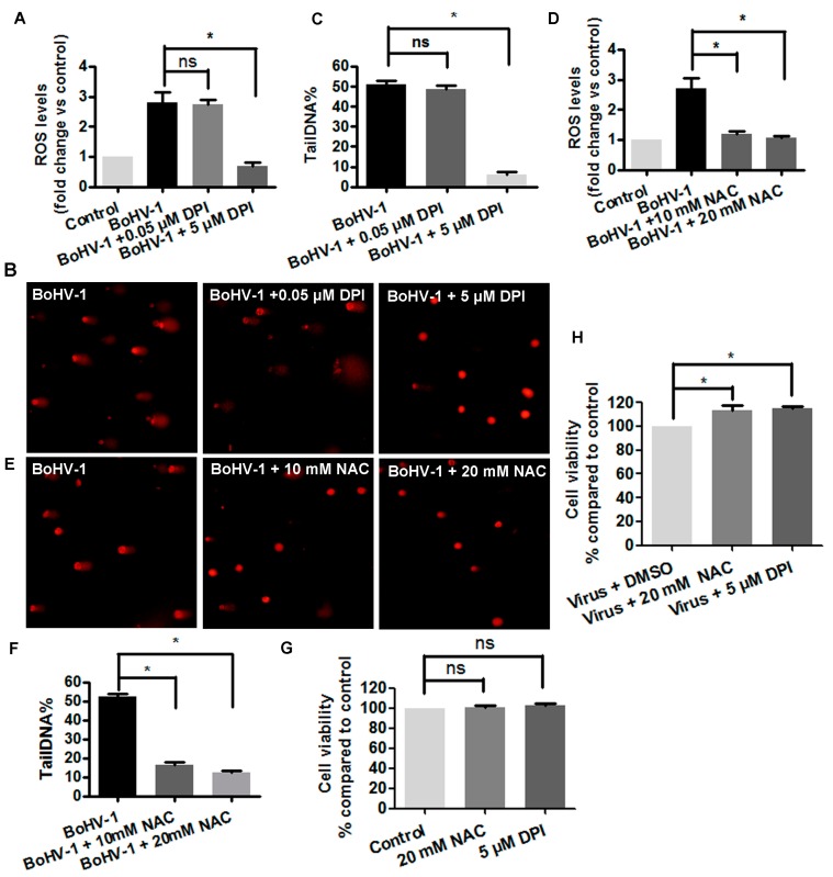
Figure 2.
Reactive oxidative species (ROS) was involved in BoHV-1-induced DNA damage in MDBK cells. (A,D) MDBK cells were infected with BoHV-1 (MOI = 0.1) and treated with DPI (A) or NAC (D) at indicated concentrations. After infection for 24 h, cellular ROS levels were determined using H2DCFDA (5 μM, 30 min) (Sigma-Aldrich, St. Louis, MO, USA) and quantitatively analyzed using software Image-pro Plus 6. *, significant differences (P < 0.05), as determined by a Student t test. (B,E) MDBK cells were infected with BoHV-1 (MOI = 0.1), and treated with DPI (B) or NAC (E) at indicated concentrations. At 24 h after infection, DNA damage in individual cells was detected with comet assay, the images were acquired under a fluorescence microscope (Magnification ×200). (C,F) Three hundred cells were randomly selected from the samples treatment with either DPI (C) or NAC (F) for the analysis of TailDNA% with software CASP. *, significant differences (P < 0.05) in tailDNA%, as determined by a Student t test. (G) MDBK cells were treated with each chemicals at indicated concentrations for 24 h, and cell viability was evaluated by Trypan-blue exclusion test. Data represent the results from three independent experiments. Significance was assessed with the student t test (*, P < 0.05). (H) MDBK cells were infected with BoHV-1 (MOI = 0.1) and treated with either NAC or DPI at indicated concentrations for 24 h, and cell viability was evaluated by Trypan-blue exclusion test. Data represent the results from three independent experiments. Significance was assessed with the student t test (*, P < 0.05).

