Abstract
As a cardiac disease caused by the inflammation of the heart muscle, viral myocarditis (VMC) causes dilated cardiomyopathy, congestive heart failure and even death. With anti-inflammatory activities, andrographolide has been used in the treatment of various human diseases. In the present study, therapeutic effects of andrographolide on VMC were investigated using a VMC mouse model. Measurement of physiological indexes and echocardiographic examination was performed to explore the effects of andrographolide on cardiac function in mice with VMC. Levels of TNF-α, hs-CRP and cTnl in serum were measured by enzyme-linked immunosorbent assay (ELISA). Effects of andrographolide on the expression of L-10, STAT3, NF-κβ p65 and NF-κβ p50 were investigated by western blot analysis. Results indicated that andrographolide treatment reduced serum levels of TNF-α, hs-CRP and cTnl, increased the expression levels of IL-10 and STAT3 and reduced the expression levels of NF-κβ p65 and NF-κβ p50 and the phosphorylation levels of phosphoinositide 3-kinase (P13K) and AKT in the heart tissues of mice with VMC. In addition, andrographolide also increased the expression level of Iκβα in heart tissue. Therefore, it was concluded that andrographolide may inhibit the progression of VMC by interacting with the IL-10/STAT3 and NF-κβ signaling pathways.
Keywords: viral myocarditis, andrographolide, IL-10/STAT3 pathway, NF-κβ pathway
Introduction
Myocarditis refers to the inflammatory condition of the heart caused by various factors (1). Clinical manifestations of myocarditis range from mild dyspnea to heart failure or even sudden death (2). Although infection with parasites, fungi and bacteria, drug-induced hypersensitivity and autoimmune disorders all induce myocarditis, viruses, including enteroviruses, influenza viruses, adenoviruses, parvoviruses, cytomegaloviruses, human immunodeficiency virus and herpes viruses, are the most common causes of myocarditis (3). High incidence of viral myocarditis (VMC) is observed in young individuals, and it has been reported that VMC is responsible for 12% sudden deaths in patients who were younger than 40 years (4,5). Besides damages of cardiac tissues directly caused by viruses, reactive inflammatory responses caused by viral reaction also significantly promote cardiac injury (6). Although, short-term outcomes of treatment of VMC are usually satisfactory, patients recovered from VMC may develop heart failure and recurrent dilated cardiomyopathy years later (2). Therefore, it would be of great clinical value to identify novel drugs to improve the treatment outcomes of VMC.
As a labdane diterpenoid isolated from Andrographispaniculata, andrographolide has been widely used in treatment of various human diseases including different types of cancers (7), obesity (8), oxidative damage (9), and so on. Andrographolide is also a safe and effective drug for treatment of various inflammatory diseases (10). A recent study reported that andrographolide improved cardiac malfunctions in mice by reducing cell apoptosis and inhibiting phosphorylation of IκB (11). In another study, andrographolide was proved to effectively improve infections of different viruses (12). In view of the pathogenesis of VMC and the functionality of andrographolide, it will be reasonable to hypothesize that andrographolide may also has certain therapeutic effects on VMC.
In this this, mice VMC model was established by Coxsackie B3m virus infection, which is a main cause of VMC in human (13). Andrographolide was used to treat mice VMC model and its effects on cardiac function was detected. In addition, interactions between andrographolide and IL-10/STAT3 pathway and phosphoinositide 3-kinase (P13K)/AKT/NF-κβ pathway were also investigated. The report is as follow:
Materials and methods
Establishment of mice VMC model and treatments
Thirty-mice (4–6 weeks old) were purchased from BALB/c Mice Guangdong Medical Laboratory Animal Center (Guangzhou, China). Mice were randomly divided into three groups including control group, model group and andrographolide group (n=10). One week before model construction, mice in Andrographolide group were intraperitoneally injected with 100 µl PBS containing Andrographolide at a dose of 1 mg/kg, while mice in control group and model group were only injected with 100 µl PBS. Mice were subjected to intraperitoneal injection of 0.2 ml PBS solution containing 1×10⁶ PFU Coxsackie B3 m virus (CVB3m, Nancy) to induce VMC. This study has been approved by the ethics committee of Jining No. 1 People's Hospital.
Measurement of physiological indexes
One week after model construction, physiological indexes including heart weight/body weight (HW/BW), heart rate (HR), mean arterial pressure (MAP), high left ventricular diastolic end pressure (LVDEP) and ventricular contractility assessment (dP/dt) were measured using conventional methods. All indexes were measured 3 times, and the data were expressed as mean ± SD.
Echocardiographic examination
Echocardiographic examination was performed at 1 week after model construction. After anesthesia with isoflurane, M-model imaging was performed through two-dimensional echocardiography (Sonos 5500; Philips Medical Systems, Inc., Bothell, WA, USA) using a 12-MHz probe to measure left ventricular systolic dimension (LVDs), left ventricular diastolic dimension (LVDd), anterior wall thickness (AWT) and posterior wall thickness (PWT). All indexes were measured 3 times, and the data were expressed as mean ± SD.
Enzyme-linked immunosorbent assay (ELISA)
Blood (about 1 ml) was collected by cutting the tail at 1 week after model construction. Blood samples were centrifuged (10,000 rpm) at room temperature for 10 min to separate serum. Levels of TNF-α, hsCRP and cTnl were measured by ELISA using kits provided by R&D Systems, Inc., (Minneapolis, MN, USA). Briefly, standard solutions were made through serial dilution of TNF-α, hsCRP and cTnl stock solutions with diluent buffer in the kits to generate standard curves. Biotin-labeled human TNF-α, hsCRP and cTnl antibodies were used at a dilution of 1:800. Avidin-peroxidase complex was sued at a dilution of 1:500. OD values at 480 nm were measured using a microplate reader. ELSIA was performed 3 times, and the data were expressed as mean ± SD.
Western blot analysis
Mice were sacrificed 1 week after model construction, and the whole heart was collected. Heart was cut into pieces and ground in liquid nitrogen. Total protein was extracted from heart tissues using conventional method, and BCA method was used to quantify protein concentration. After that, 40 µg of protein was subjected to 10% SDS-PAGE gel electrophoresis, followed by transmembrane to PVDF membrane. After blocking with 5% skimmed milk, membranes were incubated with rabbit anti-NF-κB p65 antibody (1:1,000, ab16502), rabbit anti-NF-κB p50 antibody (1:1,000, ab32360), rabbit anti-IL-10 antibody (1:1,000, ab9969), rabbit anti-STAT3 antibody (1:1,000, ab76315), rabbit anti-p-PI3K antibody (1:2,000, ab182651), rabbit anti-PI3K antibody (1:2,000, ab5451), rabbit anti-p-AKT antibody (1:2,000, ab18206), rabbit anti-AKT antibody (1:2,000, ab126811), rabbit anti-Iκβα antibody (1:1,000, ab76429), and rabbit anti-GAPDH antibody (1:1,000, ab8245; all purchased from Abcam, Cambridge, UK) overnight at 4°C. After washing with TBST 3 times, 15 min for each time, anti-rabbit IgG-HRP secondary antibody (1:1,000, MBS435036; http://www.mybiosource.com) was used to incubated with membranes at room temperature for 2 h. After washing with TBST, ECL detection reagent (Sigma-Aldrich; Merck KGaA, Darmstadt, Germany) was used to detect the signals. Band intensity within a frame covers all bands was measured using ImageJ software (National Institutes of Health, Bethesda, MD, USA). Intensity of target band was normalized to that of endogenous control. We added this information. Each experiment was performed 3 times, and the data were expressed as mean ± SD.
Statistical analysis
Statistical analyses were performed using SPSS v.19.0 (SPSS, Inc., Chicago, IL, USA). All data were expressed as mean ± standard deviation (mean ± SD). Comparisons of data among multiple groups were performed using one-way analysis of variance, followed by LSD test. P<0.05 was considered to indicate a statistically significant difference.
Results
Comparison of physiological indexes among groups
Physiological indexes were measured 1 week after model construction. As shown in Table I, HR and MAP significantly decreased, and HW/BW, LVDEP and Min dP/dt significantly increased in model group compared with control group (P<0.05), indicating the impaired cardiac function caused by VMC. Compared with model group, HR and MAP significantly decreased and HW/BW, LVDEP and Min dP/dt significantly decreased in andrographolide group (P<0.05). Those data suggest that andrographolide treatment can inhibit the reduction in cardiac function caused by VMC.
Table I.
Comparison of physiological parameters between groups.
| Items | Control | Model | Andrographolide |
|---|---|---|---|
| HR (bpm) | 391.2±5.1 | 334.1±3.9a | 377.4±9.6b |
| MAP (mmHg) | 98.4±7.7 | 71.3±4.8a | 87.5±4.6b |
| HW/BW (g/kg) | 2.4±0.1 | 3.9±0.1a | 2.7±0.1b |
| LVDEP (mmHg) | 4.4±0.7 | 6.3±0.4a | 5.0±0.5b |
| Min dP/dt (mmHg/s) | −7998.6±345.2 | −4675.8±312.5a | −6671.7±341.9b |
P<0.05 compared with the control group
P<0.05 compared with the model group. HR, heart rate; MAP, mean arterial pressure; HW/BW, heart weight/body weight; LVDEP, left ventricular diastolic end pressure; dP/dt, ventricular contractility assessment.
Comparison of echocardiographic examination results among groups
As shown in Table II, echocardiographic examination results showed that LVDd, LVDs, PWT diastole and AWT diastole were significantly increased in model group than in control group (P<0.05). Compared with model group, LVDd, LVDs, PWT diastole and AWT diastole significantly reduced in andrographolide group. Those data suggest that andrographolide treatment can significantly reduce the adverse effects of VMC on cardiac function.
Table II.
Comparison of echocardiographic examination results between groups.
| Items | Control | Model | Andrographolide |
|---|---|---|---|
| LVDd (mm) | 7.0±0.1 | 7.4±0.1a | 7.0±0.1b |
| LVDs (mm) | 3.5±0.1 | 4.7±0.2a | 3.9±0.2b |
| PWT diastole (mm) | 1.6±0.2 | 2.8±0.3a | 1.9±0.1b |
| AWT diastole (mm) | 1.5±0.1 | 2.9±0.2a | 1.8±0.2b |
P<0.05 compared with the control group
P<0.05 compared with the model group. LVDd, left ventricular diastolic dimension; LVDs, left ventricular systolic dimension; PWT, posterior wall thickness; AWT, anterior wall thickness.
Comparison of serum levels of TNF-α, hs-CRP and cTnl between groups
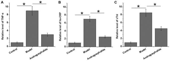
TNF-α, hs-CRP and cTnl are closely related to the development of VMC. As shown in Fig. 1, serum levels of TNF-α, hs-CRP and cTnl significantly increased in model group than in control group (P<0.05), indicating the progression of VMC in mice of model group. Compared with model group, serum levels of TNF-α, hs-CRP and cTnl were significantly reduced in andrographolide group (P<0.05). Those data suggest that andrographolide treatment can inhibit the development of VMC by reducing serum levels of TNF-α, hs-CRP and cTnl.
Figure 1.
Comparison of serum levels of TNF-α, hs-CRP and cTnl between groups. (A) Serum levels of TNF-α in three groups, (B) serum levels of hs-CRP in three groups and (C) serum levels of cTnl in three groups. *P<0.05 as indicated.
Effects of andrographolide on IL-10/STAT3 pathway
IL-10/STAT3 pathway mediates the anti-inflammatory signal transduction in various pathological processes. As shown in Fig. 2, expression levels of IL-10 protein and STAT3 protein in heart tissues were significantly reduced in model group than in control group (P<0.05), indicating that the inhibition of IL-10/STAT3 pathway is involved in the development of VMC. Compared with model group, expression levels of IL-10 protein and STAT3 protein in heart tissue significantly increased in andrographolide group (P<0.05). Those data suggest that andrographolide treatment can inhibit the development of VMC by activating IL-10/STAT3 anti-inflammatory pathway.
Figure 2.
Expression of IL-10 and STAT3 in three groups. (A) Representative western blot results, (B) relative expression level of IL-10 protein in three groups and (C) relative expression level of STAT3 protein in three groups. *P<0.05 as indicated.
Effects of andrographolide on NF-κβ pathway
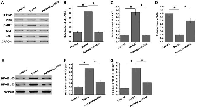
NF-κβ pathway plays pivotal roles in development of various human diseases, and PI3K/Akt signaling pathway is involved in the activation of NF-κβ pathway by regulating the degradation of Iκβα. As shown in Fig. 3A-d, no significantly differences in levels of total PI3K and Akt in heart tissue were found between groups, while phosphorylation of PI3K and Akt significantly increased in model group compared with control group (P<0.05), indicating the activation of PI3K/Akt signaling pathway. Compared with model group, phosphorylation of PI3K and Akt significantly decreased in andrographolide group. In contrast, level of Iκβα significantly decreased in model group compared with control group (P<0.05). Compared with model group, level of Iκβα significantly increased in andrographolide group. In addition, as shown in Fig. 3E-G, levels of NF-κβ p65 and NF-κβ p50 significantly increased in model group than in control group (P<0.05), indicating the involvement of NF-κβ pathway in progression of VMC. Compared with model group, levels of NF-κβ p65 and NF-κβ p50 significantly decreased in andrographolide group (P<0.05). Those data suggest that andrographolide treatment may inhibit the development of VMC by inhibiting PI3K/Akt/NF-κβ pathway.
Figure 3.
Effects of andrographolide treatment on PI3K/Akt/NF-κβ pathway. (A) Representative western blot results, (B) relative level of p-PI3K in three groups, (c) relative level of p-AKT in three groups, (d) relative level of Iκβα in three groups, (e) representative western blot results, (f) relative expression level of NF-κβ p65 in three groups and (g) relative expression level of NF-κβ p50 in three groups. *P<0.05 as indicated.
Discussion
Andrographolide is the main active ingredient of Andrographis paniculata, which is traditional herbal medicine that has been widely used in Scandinavia and Asia to treat sore throat, upper respiratory tract infections and flu (10,14). Recently, effects of andrographolide on virus infection have also been reported. Wintachai et al (15), reported that andrographolide could significantly alleviate chikungunya virus infection by inhibiting or stimulating a variety of targets. In the present study of cervical carcinoma, Ekalaksananan et al (16), found that andrographolide and its derivatives inhibited HPV16 pseudovirus infection by inhibiting the expression of viral oncogene. In another study, andrographolide was proved to inhibit the activity of hepatitis C virus by increasing the expression level of haeme oxygenase-1 through interactions with p38 MAPK/Nrf2 pathway in hepatoma cells of human (17). As a cardiac disease caused by virus infection, VMC significantly affects cardiac function. Even worse, cardiac function may be impaired after the recovery from VMC (18). Consistent with previous studies, cardiac function was significantly reduced in mice VMC model compared with control mice. However, VMC mice pretreated with andrographolide showed significantly better cardiac function compared with VMC mice without andrographolide treatment. Those data suggest that andrographolide can improve impaired cardiac function after VMC. Cardiac TNF-α, hs-CRP and cTnl play pivotal roles in development of VMC, and detection of levels of TNF-α, hs-CRP and cTnl have been proved to be an effective and accurate method to predict VMC (19). In our study, serum levels of TNF-α, hs-CRP and cTnl were significantly increased in mice VMC model, while andrographolide pretreatment significantly inhibited the increase in serum levels of TNF-α, hs-CRP and cTnl in mice with VMC. Those data suggest that andrographolide treatment can promote VMC by reducing serum levels of TNF-α, hs-CRP and cTnl.
To investigate the molecular mechanism of the therapeutic effects of andrographolide on VMC, the possible involvement of IL-10/STAT3 pathway was investigated. Cardiac injury caused by reactive inflammatory responses after viral reaction has stronger promotion effects on development of VMC compared with the cell damages directly caused by virus (6). As an anti-inflammatory factor, IL-10 inhibits various human diseases by mediating anti-inflammatory response (20). Animal studies have showed that increased levels of IL-10 protected heart cells from damages caused by acute myocarditis (21). IL-10 achieves it biological functions by interacting with its downstream STAT3, which is a transcription factor and regulates the expression of various targets (20). In our study, expression levels of IL-10 and STAT3 were significantly lower in mice with VMC than in control mice, indicating the involvement of IL-10/STAT3 in development of VMC. Compared with model group, andrographolide pretreatment significantly inhibited the decrease in expression levels of IL-10 and STAT3. Those data suggest that andrographolide may improve VMC by activating IL-10/STAT3 pathway.
NF-κβ pathway is involved in the pathogenesis of VMC through the regulation of immune responses (22), and the excessive inflammation caused by NF-κβ pathway in VMC is the main cause chronic heart failure and cardiac hypertrophy. Previous studies have shown that drug that targets NF-κβ pathway can be used to treat VMC (23). It's well accepted that activation of NF-κβ can be mediated by PI3K/Akt signaling pathway via the regulation of Iκβα degradation (24,25). In our study, phosphorylation levels of PI3K and Akt increased, while level of Iκβα decreased by VMC. In addition, expression levels of NF-κβ p65 and NF-κβ p50 also increased in VMC. Those results suggest that PI3K/Akt/NF-κβ pathway is activated in the progression of VMC. Compared with model group, andrographolide pretreatment significantly inhibited the phosphorylation of PI3K and Akt, decreased the level of Iκβα in heart tissue and downregulated the expression levels of NF-κβ p65 and NF-κβ p50. Those results suggest that andrographolide treatment may improve VMC by inhibiting PI3K/Akt/NF-κβ pathway.
In conclusion, andrographolide may improve VMC by inhibiting the increase in serum levels of TNF-α, hs-CRP and cTnl caused by VMC, activating IL-10/STAT3 anti-inflammatory pathway and inactivating PI3K/Akt/NF-κβ pathway. Further clinical studies are needed to confirm our conclusions.
Acknowledgements
Not applicable.
Funding
No funding was received.
Availability of data and materials
The datasets used and/or analyzed during the present study are available from the corresponding author on reasonable request.
Authors' contributions
YZ, YL and WD conceived and designed the study. YZ and MW performed experiments and analyzed data. YZ and YL interpreted the data. YL drafted the manuscript. All authors read and approved the final manuscript.
Ethics approval and consent to participate
The present study was approved by the Ethics Committee of Jining No. 1 People's Hospital.
Consent for publication
Not applicable.
Competing interests
The authors declare that they have no competing interests.
References
- 1.Corsten MF, Schroen B, Heymans S. Inflammation in viral myocarditis: Friend or foe? Trends Mol Med. 2012;18:426–437. doi: 10.1016/j.molmed.2012.05.005. [DOI] [PubMed] [Google Scholar]
- 2.Sagar S, Liu PP, Cooper LT., Jr Myocarditis. Lancet. 2012;379:738–747. doi: 10.1016/S0140-6736(11)60648-X. [DOI] [PMC free article] [PubMed] [Google Scholar]
- 3.Dennert R, Crijns HJ, Heymans S. Acute viral myocarditis. Eur Heart J. 2008;29:2073–2082. doi: 10.1093/eurheartj/ehn296. [DOI] [PMC free article] [PubMed] [Google Scholar]
- 4.Blauwet LA, Cooper LT. Myocarditis. Prog Cardiovasc Dis. 2010;52:274–288. doi: 10.1016/j.pcad.2009.11.006. [DOI] [PMC free article] [PubMed] [Google Scholar]
- 5.Esfandiarei M, McManus BM. Molecular biology and pathogenesis of viral myocarditis. Annu Rev Pathol. 2008;3:127–155. doi: 10.1146/annurev.pathmechdis.3.121806.151534. [DOI] [PubMed] [Google Scholar]
- 6.Shauer A, Gotsman I, Keren A, Zwas DR, Hellman Y, Durst R, Admon D. Acute viral myocarditis: Current concepts in diagnosis and treatment. Isr Med Assoc J. 2013;15:180–185. [PubMed] [Google Scholar]
- 7.Liu Y, Liang RM, Ma QP, Xu K, Liang XY, Huang W, Sutton R, Ding J, O'Neil PM, Cheng CR. Synthesis of thioether andrographolide derivatives and their inhibitory effect against cancer cells. MedChemComm. 2017;8:1268–1274. doi: 10.1039/C7MD00169J. [DOI] [PMC free article] [PubMed] [Google Scholar]
- 8.Ding L, Li J, Song B, Xiao X, Huang W, Zhang B, Tang X, Qi M, Yang Q, Yang Q, et al. Andrographolide prevents high-fat diet-induced obesity in C57BL/6 mice by suppressing the sterol regulatory element-binding protein pathway. J Pharmacol Exp Ther. 2014;351:474–483. doi: 10.1124/jpet.114.217968. [DOI] [PubMed] [Google Scholar]
- 9.Chen HW, Huang CS, Li CC, Lin AH, Huang YJ, Wang TS, Yao HT, Lii CK. Bioavailability of andrographolide and protection against carbon tetrachloride-induced oxidative damage in rats. Toxicol Appl Pharmacol. 2014;280:1–9. doi: 10.1016/j.taap.2014.07.024. [DOI] [PubMed] [Google Scholar]
- 10.Tan WSD, Liao W, Zhou S, Wong WSF. Is there a future for andrographolide to be an anti-inflammatory drug? Deciphering its major mechanisms of action. Biochem Pharmacol. 2017;139:71–81. doi: 10.1016/j.bcp.2017.03.024. [DOI] [PubMed] [Google Scholar]
- 11.Zhang J, Zhu D, Wang Y, Ju Y. Andrographolide attenuates LPS-induced cardiac malfunctions through inhibition of IκB phosphorylation and apoptosis in mice. Cell Physiol Biochem. 2015;37:1619–1628. doi: 10.1159/000438528. [DOI] [PubMed] [Google Scholar]
- 12.Liu RH, Jacob JR, Tennant B. Andrographolide derivatives to treat viral infections: U.S. Patent 8445,533 (P) 2013 [Google Scholar]
- 13.Weller AH, Simpson K, Herzum M, Van Houten N, Huber SA. Coxsackievirus-B3-induced myocarditis: Virus receptor antibodies modulate myocarditis. J Immunol. 1989;143:1843–1850. [PubMed] [Google Scholar]
- 14.Jayakumar T, Hsieh CY, Lee JJ, Sheu JR. Experimental and clinical pharmacology of andrographis paniculata and its major bioactive phytoconstituent andrographolide. Evid Based Complement Alternat Med. 2013;2013:846740. doi: 10.1155/2013/846740. [DOI] [PMC free article] [PubMed] [Google Scholar]
- 15.Wintachai P, Kaur P, Lee RC, Ramphan S, Kuadkitkan A, Wikan N, Ubol S, Roytrakul S, Chu JJ, Smith DR. Activity of andrographolide against chikungunya virus infection. Sci Rep. 2015;5:14179. doi: 10.1038/srep14179. [DOI] [PMC free article] [PubMed] [Google Scholar]
- 16.Ekalaksananan T, Sookmai W, Fangkham S, Pientong C, Aromdee C, Seubsasana S, Kongyingyoes B. Activity of andrographolide and its derivatives on HPV16 pseudovirus infection and viral oncogene expression in cervical carcinoma cells. Nutr Cancer. 2015;67:687–696. doi: 10.1080/01635581.2015.1019630. [DOI] [PubMed] [Google Scholar]
- 17.Lee JC, Tseng CK, Young KC, Sun HY, Wang SW, Chen WC, Lin CK, Wu YH. Andrographolide exerts anti-hepatitis C virus activity by up-regulating haeme oxygenase-1 via the p38 MAPK/Nrf2 pathway in human hepatoma cells. Br J Pharmacol. 2014;171:237–252. doi: 10.1111/bph.12440. [DOI] [PMC free article] [PubMed] [Google Scholar]
- 18.Becher PM, Gotzhein F, Klingel K, Escher F, Blankenberg S, Westermann D, Lindner D. Cardiac function remains impaired despite reversible cardiac remodeling after acute experimental viral myocarditis. J Immunol Res. 2017;2017:6590609. doi: 10.1155/2017/6590609. [DOI] [PMC free article] [PubMed] [Google Scholar]
- 19.Wang D, Li T, Cui H, Zhang Y. Analysis of the indicating value of cardiac troponin i, tumor necrosis factor-α, interleukin-18, Mir-1 and Mir-146b for viral myocarditis among children. Cell Physiol Biochem. 2016;40:1325–1333. doi: 10.1159/000453185. [DOI] [PubMed] [Google Scholar]
- 20.Hutchins AP, Diez D, Miranda-Saavedra D. The IL-10/STAT3-mediated anti-inflammatory response: Recent developments and future challenges. Brief Funct Genomics. 2013;12:489–498. doi: 10.1093/bfgp/elt028. [DOI] [PMC free article] [PubMed] [Google Scholar]
- 21.Roffê E, Rothfuchs AG, Santiago HC, Marino AP, Ribeiro-Gomes FL, Eckhaus M, Antonelli LR, Murphy PM. IL-10 limits parasite burden and protects against fatal myocarditis in a mouse model of Trypanosoma cruzi infection. J Immunol. 2012;188:649–660. doi: 10.4049/jimmunol.1003845. [DOI] [PMC free article] [PubMed] [Google Scholar]
- 22.Maier HJ, Schips TG, Wietelmann A, Krüger M, Brunner C, Sauter M, Klingel K, Böttger T, Braun T, Wirth T. Cardiomyocyte-specific IκB kinase (IKK)/NF-κB activation induces reversible inflammatory cardiomyopathy and heart failure. Proc Natl Acad Sci USA. 2012;109:11794–11799. doi: 10.1073/pnas.1116584109. [DOI] [PMC free article] [PubMed] [Google Scholar]
- 23.Valaperti A. Drugs targeting the canonical NF-κB pathway to treat viral and autoimmune myocarditis. Curr Pharm Des. 2016;22:440–449. doi: 10.2174/1381612822666151222160409. [DOI] [PubMed] [Google Scholar]
- 24.Hyam SR, Lee IA, Gu W, Kim KA, Jeong JJ, Jang SE, Han MJ, Kim DH. Arctigenin ameliorates inflammation in vitro and in vivo by inhibiting the PI3K/AKT pathway and polarizing M1 macrophages to M2-like macrophages. Eur J Pharmacol. 2013;708:21–29. doi: 10.1016/j.ejphar.2013.01.014. [DOI] [PubMed] [Google Scholar]
- 25.Han W, Xiong Y, Li Y, Fang W, Ma Y, Liu L, Li F, Zhu X. Anti-arthritic effects of clematichinenoside (AR-6) on PI3K/Akt signaling pathway and TNF-α associated with collagen-induced arthritis. Pharm Biol. 2013;51:13–22. doi: 10.3109/13880209.2012.698287. [DOI] [PubMed] [Google Scholar]
Associated Data
This section collects any data citations, data availability statements, or supplementary materials included in this article.
Data Availability Statement
The datasets used and/or analyzed during the present study are available from the corresponding author on reasonable request.


