Abstract
Toothpick ingestion is a rare but potentially fatal condition which may cause bowel perforation and rare complications if it migrates out of the gastrointestinal tract. This case report describes a delayed presentation of toothpick-induced small bowel injury leading to abdominal wall abscess and spondylodiscitis. A 51-year-old man was admitted twice with fever and loin pain, but repeated non-contrast CT was reported to be unremarkable. However, 5 months later, he presented with a left lower quadrant abdominal wall abscess and back pain. An updated CT showed a linear hyperdensity which was already present in previous scans, causing small bowel perforation, abdominal wall abscess and spondylodiscitis. Emergency laparotomy identified a toothpick causing small bowel perforation. The abdominal wall abscess was incised and drained, and small bowel was repaired.
Keywords: radiology, gastrointestinal surgery, foreign bodies, intestinal perforation
Background
Foreign body ingestion is not uncommon but most of it pass through the gastrointestinal tract without causing any perforation.1 2 Ingestion of sharp and pointed objects such as toothpicks, however, is often associated with bowel perforation and other devastating consequences. Most of the patients were unaware of the event of swallowing a toothpick, making diagnosis difficult.3 It was described in some case reports that toothpick can silently migrate outside gastrointestinal tract resulting in rare complications like spondylodiscitis in our case.4 We describe a case of delayed diagnosis of toothpick ingestion causing small bowel perforation, abdominal wall abscess and spondylodiscitis.
Case presentation
A 51-year-old man who was a former intravenous drug user with a history of open appendectomy and two episodes of open adhesiolysis for adhesive intestinal obstruction was initially admitted for left loin pain. A non-contrast CT was performed which was reported to be unremarkable. He was readmitted 1 week later for bilateral loin pain and septic shock. Blood culture yielded an uncommon organism, Eggerthella lenta. There were no abdominal signs, and no other septic foci were identified. He was treated with a 10-day course of intravenous piperacillin/tazobactam with a dosage of 4.5 g every 8 hours and was discharged. Three months later, he presented again with left lower quadrant pain. Another non-contrast CT was done which was again reported to be unremarkable. Five months after his first presentation, he was admitted for a left lower quadrant abdominal wall abscess and back pain.
Investigations
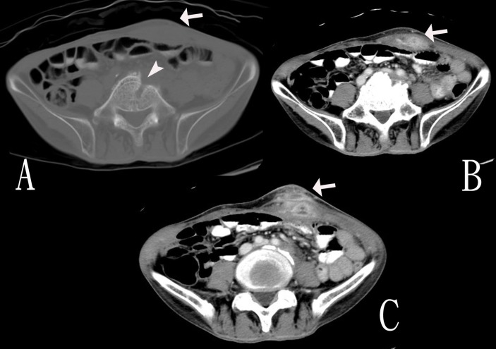
Urgent contrast CT showed a 6 cm linear hyperdensity over the left lower quadrant extending from cutaneous region into the intraperitoneal cavity abutting the small bowel with an associated abscess collection (figure 1). There was also a lytic tract with rim-enhancing fluid at L5 vertebral body suggestive of osteomyelitis. On review, the linear hyperdensity was actually present on the two previous non-contrast CT scans (video 1). The linear hyperdensity showed migrating positions in the three scans, suggesting that it was a mobile foreign body.
Figure 1.
Images of the third set of CT of the abdomen. (A) Bone window image shows a lytic tract at L5 vertebral body (arrow head). (B) Contrast-enhanced view shows the left lower abdominal wall abscess (arrow). (C) Contrast-enhanced view shows the left lower abdominal wall abscess with intraperitoneal extension (arrow).
Video 1.
Reconstruction of the consecutive transverse images of the first set of CT of the abdomen showed a linear foreign body pointing to the left side of L5 vertebra (arrow head).
Treatment
Emergency operation for incision and drainage of abdominal wall abscess and small bowel repair was done. Intraoperatively, a toothpick was found perforating a loop of small bowel, through the anterior abdominal wall to skin, resulting in a 4 cm abdominal wall abscess over the left lower quadrant. Abscess pus culture grew extended-spectrum beta-lactamases Escherichia coli and Klebsiella. He was treated with a 10-day course of intravenous ertapenem 1 g every 24 hours. The abdominal wound was treated with local therapy and subsequent secondary closure. He was discharged on day 10 postoperatively tolerating diet without further active treatment for his spondylodiscitis.
Outcome and follow-up
He was referred to orthopaedic outpatients for further management of osteomyelitis on discharge; however, he defaulted all subsequent appointments. Two years later, he was readmitted with low back pain and fever. Blood tests were unremarkable. X-ray of the lumbosacral spine showed collapsed L2 vertebra and a vague soft tissue mass over the anterior body of L4 and L5 with body destruction of L5, suspicious of L5/S1 spondylodiscitis. CT lumbosacral spine was performed, showing L5/S1 spondylodiscitis with no paraspinal collection. The location was not amenable to CT-guided fine-needle aspiration. He was treated with a 4-week course of intravenous cefuroxime 750 μg every 8 hours and cloxacillin 2 g every 6 hours, and then the antibiotic was upgraded to intravenous vancomycin 1 g every 12 hours due to persistent fever. He was discharged but unfortunately defaulted to all follow-up investigations and appointments.
Discussion
Toothpick ingestion is a rare condition, however, is usually associated with bowel perforation and other complications as it migrates outside the gastrointestinal tract. The literature suggests that the overall mortality associated with toothpick ingestion is up to 9.6%–18%.3 5 6 The presenting symptoms are mainly abdominal pain, fever and nausea.3 Bowel perforations have been reported in different segments, but most commonly in the duodenum (23%), gastric antrum (20%) and ileocecal junction (9%).3 5 There were also reports describing that the ingested toothpick migrated outside the gastrointestinal tract causing extremely rare complications such as liver abscess, pyelonephritis, pericarditis and duodenocaval fistula.7–12 To our knowledge, this is the first reported case of osteomyelitis and spondylodiscitis caused by toothpick ingestion in the literature.
Diagnosis of toothpick ingestion and its related complications is difficult as most patients do not recall the event of swallowing a toothpick. It was reported in a systemic review that 54% of patients were unaware of the event and majority (67%) of them ingested the toothpick during food intake. Eighty-five per cent of them reported that they had the habit of chewing toothpicks.3 Identification of toothpick is not easy on CT due to its long and slender structure. In our case, the presence of toothpick was overlooked in previous CT scans. It was reported that the sensitivity of CT in toothpick identification is only 42.6%.3 A recent literature review suggested that careful interpretation of CT using reformatted coronal and sagittal images may be helpful.8
Treatment of swallowed toothpicks varies between cases. Endoscopic management for perforation of the duodenum or the lower rectum is always the first-line. However, surgery should never be delayed if frank perforation or peritonitis is suspected. Migrated toothpicks should also be treated individually according to their specific location.13 14 In our case, surgery for abdominal wall abscess drainage and small bowel repair was immediately performed. Unfortunately, the management of our patient’s spondylodiscitis was delayed resulting in progression of disease. Earlier orthopaedic consultation may have facilitated obtaining an accurate microbiological diagnosis by either CT-guided or surgical biopsy, allowing for both drainage and targeted, prolonged antibiotic therapy.
In conclusion, the overall disease course of this patient was uncommon which is mainly attributable to the delayed diagnosis and the non-compliance to treatment. We would like to emphasise that for patients with recurrent abdominal pain and septicaemia from gut flora, foreign body ingestion should always be considered.
Learning points.
In cases of recurrent abdominal pain, ingested foreign bodies such as toothpick should always be considered.
When reviewed carefully, serial CT images can help to identify a small foreign body.
A patient with an enteric organism identified in blood cultures should be investigated for an intra-abdominal source.
Footnotes
Contributors: CMSL prepared the manuscript. THL collected the clinical data and supervised the work.
Funding: The authors have not declared a specific grant for this research from any funding agency in the public, commercial or not-for-profit sectors.
Competing interests: None declared.
Patient consent: Obtained.
Provenance and peer review: Not commissioned; externally peer reviewed.
References
- 1.Henderson CT, Engel J, Schlesinger P. Foreign body ingestion: review and suggested guidelines for management. Endoscopy 1987;19:68–71. 10.1055/s-2007-1018238 [DOI] [PubMed] [Google Scholar]
- 2.Velitchkov NG, Grigorov GI, Losanoff JE, et al. . Ingested foreign bodies of the gastrointestinal tract: retrospective analysis of 542 cases. World J Surg 1996;20:1001–5. 10.1007/s002689900152 [DOI] [PubMed] [Google Scholar]
- 3.Steinbach C, Stockmann M, Jara M, et al. . Accidentally ingested toothpicks causing severe gastrointestinal injury: a practical guideline for diagnosis and therapy based on 136 case reports. World J Surg 2014;38:371–7. 10.1007/s00268-013-2307-z [DOI] [PubMed] [Google Scholar]
- 4.Birindelli A, Tugnoli G, Biscardi A, et al. . Retroperitoneal colonic perforation from a foreign body--’cannibalization' effect of a toothpick--video vignette. Colorectal Dis 2016;18:319–21. 10.1111/codi.13259 [DOI] [PubMed] [Google Scholar]
- 5.Li SF, Ender K. Toothpick injury mimicking renal colic: case report and systematic review. J Emerg Med 2002;23:35–8. [DOI] [PubMed] [Google Scholar]
- 6.Zouros E, Oikonomou D, Theoharis G, et al. . Perforation of the cecum by a toothpick: report of a case and review of the literature. J Emerg Med 2014;47:e133–e137. 10.1016/j.jemermed.2014.06.065 [DOI] [PubMed] [Google Scholar]
- 7.Chong LW, Sun CK, Wu CC, Cc W, et al. . Successful treatment of liver abscess secondary to foreign body penetration of the alimentary tract: a case report and literature review. World J Gastroenterol 2014;20:3703–11. 10.3748/wjg.v20.i13.3703 [DOI] [PMC free article] [PubMed] [Google Scholar]
- 8.Izumi J, Satoh K, Iwasaki W, et al. . Small bowel obstruction caused by the ingestion of a wooden toothpick: the ct findings and a literature review. Intern Med 2017;56:657–60. 10.2169/internalmedicine.56.7463 [DOI] [PMC free article] [PubMed] [Google Scholar]
- 9.Lacroix S, Ferland A, Gilbert P, et al. . Cardiac hazard associated with eating habits. A case of infected intrapericardial foreign body due to an ingested toothpick. Can J Cardiol 2009;25:e263–e264. 10.1016/S0828-282X(09)70518-5 [DOI] [PMC free article] [PubMed] [Google Scholar]
- 10.Tüdös Z, Čtvrtlík F, Kratochvíl P, et al. . Wooden foreign body in the renal pelvis. Urology 2016;94:e7–e8. 10.1016/j.urology.2016.05.004 [DOI] [PubMed] [Google Scholar]
- 11.Justiniani FR, Wigoda L, Ortega RS. Duodenocaval fistula due to toothpick perforation. JAMA 1974;227:788–9. 10.1001/jama.1974.03230200046009 [DOI] [PubMed] [Google Scholar]
- 12.Alagiri M, Rabinovitch HH. Toothpick migration into bladder presents as abdominal pain and hematuria. Urology 1998;52:1130–1. 10.1016/S0090-4295(98)00445-2 [DOI] [PubMed] [Google Scholar]
- 13.Dente M, Santi F, Solinas L, et al. . Laparoscopic diagnosis and management of jejunal perforation resulting from accidental toothpick ingestion. Am Surg 2009;75:178–9. [PubMed] [Google Scholar]
- 14.Neumann U, Fielitz J, Ehlert HG. [Perforation of the cecum by a toothpick--a rare differential acute appendicitis diagnosis. Case report and review of the literature]. Chirurg 2000;71:1405–8. [DOI] [PubMed] [Google Scholar]


