Abstract
For gamete quality evaluation, there are innovative, rapid, and quantitative techniques that can provide useful data for aquaculture. Computerized systems for sperm analysis were developed to measure several parameters and one of the most commonly measured is the sperm motility.
Initially, this computer technology was designed for mammalian species, although it can also be used for fish sperm analysis. Fish have specific features that can affect sperm assessment such as a short motility time after activation and, in some cases, adaptation to lower temperatures. Thus, it is necessary to modify both software and hardware components to make motility analysis more efficient for fish sperm analysis. For mammalian sperm, the heating plate is used to maintain optimal temperatures of spermatozoa. However, for some fish species, it is advantageous to use a lower temperature to prolong the duration of motility, since the sperm remain active for less than 2 min. Therefore, cooling devices are necessary to refrigerate samples at constant temperature over the time of analysis, including on the optical microscope. This protocol describes the analysis of fish sperm motility using software for sperm analysis and new cooling devices to optimize the results.
Keywords: Biology, Issue 137, Reproduction, Fish sperm, Computer-assisted sperm analysis, Motility, Temperature, Frame rates, Counting chamber, Subpopulations
Introduction
The efficacy of reproduction depends on the quality of both gametes (eggs and sperm)1,2. This is the major factor that contributes to successful fertilization, allowing the development of viable offspring3,4. The convenient evaluation of gamete quality is the best tool for defining the fertility potential of a specimen.
Mixing sperm from multiple males is a common practice in the production of many aquatic commercial species4. However, the sperm variability between males can lead to sperm competition and, consequently, not all the males are equally contributing to the gene pool5. In this sense, the correct evaluation of individual ejaculate/spermatozoa features, such as motility, is fundamental to obtain discriminatory information regarding individual male fertility potential. Direct observation of sperm motility can produce inaccurate and subjective data as it requires time and experience, which leads to a lack of consistency and incompatibility of results6,7. However, there are many innovative, rapid and quantitative techniques that can provide a reliable sperm quality analysis2,4.
Computer-assisted sperm analysis was developed to offer accurate data about sperm quality8. This technology includes the development of software associated with a phase contrast microscope that allows the assessment of sperm motility. However, a limiting factor of motility parameter is the frame rate of the video camera. Individual spermatozoa trajectories are based on spermatozoa head centroid position in consecutive frames of video recordings, which is correlated with the flagellar movement patterns3,9,10,11. The main kinetic parameters measured are the straight-line velocity (VSL), curvilinear velocity (VCL) and average path velocity (VAP). VSL is the distance between the start and end-point taken by the spermatozoa divided by time. VCL is the real velocity along the precise trajectory taken by the spermatozoa. VAP is the velocity along a derived smoothed path of trajectory. These parameters allow additional kinetic information, including linearity (LIN), straightness (STR), wobble (WOB) and beating measurements like amplitude of lateral head movement (ALH) and beat-cross frequency (BCF)4,10.
The sperm analysis system was originally used for mammalian species, and one of the requirements for the system is to operate at the body temperature of the donor (about 37 °C). This software could also be used for fish species; although, it is necessary to make some adaptations to reduce the error of sperm analysis results. In some fish species, such as salmonids and eel8,12, fertilization occurs at low temperature (around 4 °C)2,4. Thus, cooling devices should be developed to avoid uncomfortable working conditions. In addition, fish spermatozoa are immotile in seminal fluid and require an osmotic shock to activate motility. For freshwater species, the activator medium should have hypotonic osmolality, while for marine species the medium should be hypertonic. However, for some species, as salmonids, the ion concentration could also be important3,4,9. After activation, fish sperm is characterized by a rapid decrease of motility (less than 2 min)13,14 and high velocity, being vital to determine the optimal frame rate to obtain reliable data15.
The objectives of this study are to design and apply refrigeration systems for fish sperm samples. In addition, this protocol defines how to determine the optimal frame rates for the establishment of standard protocols depending on the species. The use of this protocol opens new doors in the context of fish seminal evaluation, using the European eel as a model.
Protocol
Procedures involving animal subjects have been approved (2015/VSC/PEA/00064) by the General Direction of Agricultural Production and Livestock at the Universitat Politècnica de València.
1. Collect Sperm from Mature European Eels in Captivity
NOTE: Use European eel males maintained in tanks with seawater and a recirculation system at constant temperature (20 °C). Treat with hormones through weekly intraperitoneal injection (human chorionic gonadotropin (hCG); 1.5 IU per g of fish body weight). Acclimatize fish gradually starting with 3 days in freshwater followed by replacement with seawater (1/3 of the total water in the tank) every 2 days until reaching a salinity of 37.0 g/mL.
- Anesthetize the eel 24 h after hormone injection to obtain samples with better sperm quality.
- Prepare the anaesthesia beforehand: add 300 mg of benzocaine to 100 mL of 70% ethanol. Mix well and store at 4 °C.
- Dilute benzocaine in a flexible bucket with 5 L of system water to get a final concentration of 60 mg/L and mix properly.
- Transfer the fish to the bucket with system water and benzocaine. Wait a few minutes until the fish calm down.
Clear the genital area with water and dry with absorbent paper to avoid contamination of sperm samples by faeces, urine or seawater.
Apply gentle pressure in the abdominal area and collect the sperm samples into 15 mL plastic tubes using the vacuum pump. Sperm volume up to 6-7 mL, depending on the male.
Keep sperm samples at 4 °C for at least 1 h until motility analysis is carried out.
2. Refrigerate and Dilute the Sperm Samples
- Prepare the diluter solution beforehand. NOTE: The sperm samples should be diluted in the extender solution specific for each species.
- Prepare the non-activator medium for eel sperm samples (P1 medium). Add 0.42 g of sodium bicarbonate, 1.828 g of sodium chloride, 0.127 g of magnesium chloride, 0.56 g of potassium chloride and 0.037 g of calcium chloride to 250 mL of distilled water. Mix well to dissolve. Store at 4 °C.
Set the cooler block at 4 °C to maintain samples at a constant temperature. Wait until the temperature stabilizes.
- Dilute the sperm samples using the diluter solution specific for each species. NOTE: The dilution ratio is species specific and must be defined to standardize the protocol. First, estimate sperm concentration based on the concentration obtained in the pre-analysis of motility using software, as explained in steps 3 and 4. With this analysis, it is also possible to select the best sperm samples based on the percentage of motility. After this, the researcher can start to collect data for the experiment using the best samples with the correct concentration.
- Dilute eel sperm samples in the P1 medium with a ratio of 1:50. To prepare 500 µL of eel diluted sperm, add 50 µL of the fresh sperm sample and 450 µL of P1. Mix well.
3. Evaluate the Sperm Motility Parameters
- Set-up the Motility Module of the Software
- Set the cooler stage at 4 °C and wait until it stabilizes.
- Open the software and select Motility Module. Create username and password if necessary.
- Select Properties and choose the desired parameters before starting the sperm analysis.
- Click on Species | Fish.
- Select the Frames Per Second and Number of Images, depending on the best technical conditions for each species. Set both options at 120 images per second.
- Choose negative contrast. It is mandatory for accurate reconstruction of the spermatozoa trajectories16.
- Select the corresponding Counting Chamber and Scale of the video camera. Calibrate the video camera for the magnification lens used in the experiment. Set SpermTrack10 as counting chamber and 10X scale. NOTE: In general, a depth of 10 µm in the counting chamber and a magnification lens of 10X are the recommended conditions since they provide better focusing of all spermatozoa and capture the highest number of cells. However, it is recommended to make a previous study of the optimal conditions for motility analysis of each species.
- Adjust the Particle Area and Connectivity for each species. Set minimum particle area at 2 µm2 and connectivity at 7 µm. NOTE: For fish, the minimum value of particles area ranges between 2-5 µm, and connectivity depends on the spermatozoa velocity and the frame rate.
- Save the set-up.
- Select Capture.
- Analyze European Eel Sperm with Software
- Prepare the activator solution (artificial seawater) by adding 0.946 g of commercial salt to 25 mL of distilled water with 2% BSA.
- Take 500 mL of activator solution (artificial seawater) and place in the cooler block. NOTE: In general, fish sperm is activated by an osmotic shock event, although for some species the ion concentration could also be important. For freshwater species, the activator medium should have hypotonic osmolality, while for marine species the medium should be hypertonic.
- Put the counting chamber under the cooling stage and wait until the temperature stabilizes to 4 °C.
- Mix the activator and diluted sperm to activate spermatozoa and start the motility video recording between 5-10 s after activation.
- Take 4 µL of activator solution and place in the counting chamber.
- Gently homogenize the diluted sperm by shaking the microcentrifuge 3 times to avoid damage in spermatozoa cells.
- Take 0.5 µL of diluted sperm, mix with the activator, and put the cover in the counting chamber quickly. NOTE: This step should not take more than 5 s to start the analysis as soon as possible.
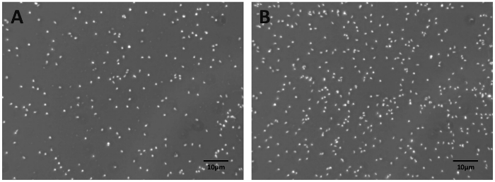
- Focus on the spermatozoa cells and find the best visual area, which is defined by a low number of spermatozoa (150 to 200 cells) to avoid interception between cells (Supplemental Figure 1A). Select Capture Video to obtain the spermatozoa tracks, which are separated according to speed.
- Select Capture to obtain 3 to 7 fields, which are the optimal interval to obtain a lower variability in the results, and proceed as before. Record videos until 120 s post-activation in order to avoid condensation on the cover of the counting chamber.
- Select Exit to have a general view of all the fields.
4. Obtain Motility Data
Select Fields | Partial | Save and choose a file name. Click Save. NOTE: The spreadsheet provides mean values of partial data, charts and images of spermatozoa tracks.
Select General Data | File Name | Save. NOTE: The data provide kinetic values for individual spermatozoa of all fields.
Representative Results
Analysis of the time effect on sperm motility
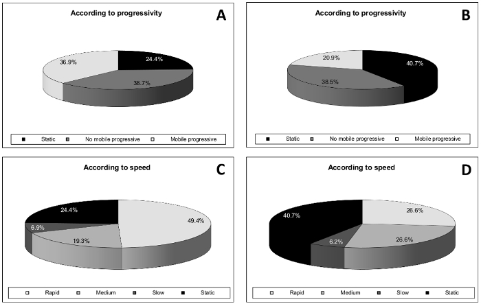
In the case of the European eel, the percentage of static spermatozoa increased from 15 s to 120 s after activation (from 24.4% to 40.7%), and the percentage of mobile progressive spermatozoa decreased (from 36.9% to 20.9%) (Figure 1A and 1B). Based on speed, spermatozoa cells showed a decrease in velocity over time (Figure 1C and 1D) decreased. The percentage of spermatozoa with rapid velocity decreased about 23% (from 49.4% to 26.6%) and the percentage of spermatozoa with medium velocity increased around 7%.

The effect of the frame rate and counting chamber
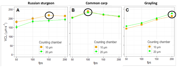
Different species require different frame rates for optimal results. Russian sturgeon requires 150 frames per second (fps; Figure 2A); common carp requires 100 fps (Figure 2B); and for the grayling, 200 fps was not enough to obtain optimal kinetic results (Figure 2C). These results were not dependent on the depth of the counting chamber tested (Figure 2A-2C).

Sperm subpopulations structure
The identification of sperm subpopulation is based on statistical analysis. The first step is the determination of principal components (principal component analysis; PCA) from the complete set of motility data. Then, the clustering procedures are performed with the sperm-derived indices obtained after the PCA.
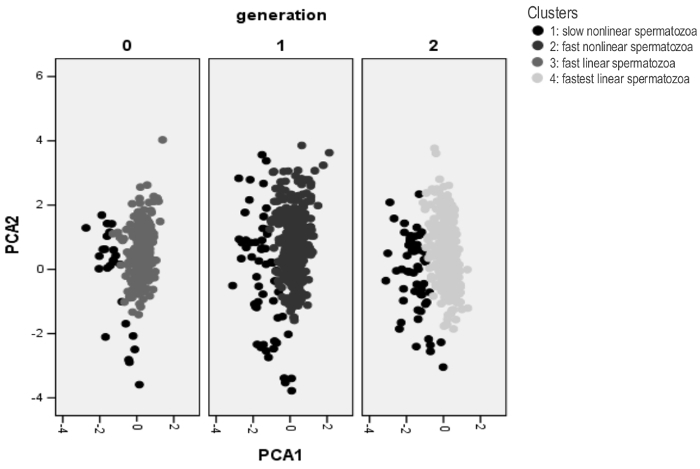
For this study, we used Atlantic salmon sperm which showed four different subpopulations, named: slow nonlinear (low LIN, STR and VCL), fast nonlinear (lower STR, high VCL), fast linear (high LIN, STR and VCL) and fastest linear (highest LIN, STR, and VCL). The distribution of these subpopulations varied along generations from wild animals to farmed ones (Figure 3).

Figure 1: Effect of time post-activation on the progressivity and motility of European eel sperm. A and B) Percentage of progressivity at 15 and 120 s, respectively; and C and D) the percentage of motility based on speed over time (15 and 120 s post-activation, respectively). The progressivity was divided in three groups: progressive motile cells (white; VCL mean value ± SD: 114.5 ± 18.2 and 101.4 ± 34.5 µm/s for 15 and 120 s, respectively), no progressive motile cells (grey; VCL mean value ± SD: 84.8 ± 17.4 µm/s for 15 s and 84.6 ± 16.5 µm/s for 120 s), and static spermatozoa (black). The spermatozoa velocity was divided into 4 groups: rapid (VCL mean value ± SD: 119.3 ± 13.7 µm/s for 15 s and 118.9 ± 13.3 µm/s for 120 s; white), medium (VCL mean value ± SD: 79.7 ± 15.5 µm/s for 15 s and 79.6 ± 15.5 µm/s for 120 s; light grey), slow (VCL mean value ± SD: 32.9 ± 6.5 µm/s for 15 s and 32.6 ± 6.6 µm/s for 120 s; dark grey) and static (black). Please click here to view a larger version of this figure.
Figure 2: The effect of the frame rate and the counting chamber in three freshwater fish species: (A) Russian sturgeon (B) Common carp and (C) Grayling. Sperm analysis was made at 50, 100, 150 and 200 fps, using two commercial counting chambers with different depths (10 and 20 µm). The black circle represents the best frame rate for each species. Please click here to view a larger version of this figure.
Figure 3: Sperm subpopulations structure of three generations of Atlantic salmon: wild males (F0), and the first two generations produced in captivity (F1 and F2). After cluster analysis, four subpopulations were observed: slow nonlinear spermatozoa (low LIN, STR and VCL; cluster 1; black), fast nonlinear spermatozoa (lower STR, high VCL; cluster 2; dark grey), fast linear spermatozoa (high LIN, STR and VCL; cluster 3; intermediate grey) and fastest linear spermatozoa (highest LIN, STR, and VCL; cluster 4; light grey). Please click here to view a larger version of this figure.
 Supplemental Figure 1. Different visual areas from the motility analysis of European eel sperm defined as (A) best and (B) worst cell concentration.
Please click here to view a larger version of this figure.
Discussion
The sperm analysis software used in this protocol has been used by researchers worldwide for different species, including fish. However, fish have some specific features that can affect the sperm assessment. Fish spermatozoa showed high speed in the moment of activation which declines quickly and leads to a short time of motility after activation. Besides, the temperature of reproduction is species-dependent and, in some cases, could be around 4 °C2,4,8,12. Thus, it is necessary to make some adaptations to improve the efficiency of a computerized system for fish sperm analysis. The basic principle of the motility parameter is the acquisition and analysis of successive images of motile spermatozoa. Most systems use standard frame rates (16, 25, 30, 50 or 60 fps) due to limitations of hardware and software17,18,19,20,21,22. However, some studies showed that a higher frame rate increases some velocity parameters, such as VCL, STR, BCF15,23,24. This is particularly important when it is necessary to evaluate the hyperactivation of spermatozoa and find the maximum velocity of spermatozoa11,17,18,19,25. In relation to the temperature of analysis, a cooling plate for the optical microscope, and a new technology to refrigerate samples at constant temperature could also improve the analysis over time. The present work offers an approach to solve two of the main problems associated with the fish sperm motility analysis and temperature sensibility. Despite the results focusing on some freshwater species, this methodology could be used for marine species, although the activation medium should be adapted for each species.
In the market, there are a range of products or even different versions of the same product. Even if the systems may have the same principle, each one has different details resulting in the incompatibility of results26,27,28. Besides, the evaluation of fish sperm quality could still have methodological and technical limitations. The sperm activation technique and the time of analysis after activation are two essential factors that affect sperm quality evaluation29,30. The time between the activation of sperm samples and the stabilization of image for video recordings should be around 5 s since the first seconds (time interval of 5-20 s) provides the most useful data2,4. The time of sperm analysis may be limited due to condensation problems on the glass slide. However, fish spermatozoa remain active with a vigorous movement for less than two minutes2. At a technical level, it is necessary to know the "optimal" frame rate to obtain detail information that gives an accurate reconstruction of the spermatozoa trajectories15. At lower frame rates, the details of the trajectory are lost, particularly for fast and nonlinear spermatozoa, whereas at higher frame rates, the information becomes redundant15,31. For some fish species, the frame rate (up to 250 fps) could not be enough to find the maximum velocity of spermatozoa for some fish species, such as Grayling and Salmon. This variation is species specific and must be defined for each species to standardize the protocol and obtain reliable results15,23,24. The depth of counting chamber may limit the movement of the large flagellum of fish and the spermatozoa could not reach maximum speed11,32,33. Besides, the software may have some limitations in the detection of the real spermatozoa when there is an intersection of cells.
Classical assessment of sperm quality is a subjective analysis based on the estimation of concentration and percentage of motility, which can produce variability on the results. However, a computerized system provides rapid, accurate and quantitative measurements of motility parameters. This objective analysis gives a large amount of data and reduces the variability of results34,35,36,37. The response of sperm motility behavior depends on the temperature of the analysis and varies among species38,39,40. Optimal temperature of the sperm analysis could provide sperm velocity and duration of the motility period similar to the natural conditions of reproduction38,40. Therefore, sperm analysis software with cooling devices improves the evaluation of fish sperm quality.
The results of sperm quality analysis could improve the knowledge about fish sperm characteristics and quality based on sperm subpopulations, which may be the future of sperm evaluation in all animal species41,42. At a practical level, this technique could be an indicator of high-quality breeders and helps to get more information about sperm selection process, seminal doses calculation for artificial insemination, sperm competition based on velocity and progressivity and fertility potential. All this knowledge can be applied to different research fields including aquaculture and conservation programs, which stimulated the interest in fish sperm storage and cryopreservation in the last decades2,13. Toxicological effects are also getting special interest due to environmental problems, as well as studies the development of phylogenetical studies based on sperm motility characteristics43,44.
This work has shown how the optimization of an automatic software for fish sperm motility analysis improves the results. Standardization of the protocol must take into account the temperature of analysis and the frame rate, which could be different depending on the species. However, the analysis of fish sperm motility has two critical steps that the researcher should pay attention. Sperm collection is the first critical step that can destroy the sample due to contaminations by faeces, urine and water (seawater or freshwater). The other critical point is associated with the moment of sperm activation and the beginning of video recordings. This step should be done as soon as possible to collect data when the spermatozoa have vigorous movement.
Disclosures
The authors have nothing to disclose.
Acknowledgments
This project has received funding from the COST Association (Food and Agriculture COST Action FA1205: AQUAGAMETE, and the European Union's Horizon 2020 research and innovation programme under the Marie Sklodowska-Curie project IMPRESS (GA No 642893). We would like to thank the scientific team of PROiSER, specifically to the student Alberto Vendrell Bernabéu, for his active participation in the video recording of this project.
References
- Kime DE, et al. Use of computer-assisted sperm analysis (CASA) for monitoring the effects of pollution on sperm quality of fish; application to the effects of heavy metals. Aquatic Toxicology. 1996;36:223–237. [Google Scholar]
- Kime DE, et al. Computer-assisted sperm analysis (CASA) as a tool for monitoring sperm quality in fish. Comparative Biochemistry and Physiology - Part C: Toxicology & Pharmacology. 2001;130:425–433. doi: 10.1016/s1532-0456(01)00270-8. [DOI] [PubMed] [Google Scholar]
- Bobe J, Labbé C. Egg and sperm quality in fish. General and Comparative Endocrinology. 2010;165:535–548. doi: 10.1016/j.ygcen.2009.02.011. [DOI] [PubMed] [Google Scholar]
- Rurangwa E, Kime DE, Ollevier F, Nash JP. The measurement of sperm motility and factors affecting sperm quality in cultured fish. Aquaculture. 2004;234:1–28. [Google Scholar]
- Bekkevold D, Hansen MM, Loeschcke V. Male reproductive competition in spawning aggregations of cod (Gadus morhua, L.) Molecular Ecology. 2002;11:91–102. doi: 10.1046/j.0962-1083.2001.01424.x. [DOI] [PubMed] [Google Scholar]
- Chong AP, Walters CA, Weinrieb SA. The neglected laboratory test: the semen analysis. Journal of Andrology. 1983;4:280–282. doi: 10.1002/j.1939-4640.1983.tb02368.x. [DOI] [PubMed] [Google Scholar]
- Overstreet JW, Katz DF, Hanson FW, Fonesca JR. Laboratory tests for human male reproductive risk assessment. Teratogenesis, Carcinogenesis, and Mutagenesis. 1984;4:67–82. doi: 10.1002/tcm.1770040108. [DOI] [PubMed] [Google Scholar]
- Gallego V, et al. Standardization of European eel (Anguilla anguilla) sperm motility evaluation by CASA software. Theriogenology. 2013;79:1034–1040. doi: 10.1016/j.theriogenology.2013.01.019. [DOI] [PubMed] [Google Scholar]
- Fauvel C, Suquet M, Cosson J. Evaluation of fish sperm quality. Journal of Applied Ichthyology. 2010;26:636–643. [Google Scholar]
- Mortimer ST, Schoëvaërt D, Swan MA, Mortimer D. Quantitative observations of flagellar motility of capacitating human spermatozoa. Human Reproduction. 1997;12:1006–1012. doi: 10.1093/humrep/12.5.1006. [DOI] [PubMed] [Google Scholar]
- Bompart D, et al. CASA-Mot technology: How results are affected by the frame rate and counting chamber. Reproduction, Fertility and Development. 2018. [DOI] [PubMed]
- Vladić T, Järvi T. Sperm motility and fertilization time span in Atlantic salmon and brown trout - the effect of water temperature. Journal of Fish Biology. 1997;50:1088–1093. [Google Scholar]
- Rurangwa E, Volckaert FAM, Huyskens G, Kime DE, Ollevier F. Quality control of refrigerated and cryopreserved semen using computer-assisted sperm analysis (CASA), viable staining and standardizes fertilisation in African catfish (Clarias gariepinus) Theriogenology. 2001;55:751–769. doi: 10.1016/s0093-691x(01)00441-1. [DOI] [PubMed] [Google Scholar]
- Cosson J, et al. Marine fish spermatozoa: racing ephemeral swimmers. Reproduction. 2008;136:277–294. doi: 10.1530/REP-07-0522. [DOI] [PubMed] [Google Scholar]
- Castellini C, Dal Bosco A, Ruggeri S, Collodel G. What is the best frame rate for evaluation of sperm motility in different species by computer-assisted sperm analysis? Fertility and Sterility. 2011;96:24–27. doi: 10.1016/j.fertnstert.2011.04.096. [DOI] [PubMed] [Google Scholar]
- Soler C, et al. A holographic solution for sperm motility analysis in boar samples. Effect of counting chamber depth. Reproduction, Fertility and Development. 2018. [DOI] [PubMed]
- Elliot FI, Sherman JK, Elliot EJ, Sullivan JJ. A photo method of measuring sperm motility. Journal of Animal Science. 1973;37:310. [Google Scholar]
- Katz DF, Dott HM. Methods of measuring swimming speed of spermatozoa. Journal of Reproduction and Fertility. 1975;45:263–272. doi: 10.1530/jrf.0.0450263. [DOI] [PubMed] [Google Scholar]
- Liu YT, Warme PK. Computerized evaluation of sperm cell motility. Computers and Biomedical Research. 1977;10:127–138. doi: 10.1016/0010-4809(77)90030-1. [DOI] [PubMed] [Google Scholar]
- Jecht EW, Russo JJ. A system for the quantitative analysis of human sperm motility. Andrologia. 1973;5:215–221. doi: 10.1111/j.1439-0272.1973.tb00908.x. [DOI] [PubMed] [Google Scholar]
- Holt WV, Palomo MJ. Optimization of a continuous real-time computerized semen analysis system for ram sperm motility assessment, and evaluation of four methods of semen preparation. Reproduction, Fertility and Development. 1996;8:219–230. doi: 10.1071/rd9960219. [DOI] [PubMed] [Google Scholar]
- Stephens DT, Hickman R, Hoskins DD. Description, validation, and performance characteristics of a new computer-automated sperm motility analysis system. Biology of Reproduction. 1988;38:577–586. doi: 10.1095/biolreprod38.3.577. [DOI] [PubMed] [Google Scholar]
- Mortimer D, Goel N, Shu MA. Evaluation of the CellSoft automated semen analysis system in a routine laboratory setting. Fertility and Sterility. 1988;50:960–968. doi: 10.1016/s0015-0282(16)60381-3. [DOI] [PubMed] [Google Scholar]
- Mortimer ST, Swan MA. Kinematics of capacitating human spermatozoa analysed at 60 Hz. Human Reproduction. 1995;10:873–879. doi: 10.1093/oxfordjournals.humrep.a136053. [DOI] [PubMed] [Google Scholar]
- Holt WV, O'Brien J, Abaigar T. Applications and interpretation of computer-assisted sperm analyses and sperm sorting methods in assisted breeding and comparative research. Reproduction, Fertility and Development. 2007;19:709–718. doi: 10.1071/rd07037. [DOI] [PubMed] [Google Scholar]
- Gill HY, Van Arsdalen K, Hypolote J, Levin R, Ruzich J. Comparative study of two computerized semen motility analyzers. Andrologia. 1988;20:433–440. [PubMed] [Google Scholar]
- Jasko DJ, Lein DH, Foote RH. A comparison of two computer-assisted semen analysis instruments for the evaluation of sperm motion characteristics in the stallion. Journal of Andrology. 1990;11:453–459. [PubMed] [Google Scholar]
- Vantman D, Koukoulis G, Dennison L, Zinaman M, Sherins R. Computer-assisted semen analysis: Evaluation of method and assessment of the influence of sperm concentration on linear velocity determination. Fertility and Sterility. 1988;49:510–515. doi: 10.1016/s0015-0282(16)59782-9. [DOI] [PubMed] [Google Scholar]
- Kime DE, et al. Computer-assisted sperm analysis (CASA) as a tool for monitoring sperm quality in fish. Comparative Biochemistry and Physiology - Part C: Toxicology & Pharmacology. 2001;130:425–433. doi: 10.1016/s1532-0456(01)00270-8. [DOI] [PubMed] [Google Scholar]
- Scherr T, et al. Microfluidics and numerical simulation as methods for standardization of zebrafish sperm cell activation. Biomedical Microdevices. 2015;17:65–75. doi: 10.1007/s10544-015-9957-6. [DOI] [PMC free article] [PubMed] [Google Scholar]
- Mortimer ST, Swan MA. Effect of image sampling frequency on established and smoothing-independent kinematic values of capacitating human spermatozoa. Human Reproduction. 1999;14:997–1004. doi: 10.1093/humrep/14.4.997. [DOI] [PubMed] [Google Scholar]
- Hoogewijs MK, et al. Influence of counting chamber type on CASA outcomes of equine semen analysis. Equine Veterinary Journal. 2012;44:542–549. doi: 10.1111/j.2042-3306.2011.00523.x. [DOI] [PubMed] [Google Scholar]
- Soler C, et al. Effect of counting chamber on seminal parameters, analyzing with the ISASv1®. Revista Internacional de Andrología. 2012;10:132–138. [Google Scholar]
- Didion BA. Computer-assisted semen analysis and its utility for profiling boar semen samples. Theriogenology. 2008;70:1374–1376. doi: 10.1016/j.theriogenology.2008.07.014. [DOI] [PubMed] [Google Scholar]
- David G, Serres C, Jouannet P. Kinematics of human spermatozoa. Gamete Research. 1981;4:83–95. [Google Scholar]
- Björndahl L. What is normal semen quality? On the use and abuse of reference limits for the interpretation of semen results. Human Fertility (Cambridge) 2011;14:179–186. doi: 10.3109/14647273.2011.580823. [DOI] [PubMed] [Google Scholar]
- Verstegen J, Iguer-ouada M, Onclin K. Computer-assisted semen analyzers in andrology research and veterinary practice. Theriogenology. 2002;57:149–179. doi: 10.1016/s0093-691x(01)00664-1. [DOI] [PubMed] [Google Scholar]
- Alavia SMH, Cosson J. Sperm motility in fishes. I. Effects of temperature and pH: a review. Cell Biology International. 2005;29:101–110. doi: 10.1016/j.cellbi.2004.11.021. [DOI] [PubMed] [Google Scholar]
- Islam MS, Akhter T. Tale of Fish Sperm and Factors Affecting Sperm Motility: A Review. Advancements in Life Sciences. 2011;1:11–19. [Google Scholar]
- Dadras H, et al. Analysis of common carp Cyprinus carpio sperm motility and lipid composition using different in vitro temperatures. Anim. Reprod. Sci. 2017;180:37–43. doi: 10.1016/j.anireprosci.2017.02.011. [DOI] [PubMed] [Google Scholar]
- Soler C, García A, Contell J, Segervall J, Sancho M. Kinematics and subpopulations' structure definition of blue fox (Alopex lagopus) sperm motility using the ISASV1 CASA system. Reproduction in Domestic Animals. 2014;49:560–567. doi: 10.1111/rda.12310. [DOI] [PubMed] [Google Scholar]
- Vásquez F, Soler C, Camps P, Valverde A, GarcíaMolina A. Spermiogram and sperm head morphometry assessed by multivariate cluster analysis results during adolescence (12-18 years) and the effect of varicocele. Asian Journal of Andrology. 2016;18:824–830. doi: 10.4103/1008-682X.186873. [DOI] [PMC free article] [PubMed] [Google Scholar]
- Soler C, et al. Dog sperm head morphometry: its diversity and evolution. Asian Journal of Andrology. 2017;19:149–153. doi: 10.4103/1008-682X.189207. [DOI] [PMC free article] [PubMed] [Google Scholar]
- Valverde A, et al. Morphometry and subpopulation structure of Holstein bull spermatozoa: variations in ejaculates and cryopreservation straws. Asian Journal of Andrology. 2016;18:851–857. doi: 10.4103/1008-682X.187579. [DOI] [PMC free article] [PubMed] [Google Scholar]


