Abstract
Acromioclavicular (AC) joint injury is a frequent diagnosis after an acute shoulder trauma – often found among athletes and people involved in contact sports.
This injury occurs five times more frequently in men than in women, with the highest incidence in the 20- to 30-year-old age group. Patients usually complain of pain and tenderness over the shoulder, particularly over the AC joint.
Depending on the degree of injury, the clavicle may become prominent on the injured site.
The original classification was described by Rockwood and Green according to the injured ligament complex and degree and direction of clavicular displacement.
Many surgical procedures have been described; among these are screws, plates, muscle transfer, ligamentoplasty procedures and ligament reconstruction using either autograft or allografts.
With the advancement of shoulder arthroscopy, surgeons are much more capable of performing mini-open or arthroscopically-assisted procedures, allowing patients an earlier return to their daily living activities. However, the results of conventional open techniques are still comparable.
The introduction of new arthroscopic equipment provides a great variety of surgical procedures, though every new technique has its own advantages and pitfalls. Currently there is no gold standard for the surgical treatment of any type of AC injury, though it should be remembered that whenever an arthroscopic technique is chosen, the surgeon’s expertise is likely to be the most significant factor affecting outcome.
Cite this article: EFORT Open Rev 2018;3:426-433. DOI: 10.1302/2058-5241.3.170027
Keywords: AC joint, coracoclavicular (CC) ligament, injury, open surgery, arthroscopically-assisted reconstruction
Introductıon
Acromioclavicular (AC) joint injury is a frequent diagnosis after acute shoulder trauma and is very common among athletic populations. It accounts for 40% to 50% of shoulder injuries in many contact sports.1 Approximately 9% of shoulder girdle injuries cause damage to the AC joint.1 They are mostly minor sprains and occur five times more frequently in men than in women, with the highest incidence in the 20- to 30-year-old age group.2 Although orthopaedic surgeons are very familiar with these kinds of injuries, there are different classification systems, diagnostic procedures, concepts of intervention, and a great variety of implants.
The AC joint is a diarthrodial joint that serves as a primary link between the axial skeleton and the upper extremity.3 The joint has dynamic and static stabilizers and it is movable in all planes so it is not a rigid structure. Its complex ligamentous structure is critical to the normal function of the shoulder girdle.4 These are mainly superior, inferior, anterior and posterior ligaments. Their main function is reinforcement of the capsule surrounding the joint. The AC and CC (coracoclavicular) ligaments are the static stabilizers whereas the deltoid and trapezoid muscles are the dynamic stabilizers.5 The normal AC joint is capable of translating 4 to 6 mm in the anterior, posterior and superior planes under 70-N loads. The joint also accommodates rotary motion of 5° to 8° during scapulothoracic motion and 40° to 45° with shoulder abduction and elevation.6
The management of a dislocation of the AC joint depends on its grade and severity.7,8 The most common mechanisms of injury to the AC joint include falling on an outstretched arm or direct trauma to the apex of the shoulder with the arm in adducted position.9 Patients commonly complain of superior shoulder pain with attempts at upper extremity elevation. There is a point tenderness over the AC joint.
The force pushes the acromion inferiorly while the clavicle maintains its anatomical position, resulting in a variable disruption of the acromioclavicular and coracoclavicular ligaments. This mechanism of injury was first described by Cadenat.10 There is a sequential pattern of injuries to the supporting structures of the AC joint during trauma. As the generated stress forces rise, first the AC ligaments are torn, followed by the coracoclavicular (CC) ligaments.11 At the end, the deltoid and trapezius muscles or their attachments fail. According to this mechanism, AC joint injury was first established by Tossy et al in 1963. About 20 years later, this classification system was refined by Rockwood et al,12,13 which is today the most widely used system.
The management of AC injuries is still a controversial matter; however, a consensus has been reached between the so-called low-energy and high-energy trauma patterns.14 Type I and type II injuries are the result of low-energy forces and they are treated by conservative methods using either a harness or a sling.15 On the other hand, type IV, V and VI injuries all result from high-energy forces and they are generally treated by operative methods.16 The treatment of type III injuries is still a matter of debate.
Numerous open or mini-open surgical fixations are currently performed, but they are associated with certain complications such as infection, failure of fixation or implant fixation.17 Recently there has been increasing interest in arthroscopic management of these injuries.17 Many techniques have been described and they are principally addressed to repair CC ligaments. However, proper management requires reconstruction of the AC ligament as well as the superior joint capsule.11 Nowadays all-arthroscopic techniques are under development and they are currently performed in many institutions, especially in the United States and many Western countries.
Classification
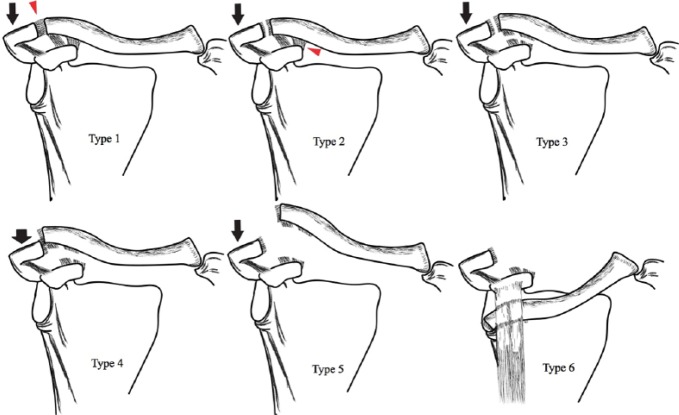
Allman and Tossy initially divided these injuries into three types depending on the severity of displacement. The original classification was modified by Rockwood and Green (Fig. 1).12,13
Fig. 1.
The classic Rockwood classification of the AC joint injury. Type I is only a sprain of the AC ligament, whereas the ligament is torn in type II injury. In type III both the AC and the CC ligaments are torn, but there is no more than 100% displacement of the distal clavicle. In type IV both ligaments are torn with posterior displacement of distal clavicle. Type V is a complex injury where the deltotrapezial fascia is stripped from its attachment, whereas in type VI injury the clavicle is moved into subcoracoid position.
Type I is a sprain injury of the AC ligament; there is no complete tear and both AC and CC ligaments are intact.
Type II is a tear of the AC ligament but not of the CC ligaments.
A type III injury involves tears of both the AC and CC ligaments, with 25% to 100% displacement of the clavicle compared with that on the contralateral side.18
In a type IV injury, both the AC and CC ligaments are torn and there is posterior displacement of the distal clavicle into the trapezius fascia.19
In a Rockwood type V injury, the AC and CC ligaments and both the origin of the deltoid and insertion of the trapezius are torn, causing extreme instability of the AC joint.20 It is a complex injury where the deltotrapezial fascia is stripped from its attachment and displacement of the clavicle is more than three times the diameter of its distal part. The CC distance is increased to 100% to 300%.
Type VI injuries are the result of inferior displacement of the distal clavicle into the subcoracoid position.
Patients with type III–VI injuries may have associated intra-articular pathologies, especially superior labral anterior posterior (SLAP) lesions.21
Clinical presentation
During the physical examination, the patient should be in the standing or sitting position, which increases the deformity due to the weight of the arm.18 Clinical examination may reveal abrasions of the shoulder and prominence of the distal clavicle. Ecchymoses and swelling may be present on the prominent distal clavicle as a result of inferior displacement of shoulder girdle. Palpation of the AC joint will reveal tenderness; shoulder range of motion is typically limited by pain.6 The presence of associated injuries should always be taken into consideration. Provocative tests for AC joint pathology (cross-arm adduction and loading of the AC joint) can be helpful to localize shoulder pain to the AC joint. These tests are especially useful in patients with type I and II (minor) injuries in which visible or palpable deformity may not be present. Selective injections with local anaesthetic may be helpful in discriminating chronic AC joint pain from other pathologies causing anterior or superior shoulder pain, but they are rarely needed for acute injuries.22
Imaging
Radiographs are the initial imaging modality of choice for diagnosis and classification of AC injuries. As soon as the patient is admitted, standard anteroposterior lateral and axillary views should be obtained as for any shoulder injury. The axillary view often helps to visualize the amount of posterior displacement of the clavicle.6 If an AC joint injury is suspected, a Zanca view is often helpful and is obtained by tilting the radiograph beam 10° to 15° cephalad compared with a standard shoulder radiograph.23 Weighted stress views of the clavicle can be obtained in order to distinguish type II and III injuries and this method was often advocated in the literature; however, this is not routinely recommended today since it does not change the course of treatment and causes patient discomfort.21 MRI is not a routine imaging method, though there is a growing interest to utilize this method to assess AC and CC ligamentous disruptions.
Management
The traditional literature supports non-operative treatment for grade I and II injuries.19 Patients with grade IV, V and VI injuries benefit from operative treatment, whereas the treatment of grade III injuries remains a controversial issue.22 Numerous surgical procedures have been described, though there is currently no gold standard for the treatment of AC injuries. The main principle of surgical therapy is accurate reduction of the AC joint in both coronal and sagittal planes.24 This is achieved either by primary repair or by reconstruction of injured ligaments and maintaining stability to protect this repair or reconstruction.25 The traditional Weaver-Dunn CA ligament transfer procedure has largely fallen into disfavour today.3
Many surgical procedures have been described; among these are screws, plates, muscle transfers, ligamentoplasty procedures and ligament reconstruction using either autograft or allografts.26 Anatomical ligament reconstruction using tendon graft can be performed by open and arthroscopic methods. Open surgery requires deltoid detachment from the clavicle and extensive soft-tissue dissection for access to the coracoid process; neurovascular structures are placed at risk because of the suboptimal visibility during tendon transfer around the coracoid.27 Most surgical procedures incorporate the use of metal hardware that could alter the biomechanics of the AC joint, which makes a second surgical intervention necessary for implant removal once the ligaments have healed.28 The hook plate, AC joint transfixion with K wires (Phemister technique) and CC fixation with a screw (Bosworth technique) are recognized as non-anatomical procedures which reduce mobility and are related to high rates of fixation failure and complications.
Recent treatment modalities for AC joint dislocation have focused on either CC ligament reconstruction or CC interval fixation.11,29 Many devices such as screws, plates, suture anchors or synthetic tapes have been used as fixation material; however, none of these methods are free of complications such as implant failure or implant migration, bony erosions and fractures of the clavicle, and recurrent dislocation. With the advancement of arthroscopic intervention techniques, the management of AC joint injuries has been shifted from open surgical procedures towards less invasive, arthroscopically-assisted or all-arthroscopic procedures.
The principle advantage of arthroscopy is that it allows the patient early release from the hospital with a shorter rehabilitation period and early return to activity.28
Arthroscopic technique
Arthroscopy allows a better, greater and clearer visibility around the coracoid, and extensive dissection of the deltotrapezial fascia is not required. This clearer visibility also puts the important neurovascular structures at less risk. The suprascapular nerve and the suprascapular artery are the structures with the closest proximity to any implanted material.24 Besides, arthroscopy makes it possible to get a straight vision of the inferior aspect of the base of the coracoid – a particularly important anatomical area, especially when placing CC fixation systems.
In any arthroscopic approach, three portals are mainly used: namely a posterior portal; an anterolateral portal for the optical device; and an operative anterosuperior portal.38 The patient is placed in the ‘beach-chair’ position and a 30° arthroscope is used to visualize the glenohumeral joint through a posterior portal. A complete diagnostic glenohumeral joint examination is performed and associated intra-articular pathologies are documented. By using an anterior portal cannula, the rotator interval tissue is removed by a motorized shaver blade until the base of the coracoid process can be visualized. At that point, a 70° arthroscope is used to get a better exposure of the base of the coracoid.26 It is very important to fully expose this area and that is best achieved again by using a motorized shaver blade or a radiofrequency device through an anterolateral portal cannula. Another option is using an anterolateral portal in line with the subcapularis tendon with the aid of an 8.5-mm arthroscopic cannula; through this portal, a radiofrequency device or a motorized shaver blade can be inserted. When the inferior base of the coracoid process is fully exposed, a supracoracoid (anteromedial) portal is established, again with the aid of an 8.5-mm arthroscopic cannula.
In the beginning, arthroscopically-assisted techniques only focused on the restoration of the CC ligament complex using strong synthetic suture material and cortical fixation buttons.31 However, it was realized later that CC repair alone does not provide proper stability; additional stability was achieved by reconstructing the AC ligament complex to a certain extent. It should be kept in mind that all described arthroscopic techniques have reported pitfalls, including implant migration, need for implant removal, failure of reduction and fracture of coracoid and clavicle.32
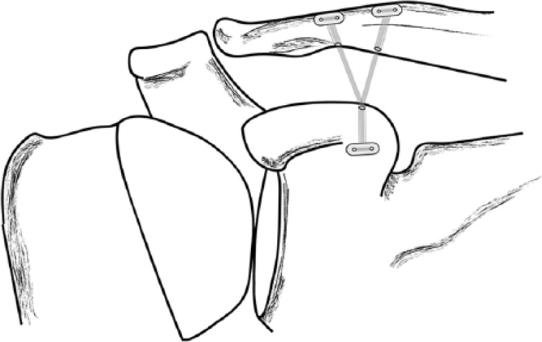
These variable arthroscopic techniques usually rely on bony tunnels and suture-graft fixation that bring some concerns into question. Use of single tunnel versus double, suture versus graft, autograft versus allograft and adjustable-length suture versus non-adjustable suture are all different variables. For instance, according to Scheibel et al, drilling two tunnels for the reconstruction of the CC complex and using an adjustable-length suture is a more anatomical method with good to excellent results.33 However, fractures of the coracoid or the clavicle remain a significant complication occurring predominantly with techniques utilizing these bony tunnels.34 Multiple passes of the drill through the clavicle during implant positioning is a predominant risk factor. It is important to create tunnels in the correct position to avoid widening a tunnel and sinking the proximal button (Fig. 2).35 In order to minimize the risk of clavicle fracture, a single drill hole through the clavicle can be opened.36
Fig. 2.

The reconstruction can be performed by opening either a single or a double tunnel from the clavicle to the coracoid. It is important to drill the bony tunnels in correct anatomical position, mimicking a native CC ligament.
Today the most commonly used arthroscopic systems are the TightRope system, suture-shuttle device and all-arthroscopic systems.
TightRope
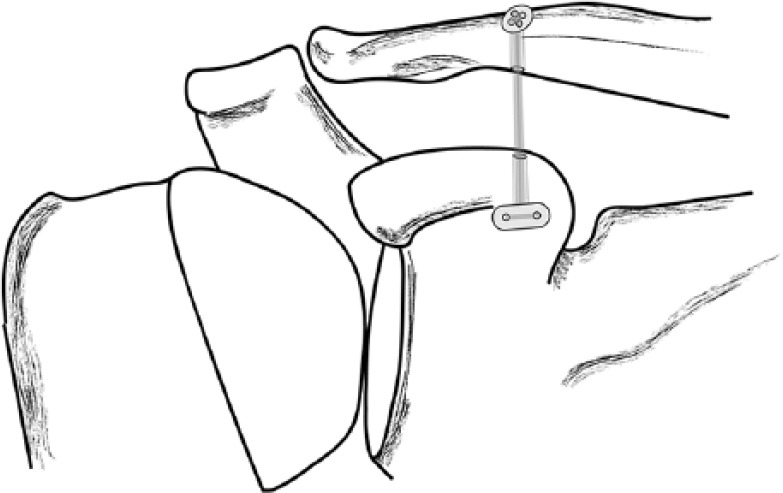
A very functional method for restoration of AC joint function is using a TightRope® (Arthrex, Naples, FL, USA) device, where a single or double drill hole through the clavicle is made. It was originally designed for syndesmotic ankle injuries and contains two titanium buttons. One button is for the clavicle and the other is for the coracoid process; they are joined by a loop of No. 5 fiberwire suture.40 This TightRope device can either be used by itself or with the augmentation of tendon allograft particularly for chronic AC joint dislocations (GraftRope, Arthrex, FL, USA). Augmentation with tendon allograft reduces the stress-rising effects of titanium buttons around the clavicle and the coracoid, so the risk of failure by suture cut-out is minimized. This procedure can also be performed with the patient in a lateral decubitus position. A 2-cm (mini-open) skin incision is made transversely along the distal clavicle and deltopectoral fascia is dissected. A bony tunnel is opened with the help of a 2.4-mm guide pin from the distal aspect of the clavicle toward the base of the coracoid. The oblong button first passes through the clavicle and then through the coracoid drill hole. Once it passes the coracoid, it is flipped to horizontal position and the AC joint is reduced by manual pressure (Fig. 3).
Fig. 3.

A very functional method for restoration of AC joint function is using a TightRope (Arthrex, Naples FL) device. This system can be augmented by using an allograft (GraftRope, Arthrex FL), particularly in chronic AC joint injuries.
Suture-shuttle (coracoid cerclage technique)
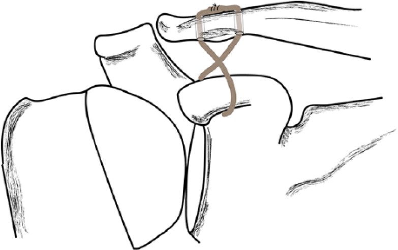
Another safe arthroscopic procedure is using a suture-shuttle device augmented by a tendon graft, especially in chronic AC injuries. A 45° suture-shuttling device can be inserted through the supracoracoid portal and placed down the medial border of the coracoid. The shuttle-suture is pulled through the anterior portal and a suture loop is established below the coracoid. When a decision is made to use a tendon graft, a tendon autograft or allograft (commonly non-irradiated) is prepared at the back table. Several locking sutures are placed at the free end of the graft. Afterwards, with the aid of a guide pin, two bony tunnels are established in the clavicle in a crossing pattern. These are the conoid and the trapezoid tunnels. The leading-end graft sutures are pulled through the conoid clavicle tunnel exiting the anterior portal. With the help of the suture loop placed under the coracoid, the leading end of the graft is shuttled from lateral to medial exiting the supracoracoid portal. Finally, the graft is shuttled through the supracoracoid portal exiting the trapezoid clavicle tunnel so at the end the whole graft is shuttled around the coracoid and through the clavicle tunnels in a crossing pattern (Fig. 4).
Fig. 4.

Repair with allograft in a crossing pattern. Again, two drill holes are opened to the clavicle.
All-arthroscopic method
The other arthroscopic procedure is the all-arthroscopic technique that is best reserved for acute cases, particularly for acute grade 4 and 5 lesions. The term ‘acute’ can be used for the surgery performed within the first six weeks after the injury. This procedure consists of two stages. In the first stage, the reconstruction of CC ligaments is performed, and in the second stage, the reconstruction of superior AC ligaments is performed. That means that this system relies on restoration of the AC joint on both the horizontal and coronal planes, a particularly important issue in terms of native joint physiodynamics.
The portals used in this procedure are named in keeping with arthroscopic shoulder portals described by Lafosse et al for the arthroscopic Latarjet operation. The so-called D portal is the main viewing portal and it is placed off the anterolateral corner of the acromion. The C portal is placed off the lateral border of the acromion and it allows working over the superior aspect of the AC joint and lateral clavicle. The standard anterior portal is the E portal that is placed immediately lateral to the tip of the coracoid process. Again, the inferior part of the coracoid is cleared using an electrocautery device to prepare a place for drilling a tunnel connecting the clavicle to the coracoid. This tunnel is used to pass two ABS TightRopes to reconstruct the coracoclavicular ligaments. Also, the superior surfaces of the clavicle and acromion are cleared to visualize reduction of the AC joint and to allow the suture bridge repair of the AC ligaments. With the aid of an aiming guide that is placed under the coracoid base, a cannulated drill is inserted through the clavicle and the coracoid. A nitinol suture-retrieving loop is retrieved through a 1-cm skin incision over the superior aspect of the clavicle in line with the coracoid using an arthroscopic grasper. This loop passes upward through the coracoid, clavicle and skin, allowing a TightRope to be pulled upward through the tunnels. After that, two self-locking ABS TightRopes are pulled through the coracoid and clavicle drill holes on a FiberWire lead suture. As the TightRopes are pulled through the tunnel, a Dog-Bone button is carefully positioned under the coracoid with the aid of an arthroscopic grasper, if required, and another Dog-Bone button is used to secure the TightRopes on top of the clavicle.
For the reconstruction of AC ligaments, a 1-cm incision is opened over the centre of the AC joint. Two Q-Fix arthroscopic anchors (Arthrocare, Austin, TX, USA) are inserted into the superior aspect of the acromion to create a suture bridge over the acromioclavicular joint and reconstruct the acromioclavicular ligaments. At the end, an eight-stranded suture-bridge construct is created over the AC joint.
In a cadaveric pilot study performed by Banaszek et al, arthroscopically-assisted techniques are all considered safe and avoid surrounding neurovascular structures,24 unlike open surgical interventions. However, the proximity of some important neurovascular structures, particularly the suprascapular nerve and the suprascapular artery, must always be remembered. On the other hand, there are also other less frequent complications, such as infection, that are usually superficial, post-operative capsulitis and pain, particularly night pain,37 irritation over the superior aspect of the implant and CC calcification.
Discussion
AC joint injuries still represent a diagnostic and therapeutic challenge for most shoulder surgeons. A rough clinical evaluation is necessary, and the surgeon should perform a detailed physical examination including all tests for AC stability (e.g. pain, translation, cross-body test). The scapular motion should also be evaluated.7 Evaluation of the neurovascular structures is also mandatory as the brachial plexus and subclavian arteries pass inferior to the medial third of the clavicle and could be jeopardized in rare instances.21 The treatment of type III injuries is controversial; however, according to recent studies, the current trend is towards conservative treatment as the results are similar with both methods.8 It has been proposed that the treatment of acute type III AC dislocation depends mainly on the demands and expectations of the patient. These are related mainly to the patient’s occupation and demands of daily living and activity in sports.39
The surgery indication for acute type IV, V and VI injuries is clear. There is no optimal surgical technique described for the treatment of these injuries yet; clinical and anatomical studies still continue for more anatomic reconstructions. The original Weaver-Dunn technique described in 1972 consists of excising the distal part of the clavicle, releasing the coracoacromial ligament from its acromial attachment and transferring it to the distal clavicle. However, recent biomechanical studies have demonstrated that anatomical reconstruction using free grafts provides better stability than other ligament transfer procedures. Holding the clavicle in a reduced position is usually sufficient as primary reconstruction by means of tension bands and K-wires applied through the AC joint or extra-articular Bosworth screws. These are all simple techniques that rely on the primary healing of the CC ligaments.40 However, they are all non-dynamic fixation methods that can result in implant failure.41 It has been reported that the CA ligament transfer, hook-plate fixation, the AC K-wire fixation and the CC screw fixation could be considered as biomechanically insufficient.42 On the other hand, the requirement of multiple drill holes in the clavicle can also create stress risers on bone that might result in implant cut-out.43 Other complications are acromial osteolysis and fracture, AC joint osteoarthritis, subacromial impingement and rotator cuff tears. The need for a second surgery for implant removal is another disadvantage that might lead to late fracture of the distal clavicle after a low-energy trauma.44
With the advancement of arthroscopic techniques, improved functional results are obtained with better cosmesis and patients are allowed an earlier return to their daily living.45
There is a variety of arthroscopic techniques divided into two groups focusing either on ligament healing or on ligament reconstruction. The repair procedure can be performed either by mini-open arthroscopically-assisted technique or by the all-arthroscopic technique. None of the mentioned techniques can be considered free of complications. Nevertheless, arthroscopic AC joint repair is overall a safe procedure that promises good results.
Among these arthroscopic techniques, the TightRope (Arthrex, Naples, FL, USA) is the most widely used one. It is a double metallic button with four FiberWire strands. Although the rehabilitation period and the time needed to return to daily activities is shorter, controversies exist because compared with other methods, some patients might need a longer rehabilitation time. Also, this method cannot be considered as cost-effective as open methods although it is a single surgical procedure. Osteolysis around the endobuttons is another complication that can occur during the follow-up period, though this is less frequent compared with open methods. The results of this double-button device are satisfactory with good functional outcome, especially in acute type III and type IV AC lesions. At the mid-term follow-up period, accurate joint congruency and ligament healing is achieved.
The reconstruction of the AC joint by the all-arthroscopic technique also relies on using the TightRope system for the CC part as well as simple suture anchors for the AC part. This all-arthroscopic method is one of the most recently introduced techniques and the results are promising; however, there is still time needed for this to be fully supported by the literature. The entry of the drill below the coracoid and its exit above the clavicle must be properly oriented, so surgeons’ arthroscopic skills are the key point for a successful outcome in this surgery. Assessment of the inferior surface of the acromion is also critical because penetration of anchors is possible and that might lead to irritation of the rotator cuff.46
Another arthroscopically-assisted technique is restoration of the CC ligament by using a double-bundle free tendon graft (autograft or allograft) and a suture-shuttling device, the so-called coracoid cerclage technique. In this procedure a conoid and a trapezoid tunnel are drilled in the clavicle and the tendon graft is shuttled in a crossing pattern (Fig. 4).
CC ligament reconstruction can also be achieved by a single-adjustable loop length suspensory fixation device; loss of reduction is common with this method as the two native components (trapezoid and conoid) of the original CC ligament are not restored.
The overall rate of serious complications following arthroscopic fixation of the AC joint including infection requiring further surgery or neurovascular compromise is low, while the rate of fracture and loss of reduction remains a concern.
In 2011, Laedermann et al conducted a retrospective study and analyzed the functional radiological and isokinetic results of 37 patients who underwent open reduction of the AC joint with the CC and AC cerclage technique.47 They obtained good to excellent results without complications linked to implant irritation, implant removal or graft source morbiditiy. Two years later, he performed a biomechanical cadaveric study where he compared the cerclage technique, TightRope, and locking plate systems with each other and found out that the cerclage technique mainly mimicked the native joint physiologically whereas the TightRope system provided the highest stability but failed earlier due to increased stiffness.48 These two studies emphasized the importance of restoring the joint structure in both the horizontal and vertical planes, an important parameter to provide native anatomy which is sometimes overlooked when introducing new materials and less invasive techniques. These studies also raised the question as to whether these new endoscopic techniques and new materials provide absolute superiority to more conventional methods.
Conclusions
There is no current gold standard for the treatment of AC joint injuries. With recent advances in arthroscopic techniques and introduction of new equipment, the treatment modalities are shifted towards less invasive and more arthroscopically-assisted methods. However, the results are still comparable with the more conventional methods. Whenever an arthroscopic approach is chosen, it should be remembered that surgeons’ arthroscopic expertise probably plays the most important role in achieving a good outcome.
Footnotes
ICMJE Conflict of interest statement: None declared.
Funding statement
No benefits in any form have been received or will be received from a commercial party related directly or indirectly to the subject of this article.
References
- 1. Mazzocca AD, Arciero RA, Bicos J. Evaluation and treatment of acromioclavicular joint injuries. Am J Sports Med 2007;35:316-329. [DOI] [PubMed] [Google Scholar]
- 2. Andreani L, Bonicoli E, Parchi P, Piolanti N, Michele L. Acromio-clavicular repair using two different techniques. Eur J Orthop Surg Traumatol 2014;24:237-242. [DOI] [PubMed] [Google Scholar]
- 3. Stucken C, Cohen SB. Management of acromioclavicular joint injuries. Orthop Clin North Am 2015;46:57-66. [DOI] [PubMed] [Google Scholar]
- 4. Cook JB, Tokish JM. Surgical management of acromioclavicular dislocations. Clin Sports Med 2014;33:721-737. [DOI] [PubMed] [Google Scholar]
- 5. Macdonald PB, Lapointe P. Acromioclavicular and sternoclavicular joint injuries. Orthop Clin North Am 2008;39:535-545. [DOI] [PubMed] [Google Scholar]
- 6. Willimon SC, Gaskill TR, Millett PJ. Acromioclavicular joint injuries: anatomy, diagnosis, and treatment. Phys Sportsmed 2011;39:116-122. [DOI] [PubMed] [Google Scholar]
- 7. Beitzel K, Mazzocca AD, Bak K, et al. ; Upper Extremity Committee of ISAKOS. ISAKOS upper extremity committee consensus statement on the need for diversification of the Rockwood classification for acromioclavicular joint injuries. Arthroscopy 2014;30:271-278. [DOI] [PubMed] [Google Scholar]
- 8. Lizaur A, Sanz-Reig J, Gonzalez-Parreño S. Long-term results of the surgical treatment of type III acromioclavicular dislocations: an update of a previous report. J Bone Joint Surg [Br] 2011;93-B:1088-1092. [DOI] [PubMed] [Google Scholar]
- 9. Hudson VJ. Evaluation, diagnosis, and treatment of shoulder injuries in athletes. Clin Sports Med 2010;29:19-32. [DOI] [PubMed] [Google Scholar]
- 10. Cadenat F. The treatment of dislocations and fractures of the outer end of the clavicle. Int Clin 1917;1:145-169. [Google Scholar]
- 11. Cutbush K, Hirpara KM. All-arthroscopic technique for reconstruction of acute acromioclavicular joint dislocations. Arthrosc Tech 2015;4:e475-e481. [DOI] [PMC free article] [PubMed] [Google Scholar]
- 12. Tossy JD, Mead NC, Sigmond HM. Acromioclavicular separations: useful and practical classification for treatment. Clin Orthop Relat Res 1963:111-119. [PubMed] [Google Scholar]
- 13. Rockwood C. Injuries to the acromioclavicular joint. In: Ca R, Green D. eds. Fractures in adults. 2nd ed. Philadelphia, PA: JB Lippincott, 1984:860-910. [Google Scholar]
- 14. Vrgoč G, Japjec M, Jurina P, et al. Operative treatment of acute acromioclavicular dislocations Rockwood III and V-Comparative study between K-wires combined with FiberTape(®) vs. TightRope System. Injury 2015;46:S107-S112. [DOI] [PubMed] [Google Scholar]
- 15. Tauber M. [Acromioclavicular injuries in professional athletes]. Orthopade 2014;43:249-255. [DOI] [PubMed] [Google Scholar]
- 16. Hastings DE, Horne JG. Anterior dislocation of the acromioclavicular joint. Injury 1979;10:285-288. [DOI] [PubMed] [Google Scholar]
- 17. Loriaut P, Casabianca L, Alkhaili J, et al. Arthroscopic treatment of acute acromioclavicular dislocations using a double button device: clinical and MRI results. Orthop Traumatol Surg Res 2015;101:895-901. [DOI] [PubMed] [Google Scholar]
- 18. Li X, Ma R, Bedi A, et al. Management of acromioclavicular joint injuries. J Bone Joint Surg [Am] 2014;96-A:73-84. [DOI] [PubMed] [Google Scholar]
- 19. Modi CS, Beazley J, Zywiel MG, Lawrence TM, Veillette CJ. Controversies relating to the management of acromioclavicular joint dislocations. Bone Joint J 2013;95-B:1595-1602. [DOI] [PubMed] [Google Scholar]
- 20. Virtanen KJ, Remes VM, Tulikoura IT, et al. Surgical treatment of Rockwood grade-V acromioclavicular joint dislocations: 50 patients followed for 15–22 years. Acta Orthop 2013;84:191-195. [DOI] [PMC free article] [PubMed] [Google Scholar]
- 21. Warth RJ, Martetschläger F, Gaskill TR, Millett PJ. Acromioclavicular joint separations. Curr Rev Musculoskelet Med 2013;6:71-78. [DOI] [PMC free article] [PubMed] [Google Scholar]
- 22. Fraser-Moodie JA, Shortt NL, Robinson CM. Injuries to the acromioclavicular joint. J Bone Joint Surg [Br] 2008;90-B:697-707. [DOI] [PubMed] [Google Scholar]
- 23. Zanca P. Shoulder pain: involvement of the acromioclavicular joint. (Analysis of 1,000 cases). Am J Roentgenol Radium Ther Nucl Med 1971;112:493-506. [DOI] [PubMed] [Google Scholar]
- 24. Banaszek D, Pickell M, Wilson E, et al. Anatomical evaluation of the proximity of neurovascular structures during arthroscopically assisted acromioclavicular joint reconstruction: a cadaveric pilot study. Arthroscopy 2017;33:75-81. [DOI] [PubMed] [Google Scholar]
- 25. Nguyen V1., Williams G, Rockwood C. Radiography of acromioclavicular dislocation and associated injuries. Crit Rev Diagn Imaging 1991;32:191-228. [PubMed] [Google Scholar]
- 26. Parnes N, Friedman D, Phillips C, Carey P. Outcome after arthroscopic reconstruction of the coracoclavicular ligaments using a double-bundle coracoid cerclage technique. Arthroscopy 2015;31:1933-1940. [DOI] [PubMed] [Google Scholar]
- 27. DeBerardino TM, Pensak MJ, Ferreira J, Mazzocca AD. Arthroscopic stabilization of acromioclavicular joint dislocation using the AC graftrope system. J Shoulder Elbow Surg 2010;19:47-52. [DOI] [PubMed] [Google Scholar]
- 28. Natera-Cisneros L, Sarasquete-Reiriz J, Escolà-Benet A, Rodriguez-Miralles J. Acute high-grade acromioclavicular joint injuries treatment: arthroscopic non- rigid coracoclavicular fixation provides better quality of life outcomes than hook plate ORIF. Orthop Traumatol Surg Res 2016;102:31-39. [DOI] [PubMed] [Google Scholar]
- 29. Shin SJ, Kim NK. Complications after arthroscopic coracoclavicular reconstruction using a single adjustable-loop-length suspensory fixation device in acute acromioclavicular joint dislocation. Arthroscopy 2015;31:816-824. [DOI] [PubMed] [Google Scholar]
- 30. Pauly S, Gerhardt C, Haas NP, Scheibel M. Prevalence of concomitant intraarticular lesions in patients treated operatively for high-grade acromioclavicular joint separations. Knee Surg Sports Traumatol Arthrosc 2009;17:513-517. [DOI] [PubMed] [Google Scholar]
- 31. Izadpanah K, Jaeger M, Ogon P, Südkamp NP, Maier D. Arthroscopically assisted reconstruction of acute acromioclavicular joint dislocations: anatomic AC ligament reconstruction with protective internal bracing-the “AC-RecoBridge” technique. Arthrosc Tech 2015;4:153-161. [DOI] [PMC free article] [PubMed] [Google Scholar]
- 32. Balog TP, Min KS, Rumley JC, Wilson DJ, Arrington ED. Arthroscopic anatomic coracoclavicular ligament repair using a 6-strand polyester suture tape and cortical button construct. Arthrosc Tech 2015;4:757-761. [DOI] [PMC free article] [PubMed] [Google Scholar]
- 33. Scheibel M, Dröschel S, Gerhardt C, Kraus N. Arthroscopically assisted stabilization of acute high-grade acromioclavicular joint separations. Am J Sports Med 2011;39:1507-1516. [DOI] [PubMed] [Google Scholar]
- 34. Woodmass JM, Esposito JG, Ono Y, et al. Complications following arthroscopic fixation of acromioclavicular separations: a systematic review of the literature. Open Access J Sports Med 2015;6:97-107. [DOI] [PMC free article] [PubMed] [Google Scholar]
- 35. Spoliti M, De Cupis M, Via AG, Oliva F. All arthroscopic stabilization of acute acromioclavicular joint dislocation with fiberwire and endobutton system. Muscles Ligaments Tendons J 2015;4:398-403. [PMC free article] [PubMed] [Google Scholar]
- 36. Ranne JO, Sarimo JJ, Rawlins MI, Heinonen OJ, Orava SY. All-arthroscopic double-bundle coracoclavicular ligament reconstruction using autogenous semitendinosus graft: a new technique. Arthrosc Tech 2012;1:e11-e14. [DOI] [PMC free article] [PubMed] [Google Scholar]
- 37. Kany J, Amaravathi RS, Guinand R, Valenti P. Arthroscopic acromioclavicular joint reconstruction using a synthetic ligament device. Eur J Orthop Surg Traumatol 2012;22:357-364. [Google Scholar]
- 38. Motta P, Bruno L, Maderni A, Tosco P, Mariotti U. Acute lateral dislocated clavicular fractures: arthroscopic stabilization with TightRope. J Shoulder Elbow Surg 2014;23:47-52. [DOI] [PubMed] [Google Scholar]
- 39. Korsten K, Gunning AC, Leenen LP. Operative or conservative treatment in patients with Rockwood type III acromioclavicular dislocation: a systematic review and update of current literature. Int Orthop 2014;38:831-838. [DOI] [PMC free article] [PubMed] [Google Scholar]
- 40. Kienast B, Thietje R, Queitsch C, Gille J, Schulz AP, Meiners J. Mid-term results after operative treatment of rockwood grade III-V acromioclavicular joint dislocations with an AC-hook-plate. Eur J Med Res 2011;16:52-56. [DOI] [PMC free article] [PubMed] [Google Scholar]
- 41. Graupe F, Dauer U, Eyssel M. [late results of surgical treatment of Tossy III acromioclavicular joint separation with the Balser plate]. Unfallchirurg 1995;98:422-426. [PubMed] [Google Scholar]
- 42. Jari R, Costic RS, Rodosky MW, Debski RE. Biomechanical function of surgical procedures for acromioclavicular joint dislocations. Arthroscopy 2004;20:237-245. [DOI] [PubMed] [Google Scholar]
- 43. Frank RM, Trenhaile SW. Arthroscopic-assisted acromioclavicular joint reconstruction using the TightRope device with allograft augmentation: surgical technique. Arthrosc Tech 2015;4:293-297. [DOI] [PMC free article] [PubMed] [Google Scholar]
- 44. Nadarajah R, Mahaluxmivala J, Amin A, Goodier DW. Clavicular hook-plate: complications of retaining the implant. Injury 2005;36:681-683. [DOI] [PubMed] [Google Scholar]
- 45. Defoort S, Verborgt O. Functional and radiological outcome after arthroscopic and open acromioclavicular stabilization using a double-button fixation system. Acta Orthop Belg 2010;76:585-591. [PubMed] [Google Scholar]
- 46. Nordin JS, Aagaard KE, Lunsjö K. Chronic acromioclavicular joint dislocations treated by the GraftRope device. Acta Orthop 2015;86:225-228. [DOI] [PMC free article] [PubMed] [Google Scholar]
- 47. Lädermann A, Grosclaude M, Lübbeke A, et al. Acromioclavicular and coracoclavicular cerclage reconstruction for acute acromioclavicular joint dislocations. J Shoulder Elbow Surg 2011;20:401-408. [DOI] [PubMed] [Google Scholar]
- 48. Lädermann A, Gueorguiev B, Stimec B, et al. Acromioclavicular joint reconstruction: a comparative biomechanical study of three techniques. J Shoulder Elbow Surg 2013;22:171-178. [DOI] [PubMed] [Google Scholar]


