

Abstract
Background
Echocardiography involves strenuous postures of the upper limbs. This study explored the physical workload in the neck and upper limbs in sonographers performing echocardiography, and the extent to which the workload differs from than in other work tasks (other sonographic examinations, and nonsonographic tasks).
Methods
The physical load was assessed by inclinometry, goniometry, and electromyography methods in 33 female sonographers during authentic work using three different echocardiography techniques and other work tasks.
Results
Echocardiography was characterized by low velocities of the head, arms, and wrists, and a low proportion of muscular resting time in the forearms, in the transducer limb, and the computer limb. The transducer limb was more elevated in one of the techniques, but this technique also involved a higher proportion of muscular resting time of the trapezius muscle. We also found a high proportion of awkward wrist postures in the transducer wrist in all three techniques; in one due to prolonged flexion, and in the others due to prolonged extension. Other work tasks were less static, and were performed with higher upper arm and wrist velocities.
Conclusion
None of the three echocardiography techniques was optimal concerning physical workload. Thus, to achieve more variation in physical load we recommend that the equipment be arranged so that the sonographer can alternate between two different techniques during the workday. We also propose alternation between echocardiography and nonsonographic tasks, in order to introduce variation in the physical workload. Clinical expertise should be used to achieve further improvements.
Keywords: Echocardiography, Ergonomics, Technical measurements, Work postures
1. Introduction
The use of sonography has increased over recent decades, both in the number of examinations and the number of hours of scanning per day for sonographers [1], [2], [3], [4]. Sonography involves strenuous postures and precise movements of the hand manipulating the transducer [5], [6], [7], which are two well-known risk factors for neck and upper extremity pain [8]. Echocardiography, sonography of the heart, is especially demanding as it involves considerable force, static postures, and monotonous movements [9]. In order to obtain the best images it is often necessary to apply a high sustained pressure with the transducer against the patient's chest [10], which is generally not needed in other types of sonography [11]. Echocardiography also includes intense work on the keyboard, which is another risk factor [12]. Most echocardiographers also perform other sonographic examinations, for example, vascular examinations, and nonsonographic tasks, for example, spirometry. It has been shown that those who perform echocardiography have a higher prevalence of elbow/hand pain than those who only perform other sonographic examinations [13].
In a recent study on Swedish echocardiographers, we identified three techniques. The most commonly used technique (by 47%) was to hold the transducer in the left hand, with the patient lying to the left of the ultrasound machine [13]. Two alternative techniques were identified. In these the examiner held the transducer in the right hand with the patient lying to the right of the ultrasound machine. Which technique was used depended on local tradition. It is not known if any one of these three techniques is more favorable than the others in terms of the physical load, or whether alternating between different techniques in echocardiography would provide a variation in workload. Furthermore, it is unknown to what extent the physical workload in echocardiography differs from that in other kinds of sonographic examinations, or nonsonographic tasks in the ward; that is, whether a variation in tasks would be favorable with respect to workload.
Previous knowledge on sonographers' physical workload is predominantly based on observational studies and surveys. Technical measurements provide quantitative exposure data, with the obvious advantage that the results are independent of the individual and the observer [14]. Technical measurements of workload in sonography have been applied in a few studies [15], [16], [17], [18], but none of these has explored differences between the three echocardiography techniques, using three different technical measurements.
The main aim of this study was, thus, to compare the physical workload in the neck and upper limbs associated with three different techniques in echocardiography We also investigated the extent to which echocardiography differs from other sonographic examinations and nonsonographic tasks, with respect to workload on the neck and upper limbs, using the most common echocardiography technique as a proxy for all types of techniques.
2. Materials and methods
2.1. Participants
Thirty-three experienced female sonographers employed at clinical physiology and cardiology departments, mean age 47 years (range, 28–66 years), participated in the study. All were right-handed. We established contact with the head of the different departments during a previous study, and at that time informed the sonographers about the technical measurements [19]. Those who were interested in participating notified their head of department, and contact was established with the researchers. The measurements were planned together with the head of each clinic. None of the participants reported musculoskeletal pain or discomfort of such intensity that it influenced their working technique. Data collection involved nine hospitals in Southern Sweden during 2011–2015. At least one sonographer and at most six per hospital participated. Technique (T)1 and T3 were represented in four hospitals and T2 in five hospitals.
2.2. Work tasks
We investigated three different types of work tasks: echocardiography, other sonographic examinations (excluding echocardiography), and nonsonographic tasks. In echocardiography, the sonographer usually sits during the examination, maneuvering a transducer connected to the ultrasound machine by a cable in one hand, controlling the keyboard, integrated to the ultrasound machine, with the other hand, and at the same time, observing the images on a screen placed on top of the ultrasound machine. The sonographer applies pressure on the transducer with the hand to achieve optimal contact [20]. The sonographers in this study used one of three techniques, denoted T1 (10 participants), T2 (13 participants), and T3 (10 participants). In T1 the table was placed on the left side of the ultrasound machine, while in T2 and T3 the table was placed on the right side. In T1 the patient faced the examiner, who held the transducer in the left hand and handled the keyboard with the right hand (Fig. 1). In T2 the patient faced the examiner, who held the transducer in the right hand and handled the keyboard with the left hand (Fig. 2). In T3 the patient faced away from the examiner, who held the transducer in the right hand and handled the keyboard with the left hand (Fig. 3).
Fig. 1.

Echocardiographic examination using Technique 1.
Fig. 2.
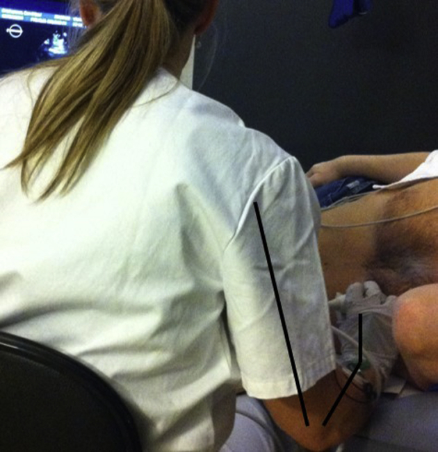
Echocardiographic examination using Technique 2.
Fig. 3.

Echocardiographic examination using Technique 3.
Among the 10 sonographers using T1, six also performed other sonographic examinations, for example, abdominal aorta scanning, mapping of veins, vascular scanning, and examination of fistulas. Their working posture varied depending on the type of examination. For example, in some examinations the sonographer could change transducer hands. In addition, these 10 sonographers also performed other nonsonographic tasks, such as computer work, booking patients, spirometry, lung scintigraphy, cleaning the equipment between examinations, and fetching the patients from the waiting room.
2.3. Recordings of physical workload
The participants carried portable data loggers that recorded and stored data. The equipment was applied in the morning, and reference postures and maximal contractions were performed. An observer followed each participant and made precise notes of the tasks that they performed. These tasks were then classified as echocardiography, other sonographic examinations, or nonsonographic tasks. We excluded data recorded during long breaks, such as lunch, from the analyses. After the recording the data were transferred to a personal computer.
To compare the different echocardiography techniques, we analyzed the data recorded while the sonographer held the transducer in her hand (transducer time). Transducer time was defined as the period from when the sonographer removed the transducer from the holder, to replacement of the transducer in the holder. The median recorded transducer time for T1 was 72 minutes (mean, 71 minutes; range, 20–169 minutes), for T2, 56 minutes (mean, 63 minutes; range, 28–94 minutes), and for T3, 87 minutes (mean, 79 minutes; range, 30–127 minutes). Data were recorded for both the transducer limb (the arm holding the transducer, i.e., the left limb in T1 and the right limb in T2 and T3), and the keyboard limb (the arm used to operate the keyboard on the ultrasound machine). For other sonographic examinations and other work tasks, only data from the right limb were analyzed, as this was the dominant limb in all participants.
In the 10 participants who used T1, the physical workload was recorded during a complete working day, and included all the work tasks performed that day. The number of examinations and other work tasks varied depending on the appointment list for that day. The examination time was defined as the time when the patient entered the room until he/she left the room. Data were recorded from at least two echocardiographic examinations per sonographer (median recording time, 159 minutes; mean, 159 minutes; range, 46–343 minutes). Data were also recorded from other sonographic examinations in six of these participants (median, 121 minutes; mean, 133 minutes; range, 64–240 minutes). Data were recorded for nonsonographic tasks in all 10 sonographers using T1 (median, 57 minutes; mean, 69 minutes; range, 1–217 minutes). For these, only data from the right limb were analyzed as this was the dominant limb in all participants, and was thus expected to have the highest exposure. We then compared the exposure of the transducer limb (i.e., the left limb) in echocardiography with that in the right limb in other tasks.
2.4. Inclinometry
Inclinometers based on triaxial accelerometers were used in combination with a data logger (Logger Teknologi HB, Åkarp, Sweden) to measure and record postures (inclination relative to the line of gravity) and movements of the head, upper back, and both upper arms [21]. The inclinometers were attached to the forehead, to the right of the spine at the C7 level (upper back), and on both upper arms just below the insertion of the deltoid muscles. A reference posture was recorded for the head and upper back (0° inclination) with the participant standing upright looking at a mark at eye level. To determine reference positions for the arms, the participant was seated with the side of the body leaning towards the back of a chair and the arm hanging vertically over the back of the chair, with a 2-kg dumbbell in the hand [22].
2.5. Goniometry
Biaxial flexible electrogoniometers (SG75; Biometrics Ltd., Cwmfelinfach, Gwent, UK) were used in combination with a data logger to measure and record postures and movements of the wrists [23]. In the first 20 recordings, a logger with a sampling rate of 20 Hz was used (Logger Teknologi HB), whereas in the remaining 13, a Mobi-8 data logger with a sampling rate of 128 Hz was used (TMS International, Oldenzaal, The Netherlands). The electrogoniometers were attached bilaterally to the wrists, one block on the third metacarpal bone and the other one at the midline between the forearm bones. The reference position (0° flexion and deviation) was defined with the forearm and hand resting on a table with the elbow flexed 90°. The hand was adjusted so that the third metacarpal bone of the middle finger and the midline between the forearm bones pointed along the same direction, with a sight line between the ulna and third metacarpal bone [24].
2.6. Electromyography
Bipolar surface electromyography (EMG) was performed with Ag/AgCl electrodes (Ambu Neuroline 720; Ambu A/S, Ballerup, Denmark), with an interactive diameter of 6 mm and a center-to-center distance of 20 mm, to record bilateral muscular activity of the trapezius muscles and the forearm extensor muscles (mm extensor carpi radialis longus and brevis) at a sampling rate of 1,024 Hz [25]. The electrodes were attached to the descending part of the upper trapezius muscle, 2 cm lateral to the line between the seventh cervical vertebra and the lateral edge of the acromion. The forearm electrodes were applied to the most prominent part of the muscles, approximately one-third of the distance from the lateral epicondyle to the ulnar styloid. The muscular activity during work was normalized to the maximum voluntary EMG activity recorded during maximal voluntary contractions [25].
2.7. Statistical analysis
2.7.1. Summary measures
The 50th percentiles of the angular distributions for work postures of the head, upper back, and both upper arms were calculated. Inclination was assessed both forwards/backwards and sideways, where positive values denote forwards and right sideways [26]. The median angular velocity distributions were obtained for the head and both upper arms, as well as the percent of the time the upper arm elevation was above 30° and 60° for both arms. The angular distributions and the median angular velocity distributions for both wrists were obtained for the 10th, 50th, and 90th percentiles. Positive values denote palmar flexion and ulnar deviation, while negative values denote dorsal extension and radial deviation [27]. Awkward postures were defined as the percent of the time exceeding 40° dorsal extension or 5° palmar flexion. The peak load was defined as the 90th percentile of the EMG amplitude distributions. The proportion of time when the muscular activity was < 0.5% of the maximum voluntary EMG activity was defined as muscular rest (% of time) [25].
2.7.2. Statistical methods
As data for some of the measures were skewed, nonparametric statistical tests were used, and group medians are therefore presented. Group means and standard deviations are also used to enable comparisons with earlier studies [26], [27]. In comparisons of independent observations, that is, of the different echocardiography techniques, the Mann–Whitney U test was used. Data recorded for the transducer limb and keyboard limb were analyzed separately. In comparisons of dependent observations, that is, echocardiography versus other sonographic examinations and nonsonographic tasks, the Wilcoxon matched-pairs signed-rank test was used. We then compared the recordings from the transducer limb, that is, the left limb, with the right limb in other work tasks (as all participants were right-handed). A p value < 0.05 was regarded as indicating statistically significant differences. We used SPSS version 22 (IBM, Armonk, NY, USA).
2.8. Ethical considerations
The study was approved by the Regional Ethics Committee in Lund, Sweden (No. 2010/19).
3. Results
3.1. Different techniques in echocardiography
3.1.1. Transducer limb
The results obtained from measurements on the transducer limb during echocardiography using three different techniques are given in Table 1. The upper arm was elevated above 30° 93% of the time in T3 (where the arm was held around the patient), which was more than twice as long as in T1 and T2. By contrast, the proportion of time the trapezius muscle was at rest was considerable higher in T3 (18%) than in the other two techniques (∼6%).
Table 1.
Physical workload in the neck and transducer limb during echocardiography transducer time in 33 female sonographers using three different techniques (T1, T2, and T3; p values from Mann–Whitney U tests∗)
| T1 |
T2 |
T1 vs T2 |
T3 |
T1 vs T3 |
T2 vs T3 |
||||
|---|---|---|---|---|---|---|---|---|---|
|
N = 10 |
N = 13 |
N = 10 |
|||||||
| Med | Mean (SD) | Med | Mean (SD) | p | Med | Mean (SD) | p | p | |
| Neck/shoulder/upper back | |||||||||
| Head | |||||||||
| Inclination (°, 50th percentile)† | –1.3 | –0.1 (7.0) | 2.8 | 2.3 (3.7) | 0.55 | 3.0 | 3.6 (4.1) | 0.25 | 0.57 |
| Sideways inclination (°, 50th percentile)‡ | 0.6 | 1.1 (2.3) | 0.7 | 0.6 (1.3) | 0.88 | 0.3 | –1.1 (2.5) | 0.12 | 0.12 |
| Velocity (°/s, 50th percentile) | 3.2 | 3.6 (1.1) | 3.4 | 3.5 (0.7) | 0.62 | 3.7 | 3.7 (1.1) | 0.81 | 0.81 |
| Upper back inclination (°, 50th percentile)‡ | 6.5 | 7.5 (5.3) | 11 | 11 (3.9) | 0.12 | 9.8 | 9.4 (4.2) | 0.35 | 0.41 |
| Upper arm | |||||||||
| Elevation (°, 50th percentile) | 29 | 28 (4.4) | 19 | 22 (12) | 0.13 | 44 | 47 (9.4) | <0.001 | 0.001 |
| Elevation above 30° (% time) | 47 | 41 (20) | 26 | 36 (32) | 0.41 | 94 | 93 (5.5) | <0.001 | 0.001 |
| Elevation above 60° (% time) | 4.9 | 5.4 (3.5) | 2.4 | 3.4 (3.2) | 0.11 | 4.8 | 15 (2.5) | 0.68 | 0.12 |
| Velocity (°/s, 50th percentile) | 3.6 | 4.1 (1.5) | 4.2 | 4.1 (0.7) | 0.50 | 4.2 | 4.3 (0.9) | 0.29 | 0.68 |
| Trapezius muscle | |||||||||
| Peak load (% MVE, 90th percentile) | 9.4 | 8.7 (3.5) | 11 | 11 (5.5) | 0.37 | 7.8 | 8.0 (2.3) | 0.57 | 0.17 |
| Rest (<0.5% MVE % time) | 4.3 | 6.1 (5.0) | 1.8 | 5.6 (10) | 0.06 | 16 | 18 (15) | 0.09 | 0.03 |
| Forearm/wrist | |||||||||
| Wrist flexion/extension§ | |||||||||
| 10th percentile (°) | –50 | –49 (5.9) | –57 | –57 (10) | 0.03 | –24 | –25 (12) | <0.001 | <0.001 |
| 50th percentile (°) | –31 | –31 (8.2) | –39 | –37 (12) | 0.22 | 10 | 8.8 (8.4) | <0.001 | <0.001 |
| 90th percentile (°) | –6.1 | –4.2 (13) | –15 | –14 (10) | 0.12 | 29 | 26 (10) | 0.001 | <0.001 |
| Velocity (°/s, 50th percentile) | 1.0 | 1.3 (0.7) | 1.0 | 1.0 (0.3) | 0.32 | 1.3 | 1.2 (0.1) | 0.15 | 0.06 |
| Awkward postures (% of time) | 44 | 46 (15) | 52 | 53 (21) | 0.39 | 65 | 81 (17) | 0.05 | 0.19 |
| Wrist deviation|| | |||||||||
| 10th percentile (°) | –16 | –16 (7.1) | –14 | –12 (5.9) | 0.12 | –18 | –21 (6.4) | 0.29 | 0.004 |
| 50th percentile (°) | –4.3 | –3.7 (6.6) | 0.1 | 0.06 (6.5) | 0.29 | –7.5 | –6.8 (6.0) | 0.26 | 0.02 |
| 90th percentile (°) | 12 | 11 (7.5) | 13 | 14 (5.7) | 0.46 | 10 | 8.7 (5.8) | 0.41 | 0.04 |
| Velocity (°/s, 50 percentile) | 0.8 | 0.8 (0.3) | 0.6 | 0.6 (0.2) | 0.14 | 0.9 | 1.0 (0.1) | 0.02 | 0.001 |
| Forearm extensor muscles | |||||||||
| Peak load (% MVE, 90th percentile) | 19 | 18 (6.4) | 15 | 17 (7.5) | 0.58 | 11 | 14 (12) | 0.02 | 0.05 |
| Rest (<0.5% MVE % time) | <0.001 | 0.22 (0.4) | <0.001 | 0.13 (0.4) | 0.85 | 0.2 | 0.6 (1.0) | 0.12 | 0.02 |
Med., median; MVE, maximum voluntary EMG activity; SD, standard deviation; T1, Technique 1; T2, Technique 2; T3, Technique 3.
Bold face denotes p < 0.05.
Positive forward, negative backward.
Positive right, negative left.
Positive palmar flexion, negative dorsal extension.
Positive ulnar deviation, negative radial deviation.
In T1 and T2, the wrist was extended during the whole examination, and in an awkward posture about 50% of the time. In T3 the wrist was flexed more than half of the examination time, and in an awkward posture 81% of the time, which was significantly more than in T1. The wrist velocities were < 2°/s and the proportion of time the forearm extensor muscles were at rest was < 1% of the time in all techniques (Table 1).
3.1.2. Keyboard limb
No major differences were found in physical workload on the keyboard limb between the three echocardiography techniques (Table 2). The upper arm was elevated above 30° approximately half of the time in all three techniques. The wrist was dorsally extended in all techniques during the whole transducer time, and most extended in T1 (−57°; 10th percentile). The time spent in awkward wrist posture was highest for T1 (46 %). The wrist velocities were < 5°/s in all three techniques, and the proportions of forearm extensor rest were < 2%.
Table 2.
Physical workload in the computer limb during echocardiography transducer time in 33 female sonographers using three different techniques (T1, T2, and T3; p values from Mann–Whitney U tests∗)
| T1 |
T2 |
T1 vs T 2 |
T3 |
T 1 vs T3 |
T 2 vs T3 |
||||
|---|---|---|---|---|---|---|---|---|---|
|
N = 10 |
N = 13 |
N = 10 |
|||||||
| Med | Mean (SD) | Med | Mean (SD) | p | Med | Mean (SD) | p | p | |
| Neck/shoulder | |||||||||
| Upper arm | |||||||||
| Elevation (°, 50th percentile) | 32 | 31 (4.2) | 28 | 28 (6.8) | 0.13 | 30 | 32 (6.0) | 0.92 | 0.37 |
| Elevation above 30° (% time) | 62 | 60 (22) | 36 | 46 (27) | 0.21 | 49 | 57 (27) | 0.85 | 0.46 |
| Elevation above 60° (% time) | 2.3 | 2.1 (1.5) | 1.03 | 1.8 (1.5) | 0.53 | 3.2 | 4.3 (3.7) | 0.21 | 0.04 |
| Velocity (°/s, 50th percentile) | 5.6 | 6.6 (2.6) | 6.8 | 7.2 (1.6) | 0.33 | 6.4 | 6.9 (2.1) | 0.56 | 0.57 |
| Trapezius muscle | |||||||||
| Peak load (% MVE, 90th percentile) | 8.3 | 8.3 (3.7) | 7.7 | 9.2 (6.2) | 0.92 | 11 | 10 (3.9) | 0.57 | 0.35 |
| Rest (<0.5% MVE % time) | 6.7 | 16 (19) | 11 | 12 (16) | 0.66 | 6.3 | 10 (9.8) | 0.81 | 0.85 |
| Forearm/wrist | |||||||||
| Wrist flexion/extension† | |||||||||
| 10th percentile (°) | –54 | –57 (12) | –46 | –43 (10) | 0.02 | –42 | –41 (12) | 0.009 | 0.71 |
| 50th percentile (°) | –36 | –38 (10) | –35 | –30 (10) | 0.26 | –27 | –25 (11) | 0.009 | 0.30 |
| 90th percentile (°) | –12 | –13 (7) | –11 | –8.5 (9.1) | 0.29 | –10 | –5.6 (11) | 0.14 | 0.53 |
| Velocity (°/s, 50 percentile) | 4.6 | 4.5 (1.3) | 3.2 | 3.2 (0.9) | 0.03 | 3.7 | 4.0 (1.7) | 0.46 | 0.37 |
| Awkward postures (% of time) | 42 | 46 (19) | 41 | 36 (14) | 0.29 | 27 | 30 (14) | 0.05 | 0.46 |
| Wrist deviation‡ | |||||||||
| 10th percentile (°) | –11 | –12 (8.6) | –11 | –10 (5.9) | 0.76 | –4.9 | –6.2 (3.9) | 0.11 | 0.07 |
| 50th percentile (°) | 0.94 | –0.6 (7.9) | 6.1 | 4.8 (6.3) | 0.08 | 7.2 | 5.4 (5.4) | 0.07 | 0.76 |
| 90th percentile (°) | 10 | 9 (6.4) | 15 | 14 (5.8) | 0.15 | 16 | 15 (5.7) | 0.04 | 0.45 |
| Velocity (°/s, 50th percentile) | 2.3 | 2.4 (0.5) | 1.8 | 1.8 (0.3) | 0.008 | 2.4 | 2.4 (0.9) | 0.65 | 0.10 |
| Forearm extensor muscles | |||||||||
| Peak load (% MVE, 90th percentile) | 21 | 20 (5.5) | 15 | 15 (8.5) | 0.13 | 15 | 17 (12) | 0.33 | 0.71 |
| Rest (<0.5% MVE % time) | 0.4 | 1.1 (2.0) | 0.9 | 1.7 (2.1) | 0.21 | 0.5 | 0.8 (0.8) | 0.89 | 0.18 |
Med., median; MVE, maximum voluntary EMG activity; SD, standard deviation; T1, Technique 1; T2, Technique 2; T3, Technique 3.
Bold face denotes p < 0.05.
Positive palmar flexion, negative dorsal extension.
Positive ulnar deviation, negative radial deviation.
3.1.3. Transducer limb versus keyboard limb
In T3, the transducer arm was elevated above 30° for a longer time (p = 0.008) and the 50th percentile (p = 0.008) for elevation was higher than in the keyboard arm (Table 1, Table 2). The opposite was found for T1 (p = 0.05) and T2, that is, the keyboard arm was elevated > 30° longer than the transducer arm. The proportion of time the trapezius muscle was at rest was higher in the transducer arm in T3 (p = 0.02) than in the keyboard arm. The wrist was less extended in the transducer limb than in the keyboard limb in all percentiles in T3 (10th: p = 0.01, 50th: p = 0.008, 90th: p = 0.008). The wrist velocity was lower in both flexion/extension in the transducer limbs than in the keyboard limbs in all techniques (T1: p = 0.005, T2: p = 0.001, T3: p = 0.008), and in deviation (T1: p = 0.005, T2: p = 0.005, T3: p = 0.005). In T3, the transducer wrist was held in an awkward posture twice as long as the keyboard wrist (p = 0.01). The proportion of time the extensor muscle was at rest was also overall lower in the transducer forearm than in the computer forearm, and significantly lower in T2 (p = 0.002).
3.2. Echocardiography versus other work tasks
Other work tasks were less static than echocardiography (Table 3). Head velocity was lower in echocardiography than in nonsonographic tasks. Upper arm velocity as well as wrist velocity were lower in echocardiography than in both other sonographic examinations and in nonsonographic tasks. The wrist was held in awkward postures nearly half of the time in both echocardiography and in other work tasks.
Table 3.
Physical workload in 10 female sonographers during different tasks. Recordings from neck and transducer limb in echocardiography (Technique 1), neck and right limb in other sonographic examinations and nonsonographic tasks (p values from Wilcoxon matched pairs test∗)
| Echocardiography |
Other sonographic examinations |
Nonsonographic tasks |
||||||
|---|---|---|---|---|---|---|---|---|
|
N = 10 |
N = 6 |
N = 10 |
||||||
| Med | Mean (SD) | Med | Mean (SD) | p vs. echo | Med | Mean (SD) | p vs. echo | |
| Neck/shoulder | ||||||||
| Head | ||||||||
| Inclination (°, 50th percentile)† | 3.3 | 4.3 (6.2) | 8.0 | 8.1 (6.5) | 0.25 | 8.3 | 7.8 (5.9) | 0.05 |
| Sideways inclination(°, 50th percentile)‡ | 0.7 | 0.9 (1.5) | 0.3 | 0.3 (0.8) | 0.35 | –0.4 | –0.3 (1.0) | 0.05 |
| Velocity (°/s, 50th percentile) | 5.3 | 5.7 (1.7) | 6.4 | 7.0 (2.8) | 0.17 | 7.3 | 8.9 (4.6) | 0.04 |
| Upper back inclination (°, 50th percentile)‡ | 8.0 | 9.1 (4.7) | 8.0 | 11 (5.6) | 0.08 | 7.3 | 10 (5.0) | 0.09 |
| Upper arm | ||||||||
| Elevation (°, 50th percentile) | 27 | 26 (3.0) | 28 | 29 (4.5) | 0.17 | 28 | 27 (4.0) | 0.89 |
| Elevation above 30° (% time) | 38 | 38 (4.9) | 43 | 46 (16) | 0.35 | 37 | 37 (14) | 0.67 |
| Elevation above 60° (% time) | 4.1 | 3.6 (1.6) | 3.5 | 3.2 (1.1) | 0.35 | 1.01 | 1.4 (1.9) | 0.07 |
| Velocity (°/s, 50th percentile) | 6.5 | 7.4 (3.1) | 12 | 14 (8.0) | 0.05 | 11 | 20 (17) | 0.05 |
| Trapezius muscle | ||||||||
| Peak load (% MVE, 90th percentile) | 7.8 | 8.6 (3.3) | 12 | 11 (2.5) | 0.50 | 10 | 9.0 (3.1) | 0.67 |
| Rest (<0.5% MVE % time) | 9.5 | 11 (8.7) | 13 | 14 (5.7) | 0.23 | 21 | 15 (13) | 0.48 |
| Forearm/wrist | ||||||||
| Wrist flexion/extension(°)§ | ||||||||
| 10th percentile (°) | –46 | –47 (4.1) | –50 | –53 (11) | 0.25 | –47 | –47 (12) | 0.87 |
| 50th percentile (°) | –21 | –22 (5.0) | –24 | –28 (11) | 0.25 | –24 | –24 (11) | 0.58 |
| 90th percentile (°) | 11 | 11 (7.4) | 5.0 | 3.8 (8.9) | 0.03 | 6.9 | 10 (13) | 0.51 |
| Velocity (°/s, 50 percentile) | 1.6 | 2.1 (1.4) | 5.1 | 6.0 (3.2) | 0.03 | 4.2 | 7.3 (6.7) | 0.007 |
| Awkward postures (% of time) | 42 | 44 (8.9) | 38 | 42 (14) | 0.75 | 40 | 41 (14) | 0.58 |
| Wrist deviation (°)|| | ||||||||
| 10th percentile (°) | –20 | –20 (6.0) | –15 | –16 (2.8) | 0.46 | –20 | –20 (8.6) | 0.79 |
| 50th percentile (°) | –3.4 | –4.0 (5.6) | –1.6 | –2.0 (2.6) | 0.75 | –6.04 | –5.9 (5.2) | 0.51 |
| 90th percentile (°) | 11 | 10 (5.8) | 9.8 | 9.9 (3.3) | 0.25 | 6.01 | 6.8 (4.6) | 0.07 |
| Velocity (°/s, 50 percentile) | 1.1 | 1.4 (0.6) | 3.0 | 3.3 (1.5) | 0.03 | 2.98 | 4.1 (2.8) | 0.005 |
| Forearm extensor muscles | ||||||||
| Peak load (% MVE, 90th percentile) | 17 | 11 (4.2) | 21 | 12 (4.2) | 0.14 | 21 | 19 (4.4) | 0.26 |
| Rest (<0.5% MVE % time) | 9.1 | 8.1 (5.5) | 3.2 | 3.1 (1.4) | 0.07 | 9.7 | 9.0 (5.2) | 0.78 |
Med., median; MVE, maximum voluntary EMG activity; SD, standard deviation; T1, Technique 1; T2, Technique 2; T3, Technique 3.
Bold face denotes p<0.05.
Positive forward, negative backward.
Positive right, negative left.
Positive palmar flexion, negative dorsal extension.
Positive ulnar deviation, negative radial deviation.
4. Discussion
In general, echocardiography is a static work task, characterized by low velocities in the head, arms, and wrists, and with low proportions of time of muscular rest, particularly in the forearm extensor muscles, also shown by Village and Trask [16], compared with other occupational groups [27]. This was true for both the transducer limb and the keyboard limb. The transducer arm was more elevated in T3 than in the other techniques, but this technique was associated with a higher proportion of time of muscular rest in the trapezius muscle. The transducer wrist was held in awkward postures a considerable proportion of the time: in T3 due to prolonged flexion, and in T1 and T2 due to prolonged extension. Other sonographic examinations and nonsonographic tasks were performed with somewhat higher upper arm and wrist velocities, and were thus less static.
4.1. Methodological considerations
Since the examination room is arranged for the technique in question and there is limited scope for variation in posture, we consider the number of measurements and recording times enough to capture possible variations. We focused on echocardiographic examinations when planning the technical measurements, which explains why all the participants performed echocardiography, but not necessarily other sonographic examinations, which was a limitation as only six sonographers performed other sonographic examinations. Sonographers who were interested participated in the study, which may have affected the number of sonographers performing other sonographic examinations and the variety of such examinations as we had a focus on echocardiography. However, since it was the same individual performing both tasks (matched pairs test), we believe that the number of participants was sufficient for reliable interpretation of the results. The extent to which nonsonographic tasks were performed was determined by hospital policies and the patient reservations on the measurement day, which was a limitation as the variation in these tasks could have been affected by these factors. However, as nonsonographic examinations were performed by all 10 sonographers, we consider the results to be reliably interpretable.
In contrast to previous measurements by our research group [28], we used the anatomical reference position of the wrist [24] instead of the functional reference. This decision was taken as the functional reference position is associated with considerable intervariability [23]. The anatomical reference position is well established and standardized [24]. Thus, we have no reason to believe that this change in reference position has had any negative effects on the validity of this study, but rather will improve comparison with future studies. However, when comparing the results in the present study to our previously published data on other occupational groups, the differences in reference position must be taken into account.
We chose to define awkward wrist postures as postures where the wrist is either in dorsal extension > 40° or in palmar flexion > 5°, for several reasons. We have measured wrist postures during work in many different occupations [27], and the mean of the group means of the 10th percentile in those was −40°, and for the 90th percentile 5° (after adjustment for differences in reference position). This is in line with the fact that a functional handgrip entails a somewhat extended wrist. The so-called functional arcs of motion have been found to be from 5°of flexion to 30° of extension [29]. O'Driscoll et al. [30] showed that the self-selected hand position was 35° of extension and 7° of ulnar deviation, when testing grip strength. We have also shown in a previous study that the risk of elbow/hand disorders increase with increasing palmar flexion [31]. We therefore suggest that the limits are not symmetrical around 0°.
4.2. Physical work load in echocardiography
It has been reported from questionnaire and observational studies that echocardiography is static [16], [32]. This was confirmed in a recent study, where we compared whole-day recordings from sonographers with those from nurses, assistant nurses, and teachers, where it was found that the sonographers had lower movement velocities than the other groups (12°/s) [19]. In the present study, a high proportion of time was spent in awkward wrist postures, and the proportion of muscular rest time in the forearm was low. Thus, low movement velocities and awkward postures are probably major reasons why echocardiographers have a high prevalence of work-related musculoskeletal disorders (WRMSDs).
Echocardiography is highly sensory demanding, requiring mental focus and control of body movements [13]. This is similar to the cases of dentists and air traffic controllers, who have also been found to experience WRMSDs [33], [34]. This supports our assumption of a causal relationship between this type of work situation and WRMSDs.
4.3. Which echocardiography technique is preferable?
Upper arm elevation was higher in T3 than in T1 and T2. By contrast, the proportion of muscular rest time was three times higher in T3 than in the other techniques, which indicates that the arm may have been supported against the patient during transducer handling. This is in accordance with another study on echocardiography [18] where arm support led to a reduction in trapezius muscle activity during scanning; T1 had the lowest proportion of trapezius muscular rest in both limbs (∼6% time), which is low in comparison to other types of work [26].
Extreme wrist extension characterized both limbs in T1 and T2 as the sonographer applies pressure with the transducer away from herself, which could explain why the transducer wrist was in an awkward extended posture about half the time in these techniques. In T3, pressure is applied towards the sonographer. Thus, the transducer wrist was in an awkward flexed wrist posture in T3 as much as 81% of the time. The direction of applied pressure with the transducer may explain the differences in forearm extensor peak load between T1/T2 and T3.
A low velocity forearm posture characterized both the transducer and keyboard limbs in all techniques. We know from observations that the wrist, but not the forearm, usually rests on the ultrasound keyboard. The keyboard wrist was more extended in T1 than in the other techniques, but we were unable to provide an explanation for this.
In summary, neither T1, T2, nor T3 was optimal, but they each had some advantages. T1 and T2 were most favorable concerning upper arm posture, whereas T3 had the advantage of the sonographer being able to support her arm against the patient. Concerning the workload on the forearm and wrist, T1 or T2 is preferable, at least for the transducer limb. The optimal solution would be to change between T1/T2 and T3 to ensure variation in wrist posture.
4.4. Is it advantageous to alternate with other work tasks?
Echocardiography was more static than other work tasks, with lower velocities in the head, upper arm, and wrist. However, the wrists were equally extended in all three work tasks, that is, more than half of the time, and held in awkward postures 40% of the time. The proportion of forearm muscular rest time was even lower in other sonographic examinations. We know from observations that some of these examinations (especially examinations of veins in the legs) were strenuous for the upper limbs as the patients were examined standing. The sonographer maneuvered the transducer with one hand and applied pressure on the vein being examined at the same time. In echocardiography, the keyboard and transducer were usually operated with the same arm/hand either right or left, while in other work tasks, alternating or two-handed operation was more common.
Echocardiography also required multitasking. The sonographer has to maneuver the transducer with one hand and the keyboard with the other, while at the same time watching the images on the screen. Nonsonographic tasks included computer work as well as several other tasks, which probably gave opportunities for more variation in posture than in echocardiography. As the physical load differs between different tasks, alternating seems favorable, especially between transducer and nontransducer tasks, that is, nonsonographic examinations.
4.5. Recommendations
We propose alternation between echocardiographic techniques, most easily accomplished between T2 and T3 (Fig. 2, Fig. 3) by placing the examination table on the right side of the ultrasound machine. Patients should be examined alternately with their heads at one end of the table or the other. As the patient lies on the left side during the examination, they will either lie facing the unit (T2) or with their back to the unit (T3). The examiner sits in front of the unit with the patient on the table on her right side, holds the transducer in her right hand, and operates the keyboard with the left hand. An adjustable table would be required, that is, one where it is easy to change head ends, with cushion drop-downs on both sides of the table, and supports for the patient. A multifunction chair will also be needed. This arrangement would provide the sonographer with two alternative ways of examining the patient.
A more flexible transducer design allowing different grips is also desirable, as forceful hand exertions have been found to be associated with carpal tunnel syndrome in a large prospective study [35]. Regarding the keyboard limb, the ultrasound keyboard should be designed so as to provide rest for a more relaxed forearm, and the keys used most often should be positioned so as to minimize arm extension. Measures taken to improve ergonomics in computer work in general have not yet been fully implemented in sonography.
As none of the three echocardiography techniques was found to be superior to the others, we recommend that as little time as possible is spent working at the ultrasound unit. This can be achieved by downloading the images to a regular computer workplace for analysis and consultation response. The computer workplace should be individually adjustable and located in an office with daylight.
As an intervention against WRMSDs, more physical variation is suggested, however, the evidence for this intervention is weak [36]. Alternation between work tasks, that is, dividing the workday into several sessions, has already been introduced in some sonography departments, and has been perceived as positive [11]. As echocardiography is static, and other sonographic and nonsonographic examinations are less so, we strongly recommend a combination of all three.
The knowledge of clinical experts is also needed. As suggested by Sommerich et al. [10], we propose that sonographers using different working techniques are brought together in focus groups for discussions, so that they can share their experiences in an effort to improve their working conditions.
5. Conclusions
Echocardiography is static, with low velocities of the head, upper arm, and wrist, awkward wrist postures, and a lack of forearm muscular rest. Both the transducer and keyboard limbs are at risk of musculoskeletal disorders. To prevent such disorders we recommend that the equipment be arranged so that the sonographer can alternate between two different techniques, which will introduce variation in physical load, although the task will still be demanding. We also recommend that other work tasks be interspersed during the workday.
Conflicts of interest
The authors have no conflicts of interest to declare.
Acknowledgments
This study was supported by AFA Insurance (AFA grant no. 130081) and the Swedish Council for Working Life and Social Research. Ms. Lothy Granqvist, Ms. Anna Larsson, and Ms. Charlotta Löfqvist provided skillful technical assistance. We are also grateful to the sonographers for their keen participation.
References
- 1.Baker J.P., Coffin C.T. The importance of an ergonomic workstation to practicing sonographers. J Ultrasound Med. 2013;32:1363–1375. doi: 10.7863/ultra.32.8.1363. [DOI] [PubMed] [Google Scholar]
- 2.Russo A., Murphy C., Lessoway V., Berkowitz J. The prevalence of musculoskeletal symptoms among British Columbia sonographers. Appl Ergon. 2002;33:385–393. doi: 10.1016/s0003-6870(02)00038-8. [DOI] [PubMed] [Google Scholar]
- 3.Brown G., Baker J. Work-related musculoskeletal disorders in sonographers. J Diagn Med Sonogr. 2004;20:85–93. [Google Scholar]
- 4.Schoenfeld A., Goverman J., Weiss D.M., Meizner I. Transducer user syndrome: an occupational hazard of the ultrasonographer. Eur J Ultrasound. 1999;10:41–45. doi: 10.1016/s0929-8266(99)00031-2. [DOI] [PubMed] [Google Scholar]
- 5.Wihlidal L.M., Kumar S. An injury profile of practicing diagnostic medical sonographers in Alberta. Int J Ind Ergon. 1997;19:205–216. [Google Scholar]
- 6.Pike I., Russo A., Berkowitz J., Baker Joan P., Lessoway Vickie A. The prevalence of musculoskeletal disorders among diagnostic medical sonographers. J Diagn Med Sonogr. 1997;13:219–227. [Google Scholar]
- 7.Kim T., Roh H. Analysis of risk factors for work-related musculoskeletal disorders in radiological technologists. J Phys Ther Sci. 2014;26:1423–1428. doi: 10.1589/jpts.26.1423. [DOI] [PMC free article] [PubMed] [Google Scholar]
- 8.Hagberg M. ABC of work related disorders. Neck and arm disorders. BMJ. 1996;313:419–422. doi: 10.1136/bmj.313.7054.419. [DOI] [PMC free article] [PubMed] [Google Scholar]
- 9.Gibbs V., Young P. A study of the experiences of participants following attendance at a workshop on methods to prevent or reduce work-related musculoskeletal disorders amongst sonographers. Radiography. 2011;17:223–229. [Google Scholar]
- 10.Sommerich C.M., Lavender S.A., Evans K., Sanders E., Joines S., Lamar S., Radin Umar R.Z., Yen W.T., Li J., Nagavarapu S., Dickerson J.A. Collaborating with cardiac sonographers to develop work-related musculoskeletal disorder interventions. Ergonomics. 2016;59:1193–1204. doi: 10.1080/00140139.2015.1116613. [DOI] [PMC free article] [PubMed] [Google Scholar]
- 11.Simonsen J.G., Gard G. Swedish Sonographers' perceptions of ergonomic problems at work and their suggestions for improvement. BMC Musculoskelet Disord 17. 2016;391:1–10. doi: 10.1186/s12891-016-1245-y. [DOI] [PMC free article] [PubMed] [Google Scholar]
- 12.Tornqvist E.W., Hagberg M., Hagman M., Risberg E.H., Toomingas A. The influence of working conditions and individual factors on the incidence of neck and upper limb symptoms among professional computer users. Int Arch Occup Environ Health. 2009;82:689–702. doi: 10.1007/s00420-009-0396-7. [DOI] [PubMed] [Google Scholar]
- 13.Gremark Simonsen J., Axmon A., Nordander C., Arvidsson I. Neck and upper extremity pain in sonographers - associations with occupational factors. Appl Ergon. 2017;58:245–253. doi: 10.1016/j.apergo.2016.06.019. [DOI] [PubMed] [Google Scholar]
- 14.Hansson G.A., Balogh I., Bystrom J.U., Ohlsson K., Nordander C., Asterland P., Sjölander S., Rylander L., Winkel J., Skerfving S., Malmö Shoulder–Neck Study Group Questionnaire versus direct technical measurements in assessing postures and movements of the head, upper back, arms and hands. Scand J Work Environ Health. 2001;27:30–40. doi: 10.5271/sjweh.584. [DOI] [PubMed] [Google Scholar]
- 15.Paschoarelli L.C., de Oliveira A.B., Cote Gil Coury H.J. Assessment of the ergonomic design of diagnostic ultrasound transducers through wrist movements and subjective evaluation. Int J Ind Ergon. 2008;38:999–1006. [Google Scholar]
- 16.Village J., Trask C. Ergonomic analysis of postural and muscular loads to diagnostic sonographers. Int J Ind Ergon. 2007;37:781–789. [Google Scholar]
- 17.Stenberg B., Elliott S.T. Can live bi-plane sonography reduce work-related musculoskeletal disorders of the wrist? J Clin Ultrasound. 2013;41:140–144. doi: 10.1002/jcu.22003. [DOI] [PubMed] [Google Scholar]
- 18.Murphey S.L., Milkowski A. Surface EMG evaluation of sonographer scanning postures. J Diagn Med Sonogr. 2006;22:298–307. [Google Scholar]
- 19.Arvidsson I., Gremark Simonsen J., Dahlqvist C., Axmon A., Karlson B., Björk J., Nordander C. Cross-sectional associations between occupational factors and musculoskeletal pain in women teachers, nurses and sonographers. BMC Musculoskelet Disord 17. 2016;35:1–15. doi: 10.1186/s12891-016-0883-4. [DOI] [PMC free article] [PubMed] [Google Scholar]
- 20.Lyon R.A., Alto P., Henderson W.R., Mesaros R., Vaughn R.M. 1999. Ultrasound transducer probe having case handle grip surfaces. US 5897503 A. [Google Scholar]
- 21.Hansson G.-Å., Asterland P., Holmer N., Skerfving S. Validity and reliability of triaxial accelerometers for inclinometry in posture analysis. Med Biol Eng Comput. 2001;39:405–413. doi: 10.1007/BF02345361. [DOI] [PubMed] [Google Scholar]
- 22.Hansson G.-Å., Arvidsson I., Ohlsson K., Nordander C., Mathiassen S.E., Skerfving S., Balogh I. Precision of measurements of physical workload during standardised manual handling. Part II: inclinometry of head, upper back, neck and upper arms. J Electromyogr Kinesiol. 2006;16:125–136. doi: 10.1016/j.jelekin.2005.06.009. [DOI] [PubMed] [Google Scholar]
- 23.Balogh I., Ohlsson K., Nordander C., Skerfving S., Hansson G.A. Precision of measurements of physical workload during standardized manual handling part III: goniometry of the wrists. J Electromyogr Kinesiol. 2009;19:1005–1012. doi: 10.1016/j.jelekin.2008.07.003. [DOI] [PubMed] [Google Scholar]
- 24.Greene W.B., Heckman J.D., editors. The clinical measurement of joint motion. American Academy of Orthopaedic Surgeons; Rosemont, IL: 1994. [Google Scholar]
- 25.Nordander C., Balogh I., Mathiassen S.E., Ohlsson K., Unge J., Skerfving S., Hansson G.-Å. Precision of measurements of physical workload during standardised manual handling. Part I: surface electromyography of m. trapezius, m. infraspinatus and the forearm extensors. J Electromyogr Kinesiol. 2004;14:443–454. doi: 10.1016/j.jelekin.2003.12.003. [DOI] [PubMed] [Google Scholar]
- 26.Hansson G.-Å., Balogh I., Ohlsson K., Granqvist L., Nordander C., Arvidsson I., Åkesson I., Unge J., Rittner R., Strömberg U., Skerfving S. Physical workload in various types of work; part II: neck, shoulder and upper arm. Int J Ind Erg. 2010;40:267–281. [Google Scholar]
- 27.Hansson G.-Å., Balogh I., Ohlsson K., Granqvist L., Nordander C., Arvidsson I., Åkesson I., Unge J., Rittner R., Strömberg U., Skerfving S. Physical workload in various types of work; part I: wrist and forearm. Int J Ind Ergon. 2009;39:221–233. [Google Scholar]
- 28.Hansson G.-Å., Balogh I., Ohlsson K., Rylander L., Skerfving S. Goniometer measurements and computer analysis of wrist angles and movements applied to occupational repetitive work. J Electromyogr Kinesiol. 1996;6:23–35. doi: 10.1016/1050-6411(95)00017-8. [DOI] [PubMed] [Google Scholar]
- 29.Palmer A.K., Werner F.W., Murphy D., Glisson R. Functional wrist motion: a biomechanical study. J Hand Surg Am. 1985;10:39–46. doi: 10.1016/s0363-5023(85)80246-x. [DOI] [PubMed] [Google Scholar]
- 30.O'Driscoll J.M., Bahia S.S., Gravina A., Di Fino S., Thompson M.M., Karthikesalingam A., Holt P.J.E., Sharma R. Transthoracic echocardiography provides important long-term prognostic information in selected patients undergoing endovascular abdominal aortic repair. Circ Cardiovasc Imaging. 2016;9:e003557. doi: 10.1161/CIRCIMAGING.115.003557. [DOI] [PubMed] [Google Scholar]
- 31.Nordander C., Ohlsson K., Åkesson I., Arvidsson I., Balogh I., Hansson G.-Å., Strömberg U., Rittner R., Skerfving S. Exposure-response relationships in work-related musculoskeletal disorders in elbows and hands – a synthesis of group-level data on exposure and response obtained using uniform methods of data collection. Appl Ergon. 2013;44:241–253. doi: 10.1016/j.apergo.2012.07.009. [DOI] [PubMed] [Google Scholar]
- 32.Claes F., Berger J., Stassijns G. Arm and neck pain in ultrasonographers. Hum Factors. 2015;57:238–245. doi: 10.1177/0018720814547872. [DOI] [PubMed] [Google Scholar]
- 33.Rolander B., Karsznia A., Jonker D., Oberg T., Bellner A.L. Perceived contra observed physical work load in Swedish dentists. Work. 2005;25:253–262. [PubMed] [Google Scholar]
- 34.Arvidsson I., Axmon A., Skerfving S. Follow-up study of musculoskeletal disorders 20 months after the introduction of a mouse-based computer system. Scand J Work Environ Health. 2008;34:374–380. doi: 10.5271/sjweh.1277. [DOI] [PubMed] [Google Scholar]
- 35.Harris-Adamson C., Eisen E.A., Kapellusch J., Garg A., Hegmann K.T., Thiese M.S., Dale A.M., Evanoff B., Burt S., Bao S., Silverstein B., Merlino L., Gerr F., Rempel D. Biomechanical risk factors for carpal tunnel syndrome: a pooled study of 2474 workers. Occup Environ Med. 2015;72:33–41. doi: 10.1136/oemed-2014-102378. [DOI] [PMC free article] [PubMed] [Google Scholar]
- 36.Mathiassen S.E. Diversity and variation in biomechanical exposure: what is it, and why would we like to know? Appl Ergon. 2006;37:419–427. doi: 10.1016/j.apergo.2006.04.006. [DOI] [PubMed] [Google Scholar]