Summary
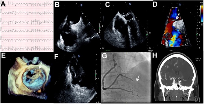
Non-atherosclerotic myocardial infarction (MI) is an important but often misdiagnosed cause of acute MI. Furthermore, non-atherosclerotic MI with concomitant acute stroke and pulmonary embolism due to in-transit thrombus across a patent foramen ovale (PFO) is a rare but potentially fatal combination (1, 2, 3). Early detection of this clinical entity can facilitate delivery of targeted therapies and avoid poor outcome (1, 2). Here, we describe a 68-year-old female with hypertension, tobacco abuse and chronic obstructive pulmonary disease presenting with facial droop, right arm weakness and aphasia. Head computed tomography (CT) without contrast was unremarkable. ECG showed an acute inferolateral ST-elevation MI (Fig. 1, Panel A). As patient presented with both an acute neurological deficit and MI, clinical suspicion of non-atherosclerotic MI was raised and the patient underwent concurrent emergency coronary angiography (CAG) and transesophageal echocardiogram (TEE). TEE revealed highly mobile masses in the left and right atrium (Fig. 1, Panel B and Video 1). The large mass (thrombus or cast of a deep venous thrombus) was caught in a PFO (Fig. 1, Panel C, D, E and Videos 2, 3). A second smaller mass/thrombus was seen on the Eustachian valve near the right atrial/inferior vena cava junction (Fig. 1, Panel F and Video 4). CAG confirmed a 100% occluded distal right posterolateral artery suggestive of an embolic phenomenon. The patient underwent successful thrombectomy, retrieving a large thrombus burden (Fig. 1, Panel G and Videos 5, 6, 7). CT angiography showed occluded internal carotid artery (Fig. 1, Panel H). Pathology from thrombectomy confirmed fibrin-rich thrombus. The patient had bilateral lower extremity deep vein thrombosis and bilateral diffuse pulmonary embolisms.
Figure 1.
(A) ECG showing an acute inferolateral ST-elevation MI; (B) TEE bicaval view revealing a highly mobile mass in the left and right atrium; (C) TEE four-chamber view showing a large thrombus across a PFO; (D) TEE bicaval with color Doppler showing a shunt across the interatrial septum; (E) 3D-TEE showing irregular shape of the thrombus; (F) TEE at lower-esophageal level showing a second smaller thrombus on the Eustachian valve; (G) CAG confirming 100% occluded distal right posterolateral artery; (H) CT angiography showing occluded internal carotid artery.
TEE bicaval revealing a highly mobile mass in both atria. View Video 1 at http://movie-usa.glencoesoftware.com/video/10.1530/ERP-18-0044/video-1.
Download Video 1 (520.5KB, mp4)
TEE four-chamber view showing the large thrombus that was caught in a PFO. View Video 2 at http://movie-usa.glencoesoftware.com/video/10.1530/ERP-18-0044/video-2.
Download Video 2 (427KB, mp4)
3D-TEE showing a large thrombus in the left atrium. View Video 3 at http://movie-usa.glencoesoftware.com/video/10.1530/ERP-18-0044/video-3.
Download Video 3 (4.3MB, mp4)
TEE showing a second smaller thrombus on the Eustachian valve. View Video 4 at http://movie-usa.glencoesoftware.com/video/10.1530/ERP-18-0044/video-4.
Download Video 4 (427.5KB, mp4)
CAG confirming 100% occluded distal right posterolateral artery. View Video 5 at http://movie-usa.glencoesoftware.com/video/10.1530/ERP-18-0044/video-5.
Download Video 5 (749.5KB, mp4)
CAG during thrombus aspiration. View Video 6 at http://movie-usa.glencoesoftware.com/video/10.1530/ERP-18-0044/video-6.
Download Video 6 (4MB, mp4)
CAG showing restored TIMI III flow in the vessel. View Video 7 at http://movie-usa.glencoesoftware.com/video/10.1530/ERP-18-0044/video-7.
Download Video 7 (10.2MB, mp4)
Declaration of interest
The authors declare that there is no conflict of interest that could be perceived as prejudicing the impartiality of this article.
Funding
This work did not receive any specific grant from any funding agency in the public, commercial, or not-for-profit sector.
Patient consent
Images are anonymized. Permission to publish was obtained from a relative.
Author contribution statement
Contribution to the patient care: Sergio Barros-Gomes; Abdallah El Sabbagh; Mackram F Eleid; Sunil V Mankad. Study design, conception and writing the manuscript: Sergio Barros-Gomes and Sunil V Mankad. Revision of the manuscript: Sergio Barros-Gomes; Abdallah, El Sabbagh; Mackram F Eleid; Sunil V Mankad.
References
- 1.Windecker S, Stortecky S, Meier B. Paradoxical embolism. Journal of the American College of Cardiology 2014. 64 403–415. ( 10.1016/j.jacc.2014.04.063) [DOI] [PubMed] [Google Scholar]
- 2.Prizel KR, Hutchins GM, Bulkley BH. Coronary artery embolism and myocardial infarction. Annals of Internal Medicine 1978. 88 155–161. [DOI] [PubMed] [Google Scholar]
- 3.Hayıroğlu Mİ, Bozbeyoğlu E, Akyüz Ş, Yıldırımtürk Ö, Bozbay M, Bakhshaliyev N, Renda E, Gök G, Eren M, Pehlivanoğlu S. Acute myocardial infarction with concomitant pulmonary embolism as a result of patent foramen ovale. American Journal of Emergency Medicine 2015. 33 984.e5–984.e7. ( 10.1016/j.ajem.2014.12.025) [DOI] [PubMed] [Google Scholar]
Associated Data
This section collects any data citations, data availability statements, or supplementary materials included in this article.
Supplementary Materials
TEE bicaval revealing a highly mobile mass in both atria. View Video 1 at http://movie-usa.glencoesoftware.com/video/10.1530/ERP-18-0044/video-1.
Download Video 1 (520.5KB, mp4)
TEE four-chamber view showing the large thrombus that was caught in a PFO. View Video 2 at http://movie-usa.glencoesoftware.com/video/10.1530/ERP-18-0044/video-2.
Download Video 2 (427KB, mp4)
3D-TEE showing a large thrombus in the left atrium. View Video 3 at http://movie-usa.glencoesoftware.com/video/10.1530/ERP-18-0044/video-3.
Download Video 3 (4.3MB, mp4)
TEE showing a second smaller thrombus on the Eustachian valve. View Video 4 at http://movie-usa.glencoesoftware.com/video/10.1530/ERP-18-0044/video-4.
Download Video 4 (427.5KB, mp4)
CAG confirming 100% occluded distal right posterolateral artery. View Video 5 at http://movie-usa.glencoesoftware.com/video/10.1530/ERP-18-0044/video-5.
Download Video 5 (749.5KB, mp4)
CAG during thrombus aspiration. View Video 6 at http://movie-usa.glencoesoftware.com/video/10.1530/ERP-18-0044/video-6.
Download Video 6 (4MB, mp4)
CAG showing restored TIMI III flow in the vessel. View Video 7 at http://movie-usa.glencoesoftware.com/video/10.1530/ERP-18-0044/video-7.
Download Video 7 (10.2MB, mp4)


 This work is licensed under a