Abstract
Context:Clinacanthus nutans Lindau (Acanthaceae) is a medicinal plant that has been reported to have anti-inflammatory, antiviral, antimicrobial and antivenom activities. In Malaysia, it has been widely claimed to be effective in various cancer treatments but scientific evidence is lacking.
Objective: This study investigates the chemical constituents, anti-proliferative, and apoptotic properties of C. nutans root extracts.
Materials and methods: The roots were subjected to solvent extraction using methanol and ethyl acetate. The anti-proliferative effects of root extracts were tested at the concentrations of 10 to 50 μg/mL on MCF-7 and HeLa by using 3-(4,5-dimethylthiazol-2-yl)-2,5-diphenyl tetrazolium bromide (MTT) assay for 72 h. Morphological changes were observed under light microscope. Pro-apoptotic effects of root extracts were examined using flow cytometric analysis and RT-PCR. The chemical compositions of root extracts were detected using GC-MS.
Results: The proliferation of MCF-7 cells was inhibited with the IC50 values of 35 and 30 μg/mL, respectively, for methanol and ethyl acetate root extracts. The average inhibition of HeLa cells was ∼25%. Induction of apoptosis in MCF-7 was supported by chromatin condensation, down-regulation of BCL2 and unaltered expression of BAX. However, only ethyl acetate extract caused the loss of mitochondrial membrane potential. GC-MS analysis revealed the roots extracts were rich with terpenoids and phytosterols.
Discussion and conclusions: The results demonstrated that root extracts promote apoptosis by suppressing BCL2 via mitochondria-dependent or independent manner. The identified compounds might work solely or cooperatively in regulating apoptosis. However, further studies are required to address this.
Keywords: Clinacanthus nutans, anti-proliferative, apoptosis, BAX, BCL2
Introduction
Despite the advancement in technology, breast cancer remains the most common occurring cancer and the leading cause of cancer death in women worldwide. It has contributed to worldwide 23% and 14% of total new cancer cases and deaths in 2008 (Jemal et al. 2011). The conventional treatments for breast cancer include surgery, radiation, chemotherapy and hormonal therapy. However, their efficacy is still unsatisfactory and prolonged use of drugs may render therapeutic resistance (Coley 2008). Resistance to the apoptotic induction is one of the multiple mechanisms evolved in cancer cells. Tremendous research has been focused on finding natural compounds that can modulate apoptosis pathway for novel drug development. Hence, various traditional plants with known medicinal properties have been widely studied in the past decades.
Apoptosis is a physiological mechanism of cell death which genetically programmed cells to undergo suicide. Apoptosis causes morphological features which can be characterized by cytoplasmic blebbing, chromatin condensation, cell shrinkage and nuclear fragmentation (Kiechle & Zhang 2002). Apoptosis pathway is composed of upstream regulators and downstream effector components (Adams & Cory 2007). It can occur via extrinsic or intrinsic pathway. The activation of the intrinsic pathway is regulated by BCL2 family proteins. They can be divided into pro-apoptotic proteins that promote apoptosis such as BAX; and anti-apoptotic proteins such as BCL2 (Gupta 2003). BCL2 arrests apoptosis by inhibiting the release of cytochrome c from mitochondria which directly inactivate caspase 3. BAX acts in an opposite manner and promotes apoptosis by facilitating the release of cytochrome c from mitochondria and thereby triggering caspase activity. Apoptosis induction is recognized as an active strategy to arrest proliferation of cancer cells (Gupta 2003). Hence, the ability of natural compound to induce apoptosis without aggravating other normal cells is therefore the key target in chemoprevention and chemotherapeutic intervention (Fulda 2010; Kuno et al. 2012).
Clinacanthus nutans Lindau (Acanthaceae) is a small shrub native to Asian countries (Sakdarat et al. 2009). In Malaysia, it is known as Sabah Snake Grass or Belalai Gajah. In folklore medicine, this plant has been used to treat diabetes mellitus, fever, diarrhoea, and dysuria, when consumed in the form of herbal tea (Uawonggul et al. 2011). In Thailand, the fresh leaves are locally used for the treatment of herpes simplex skin infection, shingles, and recommended for relieving insect bites (Thawarananth 1992; Tuntiwachwuttikul et al. 2004). In addition, this plant also shows antiviral (Jayavasu et al. 1992), immune response (Sriwanthana et al. 1996), anti-inflammatory (Wanikiat et al. 2008), antioxidant (Pannangpetch et al. 2007), and anti-proliferative activities (Yong et al. 2013). It is also effective toward antivaricella-zoster virus infection and recurrent aphthous ulcer (Buajeeb & Kraivaphan 1994; Sangkitporn et al. 1995). In Malaysia, there are emerging testimonies which claim that C. nutans is effective in treating cancer (Yong et al. 2013). However, the underlying mode of action is unclear. Furthermore, there is no report on the cytotoxic properties of the plant’s roots as all studies were done using the aerial part of the plants such as leaf and stem. Therefore, this study was carried out to examine the capability of C. nutans root extracts in growth inhibition and apoptosis induction of human breast cancer cell line. To narrow down the search of potential bioactive compound(s), we also studied the chemical compositions of the root extracts by using gas chromatography mass spectrometry (GC-MS). The research outcome will furnish our knowledge on the efficacy of using C. nutans for chemoprevention or chemotherapy.
Materials and methods
Cell lines, media and chemicals
Human breast cancer cell line (MCF-7, HTB-22), human cervical cancer cell line (HeLa, CCL-2) and mouse embryonic fibroblast cell line (NIH 3T3, CRL-1658) were obtained from the American Type Culture Collection (Rockville, MD). Roswell Park Memorial Institute (RPMI) 1640, 10,000 U/mL penicillin/10,000 μg/mL streptomycin, 1x phosphate-buffered saline (PBS) and 2.5 g/L trypsin-1 mmol/L EDTA were obtained from Nacalai Tesque (Kyoto, Japan). Fetal bovine serum (FBS) was purchased from J R Scientific (Woodland, CA). The authentic standards for compounds identification (at least 95% purity), which were squalene, campesterol, stigmasterol, sitosterol, lupeol and betulin were purchased from Sigma-Aldrich (St. Louis, MO). Solvents such as methanol and ethyl acetate (analytical and HPLC grade) were purchased from Fisher Scientific (Leicestershire, UK).
Preparation of plant extracts
Fresh plant of C. nutans was purchased from herbal supplier in Kota Kinabalu, Sabah. The plant was verified by a botanist from the Faculty of Science and Natural Resources, Dr. Berhaman Ahmad. A voucher specimen (ACCN 001/2013) was deposited in the herbarium of Universiti Malaysia Sabah. The roots were thoroughly cleaned, freeze-dried, and the dried roots were then grounded into powder by using a heavy duty blender. Plant powder was soaked in methanol and ethyl acetate with the ratio of one part of powder to ten parts of solvent. The mixtures were placed in a rotary shaker for four days at 25 °C. The mixture was filtered and concentrated under reduced pressure in a rotary evaporator. The extracts were freeze-dried and stored at −80 °C.
Cell culture
MCF-7, HeLa and NIH 3T3 cell lines were grown in RPMI 1640 media supplemented with 10% FBS and incubated at 37 °C in a humidified atmosphere containing 5% CO2.
Anti-proliferation assay
A seeding density of 1 × 10⁴ cells was grown in 96-well plates and incubated in a CO2 incubator at 37 °C for overnight. The cells were treated with root extracts at the concentrations ranging from 10 to 50 μg/mL for up to three days. MTT assay was performed using Cell Proliferation Kit I (Roche Diagnostics, Mannheim, Germany) according to manufacturer’s protocol. The optical density was measured at 550 nm using a microplate reader (Molecular Devices, Sunnyvale, CA). The 50% growth inhibitory concentration (IC50) was defined as plant extract’s concentration required for 50% inhibition of cell growth.
Methylene blue staining
After seeding, cells were treated with root extracts at the IC50 values for up to three days. The medium was discarded and cells were washed with PBS for three times. Staining was performed using 0.4% methylene blue for 20 min. Then, methylene blue was removed and cells were washed with PBS for three times. Cell morphological changes were observed under an inverted microscope (Nikon, Japan).
JC-1 staining
Apoptosis assay was performed using BDTM MitoScreen Kit (BD Biosciences, San Jose, CA) according to manufacturer’s protocol. Briefly, 3 x 10⁴ cells were seeded in 6-well plates and treated with root extracts with the concentrations of 20 to 40 μg/mL for three days. Cells were trypsinized and transferred into a sterile tube. The cells were centrifuged at 400 g for 5 min at room temperature. Supernatant was discarded and 500 μL of JC-1 solution was added. Cell was incubated at 37 °C for 15 min. After that, cells were washed twice using 1x assay buffer and centrifuged at 400 g for 5 min. Cells were resuspended with 500 μL of 1x assay buffer and analyzed using BD FACSAria flow cytometer (BD Bioscience, San Jose, CA).
RNA extraction and RT-PCR
Total RNA was extracted using RNeasy kit (Qiagen, Valencia, CA) according to the manufacturer’s protocol. RT-PCR was performed using OneStep RT-PCR kit (Qiagen, Valencia, CA). The components of RT-PCR reaction were RNA template (50 to 100 ng/μL), 0.6 μM gene specific primers, 400 μM dNTPs mix, 1x RT-PCR buffer and enzyme mix. Reverse transcription was carried out at 50 °C for 30 min. The PCR cycling conditions were 95 °C for 15 min, 95 °C for 20 sec, 55 °C to 63 °C for 30 sec, 72 °C for 20 sec, 72 °C for 5 min. The PCR was repeated for 26 to 31 cycles. After completion of PCR, the products were subjected to 2% agarose gel electrophoresis followed by ethidium bromide staining. Primers used in this study were ACTB (5′-AGAGCTACGAGCTGCCTGAC-3′,5′-GACATCCGGTTGTGTCACGA-3′), BAX (5′-TTTTCGTTCAGGGTTTCATCCA-3′, 5′-TAGAAAAGGGCGACAACCCG-3′) and BCL2 (5′-GGATAACGGAGGCTGGGATGC-3′, 5′-AACAGCCTGCAGCTTTGTTTC-3′).
GC-MS analysis
The dried root extracts were dissolved in HPLC grade methanol or ethyl acetate to appropriate concentration. The extract (1 μL) was injected into a GC-MS (GC model 7890, MS model 5975C, Agilent Technologies, Santa Clara, CA) after filtered with 0.22 μm syringe filter. GC separation was performed on a HP-5MS capillary column (Agilent Technologies, Santa Clara, CA) operating at electron impact mode at 70 eV. Pure helium gas with built-in purifier was used at a constant flow rate of 1 mL/min employed in a splitless mode with injector at 250 °C and ion source at 280 °C. The stepped temperature program was as follows: initial temperature oven was started at 220 °C and hold for 5 min and followed by a ramp to 300 °C at 5 °C/min and hold for another 15 min. A post-run of 5 min at 300 °C was sufficient for the next sample injection. Mass analyzer was used in full scan mode scanning from m/z 40 to 550 and mass spectra were taken at 70 eV. The identification of compounds was based on the comparison of their mass spectra with standards and also with the library of National Institute Standard and Technology (NIST) version 2.0, with the aid of Automated Mass Spectral Deconvolution and Identification (AMDIS) software version 2.70 by deconvoluting the chromatography peak at the corresponding retention time. Further confirmation of the identity of the chromatographic peaks was done by spiking using reference standards.
Results
Anti-proliferative effect of C. nutans root extracts
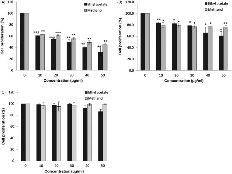
A significant growth inhibition of MCF-7 and HeLa could be seen at the concentration of 10 μg/mL for both methanol and ethyl acetate root extracts (Figure 1). The growth inhibition of MCF-7 cells was gradually increased when the concentration of both extracts increased (Figure 1(a)). However, the growth inhibition of HeLa cells caused by both root extracts did not reach 50% at the tested concentrations (Figure 1(b)). Both extracts showed no or little inhibition on NIH 3T3 normal cells (Figure 1(c)). From Figure 1(a), the IC50 values of MCF-7 cells treated with methanol and ethyl acetate extracts were 35 and 30 μg/mL, respectively. In contrast, the percentage of growth inhibition for HeLa was about 20 to 40% at the concentrations of 40 to 50 μg/mL (Figure 1(b)). This indicates that the cytotoxicity effect of root extracts is selective towards cancer cells and MCF-7 cells are more susceptible to C. nutans treatment compared to HeLa cells. Therefore, pro-apoptotic activity of root extracts was focused on MCF-7 cells only.
Figure 1.
The anti-proliferative effect of C. nutans root extracts. Treatments were performed on (a) MCF-7, (b) HeLa and (c) 3T3 cells for three days. Data represent three independent experiments performed in triplicate. Asterisks denote differences with statistical significances compared to untreated cells (*, ** and *** represent p < 0.05, p < 0.005 and p < 0.0005 respectively). p-values were obtained from a two-tailed t-test.
Nuclear morphological change
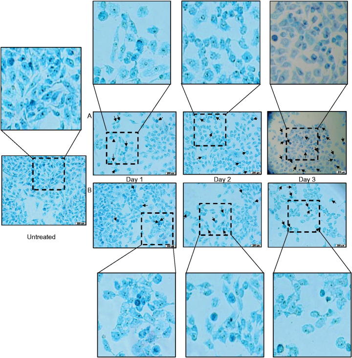
As shown in Figure 2, MCF-7 cells treated with root extracts began to exhibit peripheral nuclear membrane condensation after one day treatment compared to untreated cells. The occurrence of the morphological changes and nuclear condensation became more profound in the cell population when the duration of the treatment increased to three days. The morphological changes caused by camptothecin were depicted in Figure S1. Comparing to methanol root extracts (Figure 2(b)), ethyl acetate root extract caused more distinct morphological change after three days treatment (Figure 2(a)). Besides, cell shrinkage, loss of cell contact, and chromatin condensation were observed in the treated cells. These suggested that cells might undergo apoptosis after treatment.
Figure 2.
Morphological changes caused by C. nutans extracts on MCF-7. Cells were treated with (a) ethyl acetate and (b) methanol root extracts of C. nutans at their respective IC50 value for up to three days. Cells were stained with methylene blue and observed under microscope with 40x magnification. Arrows indicate some cells with morphological changes when compared with untreated cells.
The apoptotic effect of C. nutans root extracts
For untreated samples, about 90% of cell populations were in healthy condition but upon camptothecin treatment at the concentration of 0.35 μg/mL, about 50% of cells loss their mitochondrial membrane potential (Figure 3). Surprisingly, loss of mitochondrial membrane potential was higher in cells treated with acetyl acetate extract (∼78 to 82%) than camptothecin. In contrast, integrity of inner mitochondrial membrane was preserved in cells treated with methanol extract as the proportional of healthy and apoptotic cells were similar to untreated cells. Methanol root extract did not exhibit loss of mitochondrial potential as compared to ethyl acetate root extract, suggesting the induction of apoptosis by methanol root extract is mitochondria independent.
Figure 3.
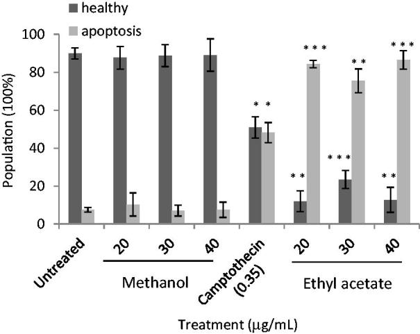
Apoptotic effects of C. nutans root extracts on MCF-7 cells. Cells were treated with C. nutans root extracts and camptothecin for three days at the indicated concentrations. Data represent three independent experiments performed in triplicate. Asterisks denote differences with statistical significances compared to untreated cells (*, ** and *** represent p < 0.05, p < 0.005 and p < 0.0005 respectively). p-values were obtained from a two-tailed t-test.
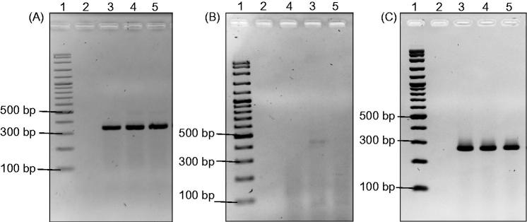
The total RNA of MCF-7 cells treated with methanol and ethyl acetate root extracts were extracted after three days treatment. The effect of root extracts on the expression of BCL2 and BAX genes was evaluated using RT-PCR. ACTB is the housekeeping gene used in this study. The expected size of the ACTB amplicon was ∼350 bp and its expression level was not altered upon treatments (Figure 4(a)). Based on Figure 4(b), BCL2 expression was only detected in untreated samples (Lane 3) with the size of ∼450 bp. On the other hand, the expression of BCL2 in samples treated with both root extracts was undetectable (Lanes 4 & 5). As for the expression of BAX, we found no alteration of band intensity (∼290 bp) in both treated and untreated MCF-7 cells (Figure 4(c)). These results indicate that C. nutans root extracts promote apoptosis by suppressing the expression of anti-apoptotic gene (BCL2) while maintaining the expression of pro-apoptotic gene (BAX) in MCF-7 cells.
Figure 4.
The effect of C. nutans root extracts on the expression of ACTB (a), BCL2 (b) and BAX (c) in MCF-7 breast cancer cell line by RT-PCR. Lane 1: 100 bp DNA marker, lane 2: negative control, lane 3: without treatment, lane 4: methanol root extract and lane 5: acetyl acetate root extract.
Chemical composition of the root extracts of C. nutans
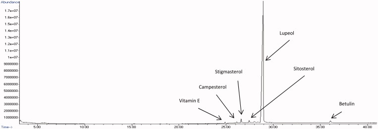
The identified bioactive compounds were characterized according to their retention time, molecular formula, molecular weight, peak area (%) and only those with reported cytotoxic effects on cancer cells were summarized in Table 1 and Table 2. The GC-MS total ion chromatograms of ethyl acetate and methanol root extracts were depicted in Figures 5 and 6, respectively. The results revealed more compounds were found in ethyl acetate root extract (Table 1) compared to methanol root extract (Table 2). The highest amount of compounds found in ethyl acetate root extract (Figure 5) was lupeol (79.05%), followed by lup-20(29)-en-3-one (2.79%), lup-20(29)-en-ol acetate (1.50%), stigmasterol (1.50%), sitosterol (1.15%), β-amyrin (1.15%), betulin (0.96%), campesterol (0.37%), squalene (0.34%), vitamin E (0.34%) and oleic acid (0.22%). Meanwhile, the methanol root extract (Figure 6) consisted of lupeol (94.21%), betulin (1.38%), stigmasterol (1.33%), sitosterol (1.01%), β-amyrin (0.82%), vitamin E (0.39%) and campesterol (0.39%) but not squalene, oleic acid and other lupeol derivatives. However, the most abundant compound found in both root extracts was lupeol.
Table 1.
Cytotoxic compounds identified in the ethyl acetate root extract of C. nutans.
| No. | Retention time (min) | Bioactive compound | Nature of compound | Peak area (%) | Molecular formula | Molecular weight | Reported cytotoxic effect on cancer cells |
|---|---|---|---|---|---|---|---|
| 1 | 3.675 | Oleic acid | Fatty acid | 0.22 | C18H34O2 | 282.46 | Nelson 2005; Menendez et al. 2006 |
| 2 | 20.374 | Squalene | Triterpenoid | 0.34 | C30H50 | 410.71 | Nakagawa et al.1985; Rao et al. 1998; De Los Reyes et al. 2015 |
| 3 | 24.956 | Vitamin E | Fat soluble vitamin | 0.34 | C29H50O2 | 430.70 | Ramdas et al. 2011 |
| 4 | 26.191 | Campesterol | Phytosterols | 0.37 | C28H48O | 400.68 | Awad et al. 2003 |
| 5 | 26.658 | Stigmasterol | Phytosterols | 1.50 | C29H48O | 412.69 | Ghosh et al. 2011; Lee et al. 2014 |
| 6 | 27.532 | gamma-Sitosterol and beta-Sitosterol | Phytosterols | 1.15 | C29H50O | 414.70 | Awad et al. 2003; Chai et al. 2008; Sundarraj et al. 2012 |
| 7 | 28.441 | Lup-20(29)-en-3-one | Triterpenoid | 1.69 | C30H48O | 424.70 | Hata et al. 2002, 2008 |
| 8 | 28.500 | Lup-20(29)-en-3-one | Triterpenoid | 1.10 | C30H48O | 424.70 | Hata et al. 2002, 2008 |
| 9 | 28.806 | beta-amyrin | Triterpenoid | 1.15 | C30H50O | 426.71 | Lee et al. 2014 |
| 10 | 28.972 | Lupeol | Triterpenoid | 26.40 | C30H50O | 426.71 | Saleem 2009; Saleem et al. 2009; Pitchai et al. 2014; Lee et al. 2014 |
| 11 | 29.065 | Lupeol | Triterpenoid | 19.50 | C30H50O | 426.71 | Saleem 2009; Saleem et al. 2009; Pitchai et al. 2014; Lee et al. 2014 |
| 12 | 29.182 | Lupeol | Triterpenoid | 33.15 | C30H50O | 426.71 | Saleem et al. 2009; Pitchai et al. 2014; Lee et al. 2014 |
| 13 | 30.621 | Lup-20(29)-en-3-ol, acetate | Triterpenoid | 1.50 | C32H52O2 | 468.75 | Lee et al. 2014 |
| 14 | 36.199 | Betulin | Triterpenoid | 0.96 | C30H50O2 | 442.73 | Kommera et al. 2011 |
Table 2.
Cytotoxic compounds identified in the methanol root extract of C. nutans.
| No. | Retention time (min) | Bioactive compound | Nature of compound | Peak area (%) | Molecular formula | Molecular weight | Reported cytotoxic effect on cancer cells |
|---|---|---|---|---|---|---|---|
| 1 | 24.898 | Vitamin E | Fat soluble vitamin | 0.39 | C29H50O2 | 430.70 | Ramdas et al. 2011 |
| 2 | 26.139 | Campesterol | Phytosterols | 0.39 | C28H48O | 400.68 | Awad et al. 2003 |
| 3 | 26.600 | Stigmasterol | Phytosterols | 1.33 | C29H48O | 412.69 | Ghosh et al. 2011; Lee et al. 2014 |
| 4 | 27.445 | gamma-Sitosterol and beta-Sitosterol | Phytosterols | 1.01 | C29H50O | 414.70 | Awad et al. 2003; Chai et al. 2008; Sundarraj et al. 2012 |
| 5 | 27.987 | beta-amyrin | Triterpenoid | 0.82 | C30H50O | 426.7174 | Lee et al. 2014 |
| 6 | 28.943 | Lupeol | Triterpenoid | 94.21 | C30H50O | 426.71 | Saleem 2009; Saleem et al. 2009; Pitchai et al. 2014; Lee et al. 2014 |
| 7 | 36.065 | Betulin | Triterpenoid | 1.38 | C30H50O2 | 442.73 | Kommera et al. 2011 |
Figure 5.
GC-MS chromatogram of ethyl acetate root extract of C. nutans.
Figure 6.
GC-MS chromatogram of methanol root extract of C. nutans.
Discussion
Although the inhibitory effect of C. nutans extracts have been observed in several other cell lines, this study is the first report on the cytotoxic and apoptotic effects of C. nutans root extracts as previous studies have focused on extracts obtained from other parts of the plant (Yong et al. 2013; Arullappan et al. 2014). As different parts of plant such as leaf, bark, stem and roots may contain different types and concentrations of compounds, the efficacy of plant extracts will be varied as well. Overall, the decrease of cell proliferation in MCF-7 and HeLa cells were observed after treatment. However, its anti-proliferative effect is cell-type dependent as inhibition is more significant in MCF-7 than HeLa. Besides, no deleterious effect was found in normal cells, NIH 3T3. The susceptibility of various cancer cell lines towards different types of extracts from various parts of this plant has been clearly reported by other researchers as well (Yong et al. 2013; Arullappan et al. 2014).
The results have demonstrated that both root extracts were capable of inducing apoptosis as substantiated by morphological changes such as peripheral nuclear condensation and cell shrinkage, as well as suppression of anti-apoptotic gene, BCL2. However, the effects on the mitochondrial membrane potential were different when compared to untreated cells. It is suggested that apoptosis induced by methanol root extract is mitochondria independent. Although mitochondrial pathway is governed by BCL-2 family proteins which regulate the release of cytochrome c, mitochondria independent apoptosis has reported before (Godefroy et al. 2004; Sinha et al. 2013). Discrepancy of both root extracts could be due to an alternative mechanism of releasing cytochrome c from mitochondria. It has been reported that this phenomenon can happen when there is functional changes of some ion transporter in the inner mitochondrial membrane (Reed et al. 1998). The increase of mitochondrial permeability through the loss of mitochondrial potential as shown in ethyl acetate root extract treated cells could promote the translocation of pro-apoptotic proteins such as BAX to mitochondria results in the release of apoptogenic proteins such as cytochrome c. This also antagonizes the effect of anti-apoptotic proteins such as BCL2 which is known to prolong cell survival (Davids & Letai 2012; Weyhenmeyer et al. 2012). Besides, similar expression profile of BAX and BCL2 but different effect on mitochondrial potential elicited by both root extracts have also indicated the possible involvement of upstream regulatory proteins such as p53, which has been shown to regulate apoptosis via a mitochondria-independent manner (Godefroy et al. 2004).
Based on GC-MS analysis, the C. nutans roots are rich with phytosterols such as stigmasterol, campesterol, β/γ-sitosterol, and triterpenes such as lupeol, β-amyrin and betulin. The detected compounds such as lupeol, stigmasterol and betulin have also been reported previously by other researchers (Dampawan et al. 1977; Lin et al. 1983; Aslam et al. 2015). Compounds previously found in various leaf or stem extracts such as C-glycosyl flavones (vitexin, isovitexin, shaftoside, isomollupentin 7-O-β-glucopyranoside, orientin and isoorientin), sulfur-containing glucosides (clinamide A-C, 2-cis entadamide A, entadamide A, entadamide C, trans-3-methylsulfinyl-2-propenol), cerebrosides and a monoacylmonogalatosylglycerol are undetectable in this study (Dampawan et al. 1977; Lin et al. 1983; Teshima et al. 1997, 1998; Tuntiwachwuttikul et al. 2004; Sakdarat et al. 2009; Tu et al. 2014; Aslam et al. 2015). However, differences in the extraction techniques, detection methods, and plant materials or parts might jeopardize the extractability and identification of some compounds. Being the most abundant compound found in this study, it is reasonable to suggest that lupeol might play role in modulating apoptosis process. Besides, this has been demonstrated in a number of in vitro and in vivo studies. For instance, lupeol was found to promote apoptosis by inhibiting Ras signalling pathway in human pancreatic adenocarcinoma cells, while Fas-mediated apoptosis was reported in prostate cancer cells (Saleem et al. 2005a,b). In addition, lupeol exhibited growth inhibition of human metastatic melanoma cells in vitro and in vivo by inducing apoptosis and G1-S phase cell cycle arrest (Saleem et al. 2008). In another study, lupeol was also shown to induce apoptosis in MCF-7 cells by down-regulating BCL2 and BCL-XL expressions (Pitchai et al. 2014).
Despite the minute amount of other identified compounds, their synergistic biological activities could not be neglected. It is worth mentioning that some of them have been reported to have apoptotic effect. For example, stigmasterol was shown to induce apoptosis in Ehrlich’s ascites carcinoma in mice through the activation of protein phosphatase 2A via ceramide (Ghosh et al. 2011). Meanwhile, γ-sitosterol was found to suppress c-MYC formation while promoting caspase activity in breast and lung cancer cells (Chai et al. 2008; Sundarraj et al. 2012). Betulin-induced apoptosis in colon cancer cells via the induction of caspase activity (Kommera et al. 2011). The low number of cancer incidence was found to be associated with the daily consumption of olive oil in Mediterranean diet. Studies have suggested that oleic acid has specifically repressed the transcriptional activity of Her-2/neu, which is commonly overexpressed in breast cancer cells (Menendez et al. 2006). Additionally, a pentacyclic triterpenoid, squalene found in olive oil and liver oil of sharks has demonstrated anti-proliferative activity against HCT-116 and MCF-7 cells with the IC50 of 4.21 and IC50 5.92 μg/mL, respectively (De Los Reyes et al. 2015). Another triterpenoid called β-amyrin also showed cytotoxicity activity against MCF-7 cells with the IC50 of 15.5 μg/mL (Lee et al. 2014). Besides these published anti-proliferative effects in various cancer cell lines, several purified and semi-purified compounds in our laboratory such as lupeol, squalene and phytosterols (mixture of stigmasterol, β-sitosterol and campesterol) were tested on MCF-7 cells to confirm their cytotoxic effect (unpublished data). Taken together, the apoptotic promoting activity of C. nutans root extracts might be associated with the high amount of lupeol and also other minute compounds such as squalene and betulin. Nonetheless, how these compound(s) modulate the underlying mechanisms will need further investigation.
Conclusions
The findings of this study showed that C. nutans root extracts are selective towards cancer cells without affecting the proliferation of normal cell line. Although both root extracts exhibited apoptotic effect based on morphological changes and the suppression of BCL2 expression. Ethyl acetate root extract promoted mitochondrial-dependent apoptosis in MCF-7 cells but this was not shown in cells treated with methanol root extract. The compounds present in both root extracts might work solely or cooperatively in regulating the apoptosis event.
Supplementary Material
Disclosure statement
The authors report no declarations of interest.
References
- Adams JM, Cory S.. 2007. Bcl-2-regulated apoptosis: mechanism and therapeutic potential. Curr Opin Immunol. 19:488–496. [DOI] [PMC free article] [PubMed] [Google Scholar]
- Arullappan S, Rajamanickam P, Thevar N, Kodimani CC.. 2014. In vitro screening of cytotoxic, antimicrobial and antioxidant activities of Clinacanthus nutans (Acanthaceae) leaf extracts. Trop J Pharmaceut Res. 13:1455–1461. [Google Scholar]
- Aslam MS, Ahmad MS, Mamat AS.. 2015. A review on phytochemical constituents and pharmacological activities of Clinacanthus nutans. Int J Pharm Pharm Sci. 7:30–33. [Google Scholar]
- Awad AB, Williams H, Fink CS.. 2003. Effect of phytosterols on cholesterol metabolism and MAP kinase in MDA-MB-231 human breast cancer cells. J Nutr Biochem. 14:111–119. [DOI] [PubMed] [Google Scholar]
- Buajeeb W, Kraivaphan P.. 1994. Clinical evaluation of Clinacanthus nutans Lindau in orabase in the treatment of recurrent aphthous stomatitis. Mahidol Dent J. 14:10–16. [Google Scholar]
- Chai JW, Kuppusamy UR, Kanthimathi MS.. 2008. beta-Sitosterol induces apoptosis in MCF-7 cells. Malaysian J Biochem Mol Biol. 16:28–30. [Google Scholar]
- Coley HM.2008. Mechanisms and strategies to overcome chemotherapy resistance in metastatic breast cancer. Cancer Treat Rev. 34:378–390. [DOI] [PubMed] [Google Scholar]
- Dampawan P, Huntrakul C, Reutrakul V, Raston CL, White AH.. 1977. Constituents of Clinacanthus nutans and the crystal structure of LUP-20 (29)-ene-3-one. J Sci Soc Thai. 3:14–26. [Google Scholar]
- Davids MS, Letai A.. 2012. Targeting the B-cell lymphoma/leukemia 2 family in cancer. J Clin Oncol. 30:3127–3135. [DOI] [PMC free article] [PubMed] [Google Scholar]
- De Los Reyes MM, Oyong GG, Ebajo VD Jr., Ng VAS, Shen CC, Ragasa CY.. 2015. Cytotoxic triterpenes and sterols from Pipturus arborescens (Link) C.B. Rob. J App Pharm Sci. 5:23–30. [Google Scholar]
- Fulda S.2010. Modulation of apoptosis by natural products for cancer therapy. Planta Med. 76:1075–1079. [DOI] [PubMed] [Google Scholar]
- Ghosh T, Maity TK, Singh J.. 2011. Evaluation of antitumor activity of stigmasterol, a constituent isolated from Bacopa monnieri Linn aerial parts against Ehrlich Ascites Carcinoma in mice. Orient Pharm Exp Med. 11:41–49. [Google Scholar]
- Godefroy N, Lemaire C, Renaud F, Rincheval V, Perez S, Parvu-Ferecatu I, Mignotte B, Vayssière J-L.. 2004. p53 can promote mitochondria- and caspase-independent apoptosis . Cell Death Differ. 11:785–787. [DOI] [PubMed] [Google Scholar]
- Gupta S.2003. Molecular signaling in death receptor and mitochondrial pathways of apoptosis. Int J Oncol. 22:15–20. [PubMed] [Google Scholar]
- Hata K, Kazuyuki H, Takahashi S.. 2002. Differentiation- and apoptosis-inducing activities by pentacyclic triterpenes on a mouse melanoma cell line. J Nat Prod. 65:645–648. [DOI] [PubMed] [Google Scholar]
- Hata K, Ogawa S, Makino M, Mukaiyama T, Hori K, Iida T, Fujimoto Y.. 2008. Lupane triterpenes with a carbonyl group at C-20 induce cancer cell apoptosis. J Nat Med. 62:332–335. [DOI] [PubMed] [Google Scholar]
- Jayavasu C, Dechatiwongse T, Balachandra K, Chavalittumrong P, Jongtrakulsiri S.. 1992. The virucidal activity of Clinacanthus nutans Lindau extracts against herpes simplex virus type-2: an in vitro study. Bull Dep Med Sci (Thailand). 234:153–158. [Google Scholar]
- Jemal A, Bray F, Center MM, Ferlay J, Ward E, Forman D.. 2011. Global cancer statistics. CA Cancer J Clin. 61:69–90. [DOI] [PubMed] [Google Scholar]
- Kiechle FL, Zhang X.. 2002. Apoptosis: biochemical aspects and clinical implications. Clin Chim Acta. 326:27–45. [DOI] [PubMed] [Google Scholar]
- Kommera H, Kaluderovic GN, Kalbitz J, Paschke R.. 2011. . Lupane triterpenoids-betulin and betulinic acid derivatives induce apoptosis in tumor cells. Invest New Drugs. 29:266–272. [DOI] [PubMed] [Google Scholar]
- Kuno T, Tsukamoto T, Hara A, Tanaka T.. 2012. Cancer chemoprevention through the induction of apoptosis by natural compounds. J Biophys Chem. 3:156–173. [Google Scholar]
- Lee YS, Tee CT, Tan SP, Awang K, Hashim NM, Nafiah MA, Ahmad K.. 2014. Cytotoxic, antibacterial and antioxidant activity of triterpenoids from Kopsia singapurensis Ridl. J Chem Pharm Res. 6:815–822. [Google Scholar]
- Lin J, Li HM, Yu JG.. 1983. Studies on the chemical constituents of niu xu hua (Clinacanthus nutans). Zhongcaoyao. 14:337–338. [Google Scholar]
- Menendez JA, Papadimitropoulou A, Vellon L, Lupu RA.. 2006. Genomic explanation connecting “Mediterranean diet”, olive oil and cancer: oleic acid, the main monounsaturated fatty acid of olive oil, induces formation of inhibitory “PEA3 transcription factor-PEA3 DNA binding site” complexes at the Her-2/neu (erbB-2) oncogene promoter in breast, ovarian and stomach cancer cells. Eur J Cancer. 42:2425–2432. [DOI] [PubMed] [Google Scholar]
- Nakagawa M, Yamaguchi T, Fukawa H, Ogata J, Komiyama S, Akiyama S, Kuwano M.. 1985. Potentiation by squalene of the cytotoxicity of anticancer agents against cultured mammalian cells and murine tumor. Jpn J Cancer Res. 76:315–320. [PubMed] [Google Scholar]
- Nelson R.2005. Oleic acid suppresses overexpression of ERBB2 oncogene. Lancet Oncol. 6:69. [DOI] [PubMed] [Google Scholar]
- Pannangpetch P, Laupattarakasem P, Kukongviriyapan V, Kukongviriyapan U, Kongyingyoes B, Aromdee C.. 2007. Antioxidant activity and protective effect against oxidative hemolysis of Clinacanthus nutans (Burm.f) Lindau. Songklanakarin J Sci Technol. 29:1–9. [Google Scholar]
- Pitchai D, Roy A, Ignatius C.. 2014. In vitro evaluation of anticancer potentials of lupeol isolated from Elephantopus scaber L. on MCF-7 cell line. J Adv Pharm Technol Res. 5:179–184. [DOI] [PMC free article] [PubMed] [Google Scholar]
- Ramdas P, Rajihuzzaman M, Veerasenan SD, Selvaduray KR, Nesaretnam K, Radhakrishnan AK.. 2011. Tocotrienol-treated MCF-7 human breast cancer cells show down-regulation of API5 and up-regulation of MIG6 genes. Cancer Genomics Proteomics. 8:19–32. [PubMed] [Google Scholar]
- Rao CV, Newmark HL, Reddy BS.. 1998. Chemopreventive effect of squalene on colon cancer. Carcinogenesis. 19:287–290. [DOI] [PubMed] [Google Scholar]
- Reed JC, Jurgensmeier JM, Matsuyama S.. 1998. Bcl-2 family proteins and mitochondria. Biochim Biophys Acta. 1366:127–137. [DOI] [PubMed] [Google Scholar]
- Sakdarat S, Shuyprom A, Pientong C, Ekalaksanan T, Thongchai S.. 2009. Bioactive constituents from the leaves of Clinacanthus nutans Lindau. Bioorg Med Chem. 17:1857–1860. [DOI] [PubMed] [Google Scholar]
- Saleem M.2009. Lupeol, a novel anti-inflammatory and anti-cancer dietary triterpene. Cancer Lett. 285:109–115. [DOI] [PMC free article] [PubMed] [Google Scholar]
- Saleem M, Kaur S, Kweon MH, Adhami VM, Afaq F, Mukhtar H.. 2005a. Lupeol, a fruit and vegetable based triterpene, induces apoptotic death of human pancreatic adenocarcinoma cells via inhibition of Ras signaling pathway. Carcinogen. 26:1956–1964. [DOI] [PubMed] [Google Scholar]
- Saleem M, Kweon MH, Yun JM, Adhami VM, Khan N, Syed DN, Mukhtar H.. 2005b. A novel dietary triterpene lupeol induces Fas-mediated apoptotic death of androgen sensitive prostate cancer cells and inhibits tumor growth in a xenograft model. Cancer Res. 65:11203–11213. [DOI] [PubMed] [Google Scholar]
- Saleem M, Maddodi N, Abu Zaid M, Khan N, Hafeez B, Asim M, Suh Y, Yun JM, Setaluri V, Mukhtar H.. 2008. Lupeol inhibits growth of highly aggressive human metastatic melanoma cells in vitro and in vivo by inducing apoptosis. Clin Cancer Res. 14:2119–2127. [DOI] [PubMed] [Google Scholar]
- Saleem M, Murtaza I, Tarapore RS, Suh Y, Adhami VM, Johnson JJ, Siddiqui IA, Khan N, Asim M, Hafeez BB, et al. 2009. Lupeol inhibits proliferation of human prostate cancer cells by targeting β-catenin signaling. Carcinogen. 30:808–817. [DOI] [PMC free article] [PubMed] [Google Scholar]
- Sangkitporn S, Chaiwat S, Balachandra K, Na-Ayundhaya TD, Bunjob M, Jayavasu C.. 1995. Treatment of herpes zoster with Clinacanthus nutans (bi phaya yaw) extract. J Med Assoc Thai. 78:624–627. [PubMed] [Google Scholar]
- Sinha K, Das J, Pal PB, Sil PC.. 2013. Oxidative stress: the mitochondria-dependent and mitochondria-independent pathways of apoptosis. Arch Toxicol. 87:1157–1180. [DOI] [PubMed] [Google Scholar]
- Sriwanthana B, Chavalittumorng P, Chompuk L.. 1996. Effect of Clinacanthus nutans on human cell-mediated immune response in vitro. Thai J Pharmaceut Sci. 20:261–267. [Google Scholar]
- Sundarraj S, Thangam R, Sreevani V, Kaveri K, Gunasekaran P, Achiraman S, Kannan S.. 2012. . γ-Sitosterol from Acacia nilotica L. induces G2/M cell cycle arrest and apoptosis through c-Myc suppression in MCF-7 and A549 cells. J Ethnopharmacol. 141:803–809. [DOI] [PubMed] [Google Scholar]
- Teshima K, Kaneko T, Ohtani K.. 1997. C-Glycosyl flavones from Clinacanthus nutans. Nat Med. 51:557. [Google Scholar]
- Teshima K, Kaneko T, Ohtani K.. 1998. Sulfur-containing glucosides from Clinacanthus nutans. Phytochem. 48:831–835. [Google Scholar]
- Thawarananth D.1992. In vitro antiviral activity of Clinacanthus nutans on Varicella zoster virus. Siriraj Med J. 44:285–291. [Google Scholar]
- Tu SF, Liu RH, Cheng YB, Hsu YM, Du YC, El-Shazly M, Wu YC, Chang FR.. 2014. Chemical constituents and bioactivities of Clinacanthus nutans aerial parts. Molecules. 19:20382–20390. [DOI] [PMC free article] [PubMed] [Google Scholar]
- Tuntiwachwuttikul P, Pootaeng-On Y, Phansa P, Taylor WC.. 2004. Cerebrosides and a monoacylmonogalactosylglycerol from Clinacanthus nutans. Chem Pharm Bull (Tokyo). 52:27–32. [DOI] [PubMed] [Google Scholar]
- Uawonggul N, Thammasirirak S, Chaveerach A, Chuachan C, Daduang J, Daduang S.. 2011. Plant extracts activities against the fibroblast cell lysis by honey bee venom. J Med Plant Res. 5:1978–1986. [Google Scholar]
- Wanikiat P, Panthong A, Sujayanon P, Yoosok C, Rossi AG, Reutrakul V.. 2008. The anti-inflammatory effects and the inhibition of neutrophil responsiveness by Barleria lupulina and Clinacanthus nutans extracts. J Ethnopharmacol. 116:234–244. [DOI] [PubMed] [Google Scholar]
- Weyhenmeyer B, Murphy AC, Prehn JHM, Murphy BM.. 2012. Targeting the anti-apoptotic BCL-2 family members for the treatment of cancer. Exp Oncol. 34:192–199. [PubMed] [Google Scholar]
- Yong YK, Tan JJ, Teh SS, Mah SH, Cheng CE, Chiong HS, Ahmad Z.. 2013. Clinacanthus nutans extracts are antioxidant with antiproliferative effect on cultured human cancer cell lines. Evid Based Complement Alternat Med. 2013:462751. doi: 10.1155/2013/462751. [DOI] [PMC free article] [PubMed] [Google Scholar]
Associated Data
This section collects any data citations, data availability statements, or supplementary materials included in this article.


