Key Clinical Message
Treating solar lentigines using picosecond‐switched lasers that selectively remove the excess pigment was combined with Kleresca® biophotonic treatment. This therapy uses fluorescent light energy to stimulate healing by increasing collagen production and reducing inflammation. Combining these therapies successfully removed solar lentigines and achieved normalized and rejuvenated treated skin.
Keywords: biophotonic, combined therapy, fluorescent light energy, laser, solar lentigines, solar lentigo
1. INTRODUCTION
Solar lentigo is a pigment change (i.e., dark brown spot) in the skin in response to exposure to ultraviolet (UV) radiation. UV radiation can cause local proliferation of melanocytes and thus an accumulation of melanin in the skin cells (keratinocytes). A solar lentigo or solar lentigines (plural) are seen primarily in elderly patients.1 Pigment changes are commonly seen on the hands but can occur almost anywhere on the body, especially in sun‐exposed areas such as the face, back, arms, feet, and shoulders. Solar lentigines can be treated with different types of lasers that emit specific wavelengths that are absorbed by melanin. Melanin is a chromophore that will transform the energy it absorbs from the laser wave into heat that will destroy the pigment in the skin.2
The development of pulsed, pigment‐specific lasers to selectively destroy the pigment in solar lentigo provides clinicians with significant improvement in treatment options. Furthermore, the treatments are associated with a relatively small number of side effects and have high patient acceptance. Various studies have shown that lasers are one of the most effective forms of treatment for this indication.3 With their short nanosecond pulse durations, Q‐switched Nd:YAG lasers have been shown to be more effective than fractional CO2 lasers for treatment of solar lentigines but are more painful and require a longer healing time.4 Furthermore, laser treatment of solar lentigines is usually the patients' preferred treatment compared with other commonly used treatments such as cryotherapy and lightening peelings, among others.
Removing solar lentigines using nanosecond and picosecond‐switched lasers is an accessible and fast method, which often only requires just one or two treatments. While these lasers selectively remove the excess pigment without affecting the surrounding skin, post‐laser treatment will create a new layer of nonpigmented skin cells, inducing a healing response. Biophotonic treatment—comprising a multi‐ light‐emitting diode (LED) lamp and specialized chromophore containing gel that together emit fluorescent light energy—has been clinically proven to enhance the overall appearance of the skin with lasting results.5 It is capable of eliminating fine lines, reducing pore size, reducing inflammation, and stimulating a healing process by increasing collagen formation.5 It therefore seems logical to combine the two techniques (i.e., laser and biophotonics) to achieve specific ablation of the pigmented spots with the laser, coupled with the rejuvenating properties of the biophotonic platform to enhance the overall appearance of the skin.
2. CASE REPORT
Facial treatment of solar lentigines was conducted by carrying out two picosecond laser treatments. The second laser treatment was conducted 4 weeks after the first treatment. Each treatment was conducted at 532 nm using a 4 mm Zoom handpiece and an energy output of 0.60 joules at 2 Hz (Picoway, Syneron‐Candela, Irvine, CA, USA). One month after the second picosecond laser treatment, four biophotonic treatments (Kleresca® Skin Rejuvenation treatment, FB Dermatology, Ireland) were carried out following the producer′s recommendation. In short, patients received 9 minutes of biophotonic treatment, once a week for 4 weeks. The patients were followed using the imaging system at all time points of the biophotonic treatment as well as at 2 months after the last biophotonic treatment.
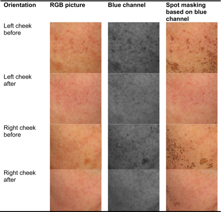
Standardized pictures obtained using a Visia camera system (Candfield, Parsippany, NJ, USA) were used to calculate the reduction of pigmentation due to the treatment. Pictures were analyzed using a free image analyzing program (ImageJ v.1.51u NIH, USA) using only the blue channel to detect spots. Subsequently, the percentage of spots were calculated in pictures of the left and right cheek both before (baseline) and after (2 months following the final biophotonic) treatment (Table 1; Figure 1).
Table 1.
Reduction in solar lentigo spots after treatment. Measured by image analysis using the free software ImageJ v.1.51u
| Orientation | Facial area with lentigo spots (%) |
|---|---|
| Left cheek before | 2.107 |
| Left cheek after | 0.036 |
| Right cheek before | 4.307 |
| Right cheek after | 0.376 |
Figure 1.

Combined treatment effect on solar lentigines visualized using image analysis before (any treatment) and after (2 months after the final biophotonic treatment)
The overall reduction in spots was effective and maintained for 3 months with almost no pigmentation changes visible after all treatments. Spot removal was due to the picosecond laser treatment. Normalization and smoothening of the post‐laser treated skin was due to the collagen buildup and anti‐inflammatory effects of the biophotonic treatment.
3. DISCUSSION
Treatment with the laser successfully removed the solar lentigines. Compared to previous cases, combining this laser treatment with the biophotonic treatment further enhanced the overall appearance of the skin. While further comparative study would support the beneficial effects of the biophotonic treatment, combining specific pigmented laser and a biophotonic tissue stimulation is a successful technique for removing solar lentigines and achieving rejuvenation of the skin. The biophotonic treatment has huge potential for possible combination with other laser and invasive therapies because of its powerful anti‐inflammatory effects, which can normalize the skin, as well as build up collagen to ensure skin rejuvenation. Furthermore, it is a year‐round treatment as it does not cause photosensitivity if used during the summer months.
AUTHORSHIP
GS and MCEN: responsible for the concept and writing. MWD: carried out the image analysis.
ACKNOWLEDGMENTS
We would like to thank Deirdre Edge for her help in improving the manuscript and Anne Downes for copy‐editing and proofreading it.
CONFLICT OF INTEREST
G. Scarcella has served as an investigator, consultant, and speaker for FB Dermatology/Kleresca®. He has received honoraria for membership of advisory board, travel, and research funding. M.C.E. Nielsen is an employee of FB Dermatology Kleresca®.
Scarcella G, Dethlefsen MW, Nielsen MCE. Treatment of solar lentigines using a combination of picosecond laser and biophotonic treatment. Clin Case Rep. 2018;6:1868–1870. 10.1002/ccr3.1749
REFERENCES
- 1. Larosa CL, Foulke GT, Feigenbaum DF, Cordoro KM, Zaenglein AL. Lentigines in resolving psoriatic plaques: rarely reported sequelae in pediatric cases. Pediatr Dermatol. 2015;32(3):e114‐e117. [DOI] [PubMed] [Google Scholar]
- 2. Polder KD, Landau JM, Vergilis‐Kalner IJ, Goldberg LH, Friedman PM, Bruce S. Laser eradication of pigmented lesions: a review. Dermatol Surg. 2011;37(5):572‐595. [DOI] [PubMed] [Google Scholar]
- 3. Schoenewolf NL, Hafner J, Dummer R, Bogdan Allemann I. Laser treatment of solar lentigines on dorsum of hands: QS ruby laser versus ablative CO2 fractional laser – a randomized controlled trial. Eur J Dermatol. 2015;25(2):122‐126. [DOI] [PubMed] [Google Scholar]
- 4. Vachiramon V, Panmanee W, Techapichetvanich T, Chanprapaph K. Comparison of Q‐switched Nd: YAG laser and fractional carbon dioxide laser for the treatment of solar lentigines in Asians. Laser Surg Med. 2016;48(4):354‐359. [DOI] [PubMed] [Google Scholar]
- 5. Nikolis A, Fauverghe S, Scapagnini G, et al. An extension of a multicenter, randomized, split‐face clinical trial evaluating the efficacy and safety of chromophore gel‐assisted blue light phototherapy for the treatment of acne. Int J Dermatol. 2018;57(1):94‐103. [DOI] [PubMed] [Google Scholar]


