Abstract
Background:
The anatomy of the articularis genus muscle has prompted speculation that it elevates the suprapatellar bursa during extension of the knee joint. However, its architectural parameters indicate that this muscle is not capable of generating enough force to fulfill this function. The purpose of the present study was to investigate the anatomy of the articularis genus, with special emphasis on its relationship with the adjacent vastus intermedius and vastus medialis muscles.
Methods:
The articularis genus muscle was investigated in 18 human cadaveric lower limbs with use of macrodissection techniques. All components of the quadriceps muscle group were traced from origin to insertion, and their affiliations were determined. Six limbs were cut transversely in the middle third of the thigh. The modes of origin and insertion of the articularis genus, its nerve supply, and its connections with the vastus intermedius and vastus medialis were studied.
Results:
The muscle bundles of the articularis genus were organized into 3 main layers: superficial, intermediate, and deep. The bundles of the superficial layer and, in 60% of the specimens, the bundles of the intermediate layer originated from both the vastus intermedius and the anterior and anterolateral surfaces of the femur. The bundles of the deep layer and, in 40% of the specimens, the bundles of the intermediate layer arose solely from the anterior surface of the femur. The distal insertion sites included different levels of the suprapatellar bursa and the joint capsule. A number of connections between the articularis genus and the vastus intermedius were found. While the vastus medialis inserted into the whole length of the vastus intermedius aponeurosis, it included muscle fibers of the articularis genus, building an intricate muscle system supplied by nerve branches of the same medial deep division of the femoral nerve.
Conclusions:
The articularis genus, vastus medialis, and vastus intermedius have a complex, interacting architecture, suggesting that the articularis genus most likely does not act as an independent muscle. With support of the vastus intermedius and vastus medialis, the articularis genus might be able to function as a retractor of the suprapatellar bursa. The finding of likely interplay between the articularis genus, vastus intermedius, and vastus medialis is supported by their concurrent innervation.
Clinical Relevance:
The association between the articularis genus, vastus medialis, and vastus intermedius may be more complex than previously believed, and this close anatomical connection could have functional implications for knee surgery. Dysfunction, scarring, or postoperative arthrofibrosis of the sophisticated interactive mechanism needs further investigation.
The articularis genus comprises multiple layered muscle bundles that originate from the anterior surface of the distal third of the femur1-3. Distal attachment sites include the proximal and posterior walls of the suprapatellar bursa and the synovial membrane of the medial and lateral aspects of the knee joint capsule1,4. The articularis genus has been examined in various age groups (including fetuses)1-3,5-7, and, although its anatomy has been described thoroughly, its function is not fully understood. Previous investigators have suggested that the articularis genus coordinates the movement of the suprapatellar bursa during flexion and extension2,8-11 and also that it has a proprioceptive function2. There is some evidence that the articularis genus retracts the suprapatellar bursa during knee extension and prevents entrapment of the bursa between the patella and the femur1,2,5,6,8-11; however, its architectural parameters indicate that the muscle might not be capable of generating enough force to fulfil this function2. Mechanical interactions between synergistic muscles can be ascribed to changes in the position of 1 muscle relative to the other12, and, while the articularis genus has a complex interaction with its adjoining muscles1, its function relative to the vastus intermedius and vastus medialis is not fully understood2. We hypothesized that the articularis genus, supported by its close anatomical and neural connections with the vastus medialis and vastus intermedius, might be able to function as a retractor of the suprapatellar bursa. Therefore, the purpose of the present study was to further investigate the anatomy of the articularis genus, with special emphasis on its relation to and interaction with the vastus intermedius and vastus medialis.
Materials and Methods
Using macrodissection techniques, we investigated 18 cadaveric lower limbs (12 paired and 6 unpaired) from 12 donors (8 male and 4 female) who had had a mean age of 77 years (range, 67 to 86 years) at the time of death. The cadaveric specimens were obtained from the institutional body donation program (http://www.anatom.uzh.ch/Bodydo-nation.html) according to the ethical guidelines of The Academy of Medical Sciences13. None of the cadavers showed any evidence of previous trauma or surgery involving the femur, hip, or knee. All lower limbs were embalmed in a formalin-based solution. All thighs were examined by 2 of us (K.G. and M.M.) with use of a standardized dissection protocol. Each lower limb was placed supine on a dissection table. After the removal of skin, an anterior approach to the hip joint14 and an ilioinguinal approach to the pelvis15 were performed. The bellies of the anterior thigh muscles, including the tensor fasciae latae, sartorius, and all components of the quadriceps muscle group, were identified. After resection of the hip joint capsule, the proximal origin of the components of the quadriceps muscle group were located, and each muscle, with its aponeurosis, was traced from proximal to distal. The anatomical relationship and connections between the articularis genus, vastus medialis, and vastus intermedius were studied from origin to insertion.
Finally, the vastus medialis was released from its medial attachments at the medial intermuscular septum, the medial lip of the linea aspera, the tendon of the adductor magnus, the adductor canal, the aponeurosis of the adductor longus, and the periarterial connective tissue of the groove for the femoral vessels16 and then was lifted laterally to allow viewing of the medial surface of the femur, the deep distal aspect of the quadriceps muscle group, and the capsule and cavity of the knee joint. The articularis genus, vastus intermedius, and vastus medialis were dissected separately. Special attention was given to the anatomy and architecture of the articularis genus and its anatomical relation to the surrounding muscles. The number of articularis genus muscle bundles, the layers of the muscle, and the arrangement, origins and insertions, and innervation of the muscle components were investigated. Finally, after the preliminary dissection, 6 of the 18 limbs (4 paired and 2 unpaired) were cut transversely through the middle third of the thigh.
Proximal to the inguinal ligament, the femoral nerve and its muscular branches were dissected and traced distally. The saphenous nerve was traced separately until it exited the adductor canal. All branches of the lateral femoral circumflex artery and all muscle branches of the femoral nerve to the quadriceps muscle group and the articularis genus were identified.
Results
Origin, Insertion, and Architecture of the Articularis Genus Muscle
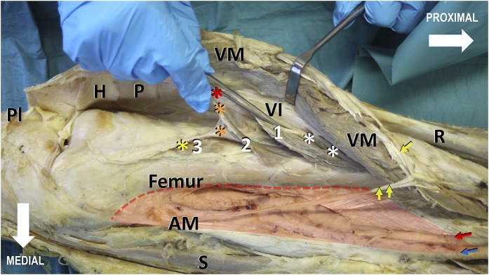
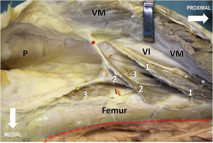
The articularis genus muscle was present in all specimens and showed a variable architecture consisting of 3 to 6 muscle bundles (Table I). The number of bundles was comparable between sexes and overall between the left and right legs in paired specimens. The muscle bundles of the articularis genus were organized into 3 layers (Fig. 1), with the proximal bundles forming the most superficial layer and the distal bundles forming the deepest layer. Proximally, the bundles of the superficial layer and, in 60% of the specimens, the bundles of the intermediate layer originated from both the vastus intermedius and the anterior and anterolateral surfaces of the femur. The bundles of the deep layer and, in 40% of the specimens, the bundles of the intermediate layer arose solely from the anterior surface of the femur (Fig. 2). Proximally, there was no clear separation between the articularis genus and the vastus intermedius, with the articularis genus muscle bundles being in continuity with the deep muscle fibers of the vastus intermedius. Distally, the deep and intermediate layers of the articularis genus muscle bundles were partially separated by fatty tissue (Fig. 2).
Fig. 1.
Photograph showing the medial view of the distal two-thirds of a right thigh. The vastus medialis (VM), together with its insertion into the vastus intermedius (VI), is lifted from its hammock-like origin (red transparent shading), the medial lip of the linea aspera, and the medial supracondylar line (red dotted line), thereby revealing the knee joint cavity, the articularis genus (indicated by the white numbers), the vastus intermedius aponeurosis, and the medial surface of the distal part of the femur. The articularis genus is arranged in 3 layers: superficial (indicated by the number 1), intermediate (indicated by the number 2), and deep (indicated by the number 3). Distally, the articularis genus bundles insert gradually into the suprapatellar bursa (asterisks) and the joint capsule. The bundles of the superficial layer insert into the synovial membrane adjacent to the quadriceps tendon (red asterisk), the bundles of the intermediate layer insert into the middle section of the suprapatellar bursa (orange asterisks), and the muscles of the deep layer insert into the synovial membrane (yellow asterisk) facing the femur. Distal deep muscle bundles of the vastus intermedius (white asterisks) contribute to the vastus intermedius aponeurosis and finally merge with the quadriceps tendon, inserting into the base of the patella (P). The long nerve branch to the vastus medialis (single yellow arrow), lifted with the vastus medialis muscle, courses distally along the anteromedial border of the muscle and, in contrast to the saphenous nerve (double yellow arrows), remains lateral to its superficial aponeurosis in a separate fibrotic tunnel. Red and blue arrows = superficial femoral vessels, H = Hoffa fat pad, PI = transected patellar tendon, R = rectus femoris, AM = tendon of the adductor magnus, and S = sartorius.
Fig. 2.
Enlarged view of Figure 1, showing the origin and insertion of the articularis genus. The bundles of the superficial layer (indicated by the number 1) and, in 60% of the specimens, the bundles of the intermediate layer (indicated by the number 2) originated from both the deep surface of the vastus intermedius (VI) aponeurosis and from the anterior and anterolateral surfaces of the femur. The bundles of the deep layer (indicated by the number 3) and, in 40% of the specimens, the bundles of the intermediate layer originated entirely from the anterior surface of the femur. Distally, the articularis genus bundles inserted gradually into the suprapatellar bursa and the joint capsule (asterisks), with the bundles of the superficial layer inserting into the synovial membrane adjacent to the quadriceps tendon (red asterisk), the bundles of the intermediate layer inserting into the middle section of the suprapatellar bursa (orange asterisks), and the bundles of the deep layer inserting into the synovial membrane facing the femur (yellow asterisk). Proximally, no distinct investing fascia separated the articularis genus from the vastus intermedius; in that area, the articularis genus muscle bundles were in continuity with the deep muscle fibers of the vastus intermedius (white asterisks). Distally, the deep and intermediate articularis genus muscle bundles were partially separated by or imbedded in a considerable amount of fatty tissue (orange arrow). For better visualization of the articularis genus muscle bundles, the fatty tissue was partially removed. VM = vastus medialis. P = Patella.
TABLE I.
Characteristics of Specimens and Number of Articularis Genus Muscle Bundles and Layers
| Specimen | Case | Side | Sex | Layers | Articularis Genus Bundles |
| 1 | 1 | Right | Male | 3 | 4 |
| 2 | 1′ | Left | Male | 3 | 6 |
| 3 | 2 | Right | Male | 3 | 4 |
| 4 | 2′ | Left | Male | 3 | 5 |
| 5 | 3 | Right | Male | 3 | 6 |
| 6 | 3′ | Left | Male | 3 | 5 |
| 7 | 4 | Right | Male | 3 | 6 |
| 8 | 4′ | Left | Male | 3 | 6 |
| 9 | 5 | Right | Female | 3 | 3 |
| 10 | 5′ | Left | Female | 3 | 4 |
| 11 | 6 | Right | Female | 3 | 6 |
| 12 | 6′ | Left | Female | 3 | 5 |
| 13 | 7 | Left | Female | 3 | 6 |
| 14 | 8 | Right | Female | 3 | 5 |
| 15 | 9 | Right | Male | 3 | 4 |
| 16 | 10 | Left | Male | 3 | 6 |
| 17 | 11 | Right | Male | 3 | 5 |
| 18 | 12 | Right | Male | 3 | 5 |
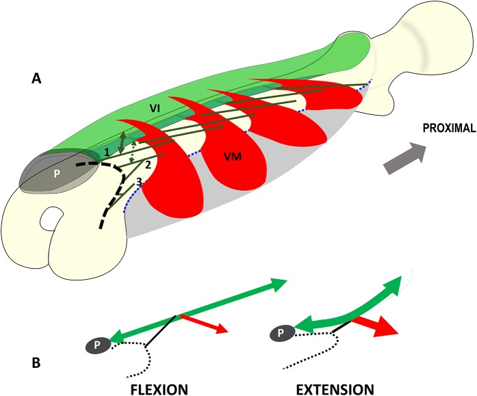
While the deep layer of the vastus intermedius always corresponded with the muscle bundles that finally continued as the articularis genus to the knee joint, the superficial layer of the vastus intermedius contributed to the layers of the quadriceps tendon and finally inserted into the base of the patella. Distally, where the articularis genus inserted into the suprapatellar bursa and the knee joint capsule, connections between the superficial bundles of the articularis genus and the deep muscle fibers of the vastus medialis could be observed. Conversely, the insertion of the vastus medialis expanded from the joint capsule and the patella to the medial edge of the rectus femoris and the aponeurosis of the vastus intermedius (Fig. 3).
Fig. 3.
Figs. 3-A and 3-B Schematic drawings showing the anatomy and mode of operation of the interacting muscle complex consisting of the articularis genus, vastus intermedius (VI), and vastus medialis (VM). Fig. 3-A Schematic illustration depicting the hammock-like origin (gray) and the fleshy clip-like double-insertion of the vastus medialis units (red) into the entire vastus intermedius aponeurosis (green). The blue dotted line corresponds to the origin of the vastus medialis at the medial lip of the linea aspera. The vastus medialis clamps the vastus intermedius aponeurosis like a clip holding a sheet. The deep layer of the vastus intermedius (green lines) corresponds with the muscle bundles that continue as the articularis genus to the knee joint. The superficial layer of the vastus intermedius contributes to the layers of the quadriceps tendon and finally inserts into the base of the patella (P). In the present study, the superficial articularis genus muscle bundles (indicated by the number 1) always originated from both the anterior surface of the femur and the vastus intermedius (double arrows). The deep articularis genus muscle bundles (indicated by the number 3) arose solely from the femur. The intermediate articularis genus muscle bundles (indicated by the number 2) always arose from the anterior surface of the femur. However, in 60% of the specimens, muscle fibers also originated from the vastus intermedius aponeurosis. All muscle bundles of the articularis genus inserted into the synovial membrane of the joint capsule and the suprapatellar bursa (black dashed line). Fig. 3-B Schematic illustration depicting the mode of operation of the muscle complex consisting of the articularis genus (black lines), vastus intermedius (green double arrows), and vastus medialis (red arrows). As a derivative of the vastus intermedius, the articularis genus consists of a few muscle bundles that arise from the deep part of the vastus intermedius. Therefore, the articularis genus does not act as an independent entity. The muscle bundles of the articularis genus connect with the neighboring vastus intermedius and vastus medialis. With their support, the articularis genus retracts or elevates the suprapatellar bursa (black dotted lines) during extension of the knee, preventing entrapment of the bursa between the patella (P) and the femur.
Innervation of the Articularis Genus, Vastus Intermedius, and Vastus Medialis Muscles
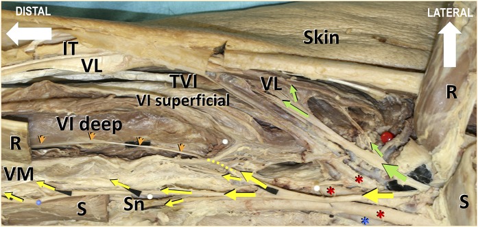
The articularis genus, the vastus medialis, and the medial part of the vastus intermedius were always supplied by the same medial deep division of the femoral nerve, which subdivided into 3 to 4 branches. The most medial branch continued as the saphenous nerve. The second branch supplied the vastus medialis muscle, coursing distally along the anteromedial border of the muscle and, in contrast with the saphenous nerve, remaining lateral to its superficial aponeurosis in a separate fibrotic tunnel. The third and fourth branches innervated the proximal parts of the vastus medialis and the medial parts of the vastus intermedius (Fig. 4). At first glance, these branches appeared to have short courses and were often hidden by branches of the lateral circumflex femoral artery; however, 1 of these terminal branches crossed the medial portion of the vastus intermedius, coursed distally above the deep layer of the vastus intermedius, and branched to the articularis genus, finally inserting into the upper part of the synovial pouch of the knee joint. This terminal nerve branch formed a guide for (1) separating the vastus medialis from the vastus intermedius proximally and (2) separating the articularis genus and the deep layer of the vastus intermedius from the superficial layer of the vastus intermedius.
Fig. 4.
Photograph showing the anterior view of the proximal part of a right thigh along with the nerve supply to the extensor apparatus of the knee joint. For better visualization of the femoral nerve branches, the sartorius (S) and rectus femoris (R) muscles were transected and elevated. Some nerve branches are identified with black paper. The red pinhead on the right side of the image indicates the middle of the neck of the femur on the intertrochanteric line. The vastus medialis (VM), the medial part of the vastus intermedius (VI), and the articularis genus are innervated by the medial division of the femoral nerve (yellow arrows). One of these terminal branches crosses the medial portion of the vastus intermedius (yellow dotted line), courses distally above the deep layer of the vastus intermedius, and finally branches to the articularis genus and the synovial pouch of the knee joint. This nerve branch (orange arrowheads) is a guide for separating the vastus medialis from the vastus intermedius proximally and the articularis genus inclusive of the deep layer of the vastus intermedius from the superficial layer of the vastus intermedius distally. The nerve branch to the vastus medialis runs along the anteromedial border of the muscle. It separates from the saphenous nerve (Sn) proximally. The green arrows indicate the lateral division of the femoral nerve to the lateral parts of the vastus intermedius, the vastus lateralis (VL), and the tensor vastus intermedius (TVI). Red asterisks = superficial, deep, and lateral circumflex femoral arteries; blue asterisk = superficial femoral vein; and IT = iliotibial tract.
Discussion
The present study revealed that the articularis genus is part of a complex and intricate muscular system consisting of the articularis genus, vastus intermedius, and vastus medialis. These 3 structures share the same innervation pattern and work in concert as a functional unit.
The articularis genus consists of 3 to 6 muscle bundles and is arranged in deep, intermediate, and superficial layers. Each layer spans a different section of the suprapatellar bursa (Figs. 1 and 2). The superficial bundles fix the suprapatellar bursa firmly to the quadriceps tendon. Although the distal bundles of the articularis genus are visibly separate from the vastus intermedius, this distinction is less clear more proximally. No fascia separates the articularis genus from the vastus intermedius; this finding is consistent with those of previous studies1,2,17. Hence, the muscle bundles of the articularis genus could be considered to be an extension of the deep layer of the vastus intermedius (Fig. 3); this view is in agreement with previous studies in which it was assumed that the articularis genus derived from the vastus intermedius1-3,5. Some authors consider the articularis genus muscle to include only the muscle bundles that arise as an independent entity from the anterior surface of the distal femoral shaft and blend with the joint capsule18; this simplified definition would correspond with the isolated deep layer of the articularis genus in the present study (Fig. 2). Others believe that it includes all distal muscle fibers that blend with the vastus intermedius as well as the bundles that expand between the femur and the vastus intermedius tendon2. We consider the muscle strands between the femur and the vastus intermedius tendon to be deep vastus intermedius fibers rather than being part of the articularis genus. However, we consider muscle bundles that insert into the synovial membrane of the joint capsule and into the suprapatellar bursa as being part of the articularis genus, regardless of the origin of the muscle bundles. The lack of a uniform definition of the articularis genus muscle in the literature might explain the wide variability in the number of articularis genus muscle bundles (range, 2 to 10) as reported in previous studies1-3,5,19-22.
An important finding of the present study is that the vastus intermedius aponeurosis represents a point of origin of the articularis genus and an important insertion site for the vastus medialis. The insertion of the vastus medialis expands from the patella, the medial edge of the rectus femoris, and the strong aponeurosis of the vastus intermedius16. Some muscle fibers of the vastus medialis also merge with the superficial bundles of the articularis genus. This close connection suggests that the vastus medialis holds the vastus intermedius medially, like a paper clip holding a sheet of paper, with the vastus medialis muscle fibers wrapped firmly around the aponeurosis of the vastus intermedius (Fig. 3-A). This clip-like double insertion of the vastus medialis into the aponeurosis of the vastus intermedius leads to the impression of the existence of an aponeurosis on the deep surface of the vastus medialis, when in fact this aponeurosis belongs to the vastus intermedius9,19,20,23,24.
Many investigators have attempted to clarify the functional status of the articularis genus muscle and its relationship to the range of motion of the knee1-3,5-7. The insertion of this muscle has prompted speculation that the articularis genus retracts or elevates the suprapatellar bursa during extension of the knee joint and prevents impingement of the synovial membrane between the patella and the femur1,2,8-11. However, measurements have revealed that the cross-sectional area of the articularis genus is minute2, raising the question as to whether this small muscle is capable of retracting the bursa during extension of the knee joint; direct evidence for this function is limited2,5,11,25. Our findings suggest that this function might be performed by an anatomical complex consisting of the articularis genus, vastus intermedius, and vastus medialis. The vastus intermedius, together with the vastus medialis, regulates and enhances the power of the articularis genus during the entire range of motion of the knee. While this assistance to the articularis genus is especially important in full contraction during the terminal phase of leg extension, the articularis genus should be fully relaxed (and unassisted) when the knee is in full flexion. Between maximum flexion and maximum extension, the tension of the vastus intermedius, and therefore the articularis genus, must be adjusted continuously by the medial-oblique pull of the vastus medialis on the longitudinally orientated vastus intermedius aponeurosis (Fig. 3-B)16. Thus, it can be hypothesized that the vastus medialis pretensions and consequently triggers the vastus intermedius and the articularis genus at full extension; this hypothesis agrees with the observation that the vastus medialis is mostly active during the terminal range of extension, just in time for the articularis genus to fulfill its function as a retractor of the suprapatellar bursa26,27. In other words, the articularis genus functionally linked with the vastus intermedius and vastus medialis. This interpretation is in contrast with classic anatomy textbooks, which tend to define each muscle as a separate entity with a unique function at the joint it spans. The vastus medialis, vastus intermedius, and articularis genus cannot be seen as mechanically independent actuators. The potential of force transmission between synergistic skeletal muscles through connective tissue linkage has been highlighted in several recent studies12,28,29. The close interaction of the articularis genus, vastus intermedius, and vastus medialis is also underlined by the fact that the articularis genus, the vastus medialis, and the medial half of the vastus intermedius are supplied by the same deep medial branch of the femoral nerve (Fig. 4).
Deterioration of the vastus medialis, vastus intermedius, or articularis genus in isolation is unlikely. Rather, scarring or malfunction may lead to disruption of the timing of muscle interaction. An effusion may increase the tension of the articularis genus-vastus intermedius complex and subsequently affect vastus medialis activation. Additionally, trauma or surgery involving the knee joint probably affects not only the vastus medialis but also the functionally linked vastus intermedius and articularis genus. Injury to the articularis genus muscle during the surgical approach to the knee joint (e.g., during synovectomy for total knee arthroplasty) could be the reason for heterotopic ossification manifested as bone spurs30, which has been correlated with the presence of stiffness following total knee arthroplasty and fracture in the supracondylar region of the femur31-35. A decrease in the mass of the articularis genus and vastus intermedius is difficult to diagnose clinically27,36,37. However, in the study by Saito et al., atrophic changes of the articularis genus muscle were documented with ultrasound, and these findings were found to be correlated with decreased range of motion of the knee and knee pain11.
An important limitation of the present study is that all donors were elderly (mean age, 77 years). Age-related muscle atrophy may have distorted the results, which therefore might not be representative for young individuals. Embalmed tissue also has been reported to shrink by 2.2% to 12%38,39. Finally, the number of specimens used in the present study was limited.
In conclusion, the anatomical construct consisting of the articularis genus, vastus medialis, and vastus intermedius presents a more complex interacting architecture than has been described in the current literature4,8,9,40-42. The articularis genus consists of muscle fibers that arise from the femur but also from the deep part of the vastus intermedius. The articularis genus does not act independently as its muscle bundles are strongly linked to the vastus intermedius and, subsequently, the vastus medialis.
Disclosure of Potential Conflicts of Interest
Footnotes
Investigation performed at Department of Anatomy, University of Zürich-Irchel, Zürich, Switzerland
Disclosure: No external funding was used in this study. On the Disclosure of Potential Conflicts of Interest forms, which are provided with the online version of the article, one or more of the authors checked “yes” to indicate that the author had a relevant financial relationship in the biomedical arena outside the submitted work (http://links.lww.com/JBJSOA/A25).
References
- 1.Kimura K, Takahashi Y. M. articularis genus. Observations on arrangement and consideration of function. Surg Radiol Anat. 1987;9(3):231-9. [DOI] [PubMed] [Google Scholar]
- 2.Woodley SJ, Latimer CP, Meikle GR, Stringer MD. Articularis genus: an anatomic and MRI study in cadavers. J Bone Joint Surg Am. 2012. January 4;94(1):59-67. [DOI] [PubMed] [Google Scholar]
- 3.Puig S, Dupuy DE, Sarmiento A, Boland GW, Grigoris P, Greene R. Articular muscle of the knee: a muscle seldom recognized on MR imaging. AJR Am J Roentgenol. 1996. May;166(5):1057-60. [DOI] [PubMed] [Google Scholar]
- 4.Platzer W. Taschenatlas anatomie. Bewegungsapparat. 11th ed. New York: Thieme; 2013. p 248-9. [Google Scholar]
- 5.Ahmad I. Articular muscle of the knee—articularis genus. Bull Hosp Joint Dis. 1975. April;36(1):58-60. [PubMed] [Google Scholar]
- 6.DiDio LJ, Zappalá A, Carney WP. Anatomico-functional aspects of the musculus articularis genus in man. Acta Anat (Basel). 1967;67(1):1-23. [DOI] [PubMed] [Google Scholar]
- 7.DiDio LJ, Zappalá A, Cardoso AD, Diaz RA. Musculus articularis genus in human fetuses, newborn and young individuals. Anat Anz. 1969;124(2):121-32. [PubMed] [Google Scholar]
- 8.Moore KL, Agur AMR, Dalley AF. Clinically oriented anatomy. 7th ed. Philadelphia: Lippincott Williams & Wilkins; 2014. p 545-8. [Google Scholar]
- 9.Drake RL, Vogl AW, Mitchell AWM. Gray’s anatomy for students. 3rd ed. Philadelphia: Churchill Livingstone Elsevier; 2014. p 592-3. [Google Scholar]
- 10.Sakuma E, Sasaki Y, Yamada N, Wada I, Soji T. Morphological characteristics of the deep layer of articularis genus muscle. Folia Morphol (Warsz). 2014. August;73(3):309-13. [DOI] [PubMed] [Google Scholar]
- 11.Saito A, Okada K, Saito I, Kinoshita K, Seto A, Takahashi Y, Shibata K, Sato H, Wakasa M. Functional status of the articularis genus muscle in individuals with knee osteoarthritis. J Musculoskelet Neuronal Interact. 2016. December 14;16(4):348-54. [PMC free article] [PubMed] [Google Scholar]
- 12.Maas H, Sandercock TG. Force transmission between synergistic skeletal muscles through connective tissue linkages. J Biomed Biotechnol. 2010;2010:575672 Epub 2010 Apr 12. [DOI] [PMC free article] [PubMed] [Google Scholar]
- 13.Universität Zürich. Körperspende. 2017. September 11 http://www.anatomy.uzh.ch/de/koerperspende.hml. Accessed 2017 Oct 11.
- 14.Smith-Petersen MN. Approach to and exposure of the hip joint for mold arthroplasty. J Bone Joint Surg Am. 1949. January;31A(1):40-6. [PubMed] [Google Scholar]
- 15.Gänsslen A, Grechenig ST, Nerlich M, Müller M, Grechenig W. [Standard approaches to the acetabulum part 2: ilioinguinal approach]. Acta Chir Orthop Traumatol Cech. 2016;83(4):217-22. [PubMed] [Google Scholar]
- 16.Grob K, Manestar M, Filgueira L, Kuster MS, Gilbey H, Ackland T. The interaction between the vastus medialis and vastus intermedius and its influence on the extensor apparatus of the knee joint. Knee Surg Sports Traumatol Arthrosc. 2017. January 25 Jan 25. Epub 2017 Jan 25. [DOI] [PubMed] [Google Scholar]
- 17.Reider B, Marshall JL, Koslin B, Ring B, Girgis FG. The anterior aspect of the knee joint. J Bone Joint Surg Am. 1981March;63(3):351-6. [PubMed] [Google Scholar]
- 18.Poirier P, Charpy A. Traité d’anatomie humaine. 12th ed. Paris: Masson et Co; 1912. p 514. [Google Scholar]
- 19.Gegenbaur C. Lehrbuch der anatomie des menschen. 7th ed. Leipzig: Wilhelm Engelmann; 1899. p 446-50. [Google Scholar]
- 20.Warwick R, Williams PL. Gray’s anatomy. 35th ed. Philadelphia: Longmans; 1973. p 562-4. [Google Scholar]
- 21.Bryce TH. Myology. Quain’s elements of anatomy. 11th ed, vol. 4, pt 2. Longmans, Green; 1923. p 235-41. [Google Scholar]
- 22.Merkel FJ. Henle’s grundgriss der anatomie des menschen. Braunschweig: Friedrich Vieweg and Sons; 1901. p 209. [Google Scholar]
- 23.Testut L. Traité d’anatomie humaine. Paris: Gaston Doin; 1893. p 170-5. [Google Scholar]
- 24.Thiranagama R. Nerve supply of the human vastus medialis muscle. J Anat. 1990. June;170:193-8. [PMC free article] [PubMed] [Google Scholar]
- 25.Becker I, Woodley SJ, Baxter GD. Gross morphology of the vastus lateralis muscle: An anatomical review. Clin Anat. 2009. May;22(4):436-50. [DOI] [PubMed] [Google Scholar]
- 26.Lieb FJ, Perry J. Quadriceps function. An anatomical and mechanical study using amputated limbs. J Bone Joint Surg Am. 1968. December;50(8):1535-48. [PubMed] [Google Scholar]
- 27.Lieb FJ, Perry J. Quadriceps function. An electromyographic study under isometric conditions. J Bone Joint Surg Am. 1971. June;53(4):749-58. [PubMed] [Google Scholar]
- 28.Huijing PA. Epimuscular myofascial force transmission: a historical review and implications for new research. International Society of Biomechanics Muybridge Award Lecture, Taipei, 2007. J Biomech. 2009. January 5;42(1):9-21. Epub 2008 Nov 29. [DOI] [PubMed] [Google Scholar]
- 29.Yucesoy CA, Baan GC, Koopman BHFJM, Grootenboer HJ, Huijing PA. Pre-strained epimuscular connections cause muscular myofascial force transmission to affect properties of synergistic EHL and EDL muscles of the rat. J Biomech Eng. 2005. October;127(5):819-28. [DOI] [PubMed] [Google Scholar]
- 30.Manrique J, Gomez MM, Parvizi J. Stiffness after total knee arthroplasty. J Knee Surg. 2015. April;28(2):119-26. Epub 2014 Dec 16. [DOI] [PubMed] [Google Scholar]
- 31.Moya-Angeler J, Bas MA, Cooper HJ, Hepinstall MS, Rodriguez JA, Scuderi GR. Revision arthroplasty for the management of stiffness after primary TKA. J Arthroplasty. 2017. June;32(6):1935-9. Epub 2017 Jan 19. Epub ahead of print. [DOI] [PubMed] [Google Scholar]
- 32.Hahn SB, Choi YR, Kang HJ, Lee SH. Prognostic factors and long-term outcomes following a modified Thompson’s quadricepsplasty for severely stiff knees. J Bone Joint Surg Br. 2010. February;92(2):217-21. [DOI] [PubMed] [Google Scholar]
- 33.Shang P, Liu HX, Zhang Y, Xue EX, Pan XY. A mini-invasive technique for severe arthrofibrosis of the knee: A technical note. Injury. 2016. August;47(8):1867-70. Epub 2016 Jun 11. [DOI] [PubMed] [Google Scholar]
- 34.Gittings D, Hesketh P, Dattilo J, Zgonis M, Kelly J, Mehta S. Arthroscopic lysis of adhesions improves knee range of motion after fixation of intra-articular fractures about the knee. Arch Orthop Trauma Surg. 2016. December;136(12):1631-5. Epub 2016 Aug 30. [DOI] [PubMed] [Google Scholar]
- 35.Cheuy VA, Foran JRH, Paxton RJ, Bade MJ, Zeni JA, Stevens-Lapsley JE. Arthrofibrosis associated with total knee arthroplasty. J Arthroplasty. 2017. August;32(8):2604-11. Epub 2017 Feb 14. [DOI] [PubMed] [Google Scholar]
- 36.Giles LS, Webster KE, McClelland JA, Cook J. Atrophy of the quadriceps is not isolated to the vastus medialis oblique in individuals with patellofemoral pain. J Orthop Sports Phys Ther. 2015. August;45(8):613-9. Epub 2015 Jun 25. [DOI] [PubMed] [Google Scholar]
- 37.Pattyn E, Verdonk P, Steyaert A, Vanden Bossche L, Van den Broecke W, Thijs Y, Witvrouw E. Vastus medialis obliquus atrophy: does it exist in patellofemoral pain syndrome? Am J Sports Med. 2011. July;39(7):1450-5. Epub 2011 Apr 12. [DOI] [PubMed] [Google Scholar]
- 38.Cutts A. Shrinkage of muscle fibres during the fixation of cadaveric tissue. J Anat. 1988. October;160:75-8. [PMC free article] [PubMed] [Google Scholar]
- 39.Friederich JA, Brand RA. Muscle fiber architecture in the human lower limb. J Biomech. 1990;23(1):91-5. [DOI] [PubMed] [Google Scholar]
- 40.Putz R, Pabst R. Sobotta atlas of human anatomy. 14th ed. München: Elsvier/Urban & Fischer; 2008. p 302-5. [Google Scholar]
- 41.Schünke M, Schulte E, Schumacher U. Prometheus lernatlas der anatomie: allgemeine anatomie und bewegungssystem. Stuttgart: Thieme; 2011. p 476-84. [Google Scholar]
- 42.Netter FH. Atlas of human anatomy. 5th ed. Philadelphia: Saunders Elsevier; 2011. p 398-401. [Google Scholar]
Associated Data
This section collects any data citations, data availability statements, or supplementary materials included in this article.


