Abstract
Honeybees directly transfer plant compounds from nectar into honey. Each plant species possesses a specific metabolic profile, the amount and the typology of plant molecules that may be detected in honey vary according to their botanical origin. Aim of the present work was the spectrophotometrical determination of concentration ranges of simple phenols and flavonoids in 460 several Italian monofloral honeys, in order to individuate specific intervals of plant metabolites for each typology of honey. Moreover, an LC–MS analysis was performed to determine amount of various secondary metabolites in the samples, with the purpose to use them as potential molecular markers in support to honey melissopalynological classification. As plant molecules have a strong reducing power, the antioxidant activity of the honeys was evaluated by two antiradical assays, DPPH and FRAP. The free radical scavenging effect of each monofloral group was correlated to the concentration of simple phenols and flavonoids, with the aim to deduce the existence of possible relationships between these parameters. In conclusion, dark honeys (Castanea sativa, honeydew, Erica sp. and Eucalyptus sp.) appeared to be the richest in secondary metabolites and, consequently, showed higher antioxidant activity. However, all analyzed monofloral honeys showed to be good sources of antioxidants.
Keywords: Secondary metabolites, Monofloral honeys, DPPH, FRAP, LC–MS
Introduction
Plant organisms are the only living entities able to produce secondary metabolites. Indeed, these molecules are specifically synthesized by plant biochemical pathways which are absent in animal cells (Kaufman et al. 1999).
In nature, more than 200,000 different plant compounds were evolved, in order to regulate the interaction between plants and environment (Fiehn 2002). In particular, they provide for plants advantageous devices against biotic (competitive species, predators, pathogens) and abiotic (UV radiations, desiccation, thermal shock) stresses and promote the attraction of pollinators, the dispersal of seeds and the allelopathic phenomenon (Dudareva and Pichersky 2008). On the other hand, plant secondary metabolites have also a positive effect on animal systems, carrying out a strong antioxidant activity (Bertoncelj et al. 2007). For this reason, in the last decades, medicinal plants, or rather vegetal organisms containing high doses of bioactive molecules, were widely studied for their potential beneficial role on human health, especially in the reduction of cell damages induced by radical species (Gismondi et al. 2013, 2014). In fact, plant compounds are considered excellent free radical scavenging molecules, thanks to their peculiar chemical structure (Gismondi et al. 2017a, b), and were also proposed as natural drugs by a lot of pharmaceutical industries (Raskin et al. 2002).
Plant antioxidants, introduced by diet and presenting a great bioactivity and molecular diversity, were demonstrated to be able to act on different cell targets, influencing consumers’ health (Raskin et al. 2002; Crozier et al. 2008). In general, they are employed to restore the tissue redox equilibrium unbalanced by high amount of reactive species, naturally produced by metabolism or resulting from bad life conditions and environmental pollution. Among plant metabolites, phenolics (i.e. simple phenols, flavonoids) are the most abundant and antiradical (Ferreres et al. 2008; Can et al. 2015).
Nowadays, it is well-known that such type of natural compounds is also present in honey (Tahir et al. 2017). Indeed, since honey is produced by bees (Apis mellifera ligustica Spinola) starting from floral nectar or plant secretions, secondary metabolites are directly transferred and accumulated from plants into this food. As consequence, honey composition, including physico-chemical, organoleptic and nutraceutical properties, is directly linked with the environmental characteristics of the areas where it is produced (Di Marco et al. 2016). Consequently, the concentration of phenolic compounds in honey determines the antioxidant power of this natural product and its potential biological activity on humans (Vela et al. 2007). Indeed, Can et al. (2015) and Tahir et al. (2017) correlated the antiradical capacity of different honeys with their total phenol content, while Jaganathan and Mandal (2009), Pichichero et al. (2010, 2011) reported the antineoplastic effect of honey plant metabolites on in vitro mammalian systems, suggesting the potential role of honey antioxidants in human disease prevention and health promotion (Bogdanov et al. 2008).
Honey has an important role in human nutrition since prehistoric time. Over its use as sweetener, honey is known for curative properties (Yaniv and Rudich 1997). In particular, a lot of scientific works demonstrated the effect of honey treatments on human health (Bogdanov et al. 2008; Muhammad et al. 2016). Moreover, since honey quality and chemical content reflect the source of nectar, honeys with different botanical origin would show dissimilar characteristics and properties (Pichichero et al. 2009).
Based on this evidence, the present research aimed to identify the existence of specific ranges of biological parameters (i.e. plant secondary metabolite amount) which could significantly distinguish the various existing monofloral honeys. In fact, beyond the pollen spectrum, other parameters (i.e. electrical conductivity, ashes) are necessary to corroborate or support honey melissopalynological classification (Di Marco et al. 2016). Moreover, the antioxidant activity associated to each typology of honey was measured, by two antiradical assays, in order to investigate the putative correlation between free radical scavenging power of the samples and their levels of plant antioxidant compounds. According to all previous observations, main objective of the present work was the characterization of the metabolic profile and antioxidant power of 460 Italian monofloral honeys, whose botanical origin was previously identified by Di Marco et al. (2016), through determination of principal physico-chemical parameters and melissopalynological study. In particular, by spectrophotometric and chromatographic approaches, for each honey, simple phenol and flavonoid content, concentration of specific secondary metabolites and antiradical property were quantified. All results, obtained for each monofloral typology, were analyzed to determine the existence of a possible correlation between nutraceutical value and botanical origin of the honeys.
Materials and methods
Honey samples
Four hundred and sixty monofloral honeys, collected in the whole Italian territory, were studied. The botanical origin of the samples was determined in Di Marco et al. (2016) as follows: 140 acacia honeys (Robinia pseudoacacia L.), 70 chestnut honeys (Castanea sativa Mill.), 10 rhododendron honeys (Rhododendron sp.), 15 dandelion honeys (Taraxacum officinale Web.), 10 lime-blossom honeys (Tilia sp.), 10 thyme honeys (Thymus sp.), 20 heather honey (Erica sp.), 45 eucalyptus honeys (Eucalyptus sp.), 35 hedysarum honeys (Hedysarum coronarium, L.), 45 orange blossom honeys (Citrus sp.) and 60 honeydew honeys.
Secondary metabolite determination: simple phenols and flavonoids
The quantitation of simple phenols and flavonoids was performed by spectrophotometric assays, using a spectrophotometer CARY 50 BIO model (Varian, Italy), according to Di Marco et al. (2014) method with some modifications. For simple phenol determination, 1 g of honey was diluted in 10 mL of bi-distilled water and sonicated for 20 min. To 0.5 mL of honey solution, 2.5 mL of 0.2 N Folin-Ciocalteu reagent and 2.0 mL of Sodium Carbonate 0.7 mol L−1 were added. Then, sample was incubated for 2 h at room temperature in the dark. Absorbance of the solution was read at 760 nm with respect to a blank control of bi-distillate water. A calibration curve with gallic acid (0–1000 mg L−1) was made to extrapolate simple phenol concentration in the samples. Results were expressed as mg of gallic acid equivalent (GAE) per kg of honey. For flavonoid determination, 1 g of honey was dissolved in 1 mL of methanol and sonicated for 20 min. Then, 0.25 mL of honey extract was mixed with 0.75 mL of 5% sodium nitrite, 0.15 mL of 2% aluminium chloride, 0.5 mL of 1 mol L−1 sodium hydroxide and 0.25 mL of distillate water. Sample absorption reading at 510 nm was taken after 10 min of incubation in the dark, with respect to a methanol control. Total flavonoid content was determined using a calibration curve obtained with increasing concentrations of quercetin (0–1000 mg L−1). Data were expressed as mg of quercetin equivalent (QE) per kg honey.
Secondary metabolite analysis by LC–MS
The biochemical profile of honey samples was performed, as described in Impei et al. (2015), by a liquid chromatographic system associated with an LC-20 AD pump, a CBM-20A controller, a SIL-20A HT auto sampler and a single quadrupole mass spectrometer (Shimadzu, Tokyo, Japan). The instrument operated using electro-spray ionization (ESI) source, in positive and negative ion modes. Data were acquired by Lab solution software (Shimadzu). During the analyses, mass spectrometer parameters were set as follows: capillary voltage 3.0 kV, interface voltage 4.5 kV, heat block 200 °C, DL temperature 250 °C, nebulising gas 1.5 L min−1 and drying gas flow 15 L min−1 (N2). Qualitative and quantitative determination of specific secondary metabolites present in honey samples was carried out on the basis of retention time (min), molecular weight (kDa) and mass spectrum with molecular ion of reference (mass-to-charge ratio; m/z), of increasing concentration of adequate standards (Sigma-Aldrich, Milan, Italy). Results were expressed as mg of standard equivalent per Kg of honey. For LC–MS study, secondary metabolites were extracted from honeys as described in Bertoncelj et al. (2011) with appropriate modifications. Ten g of honey were dissolved in 15 mL of acidified water with chloridric acid (pH 2.00). Then, the solution was purified through DSC-C18 cartridges (Sigma-Aldrich), previously conditioned with 3 mL of methanol and 3 mL of distillate water. Columns were washed with 5 mL of acidified water and 15 mL of bi-distillate water. Plant metabolites were eluted with 3 mL of methanol/acetonitrile (2:1; v/v) which were then diluted with 0.1 mL of 10 mmol L−1 sulfuric acid. 10 µL of purified extract were loaded into the instrument. Metabolite separation was obtained using a Reprosil_pur ODS-3 FAST column (100 mm × 2 mm × 3 µm) (Dr Maisch, Germany) set at 30 °C. Total run time was of 32 min and mobile phases were 0.1% formic acid (phase A) and acetonitrile (phase B). The analysis was performed at constant flow rate (0.3 mL min−1) and setting the elution gradient as reported: 0–3rd min at 85% A–15% B; 6th min at 70% A–30% B; 18th min at 65% A–35% B; 25th–29th min at 30% A–70% B; 31st–32nd min 85% A–15% B.
Antioxidant activity of honeys: DPPH and FRAP assays
The radical DPPH (DPPH·; 2,2-diphenyl-1-pycrilhydrazyl) is commonly used to evaluate the antioxidant activity of different food matrixes, since it is highly reactively toward the reducing species. The DPPH solution has a violet coloration that changes to yellow in presence of the antioxidant scavenger, developing its reduced form (DPPH-H). The antioxidant activity was estimated by spectrophotometric analysis (spectrophotometer CARY 50 BIO model—Varian, Italy), according to Beretta et al. (2005) method, adequately modified as follows. Honey samples were dissolved in bi-distilled water, with the aim to prepared different sample concentrations (20, 100, 350 and 800 mg mL−1). One hundred µL of each honey dilution was mixed with 1.9 mL of 130 µmol L−1 DPPH and 1 mL of 0.1 mol L−1 Acetate buffer (pH 5.5); samples were vortexed and, then, incubated for 90 min at 37 °C in the dark. The spectrophotometric readings of the different samples and relative blanks (not containing DPPH·) were used to create a calibration curve, in order to calculate the IC50 values, that is the amount of honey (mg) which reduces the 50% of one mL of 130 μmol L−1 DPPH· radical solution. The FRAP (Ferric Reducing Antioxidant Power) assay is a direct colorimetric test used to determine the antioxidant power of food and plant extracts (Benzie and Strain 1996). This test evaluates the absorbance variation, at 593 nm, caused by the formation of blue-colored Fe2+-TPTZ from colorless oxidized Fe3+-TPTZ, by the action of electron-donating antioxidants. It was carried out according to Bertoncelj et al. (2007), with some modifications. Honey samples were dissolved in bi-distillated water at the final concentration of 1 g mL−1. Two hundred µL of honey solution was added to 1.8 mL of FRAP reagent (TPTZ 10 mmol L−1 in HCl 40 mmol L−1, FeCl3 20 mmol L−1, acetate buffer 0.3 mol L−1 pH 3.6 in a ratio 1:1:10 v/v/v) and incubated 10 min at 37 °C. Absorbance readings at 593 nm were performed by a spectrophotometer CARY 50 BIO model (Varian, Italy). Results were expressed as µmol L−1 of ascorbic acid equivalents per g of honey (µmol L−1 AAE g−1), according to a calibration curve adequately created with pure ascorbic acid (20–700 µmol L−1).
Statistics
All experiments were repeated in triplicate. Data were reported as mean ± standard deviation (sd) of the three independent analyses. Significance was calculated by one-way ANOVA test through PAST software. p values ≤ 0.05 were considered significant; in particular, they were reported as: a (p values ≤ 0.05) or b (p values ≤ 0.01). R statistics software (R version 3.3.3) was used to produce 3D-diagrams of honey data. A Multivariate Analysis (Principal Components) of the correlation between phenols, flavonoids, DPPH results and FRAP data was performed by PAST software.
Results and discussion
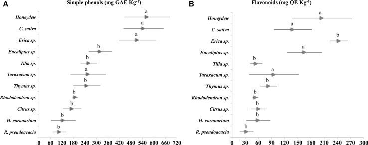
The nutraceutical potential of honey samples was determined by studying their content in secondary metabolites. Therefore, total simple phenols and flavonoids were quantified (Fig. 1).
Fig. 1.
Secondary metabolite quantification. Concentration ranges of simple phenols (A) and flavonoids (B) detected in different monofloral honeys were shown. Results, expressed in mg of standard equivalent (GAE and QE) per kg of sample, represent the mean ± SD of three independent measurements
As indicated in Fig. 1A, on average, honeydew, C. sativa and Erica sp. honeys showed the highest quantity of phenols, presenting 564.2 ± 120.6, 545.7 ± 104.7 and 475.0 ± 110.3 mg GAE kg−1, respectively. Lower values were found in Eucalyptus sp. (320.0 ± 60.2 mg GAE kg−1), Tilia sp. (260.0 ± 42.4 mg GAE kg−1), Taraxacum sp. (256.7 ± 92.9 mg GAE kg−1), Thymus sp. (250.0 ± 70.7 mg GAE kg−1), Rhododendron sp. (190.0 ± 14.1 mg GAE kg−1), Citrus sp. (167.8 ± 18.8 mg GAE kg−1), R. pseudoacacia (107.2 ± 35.7 mg GAE kg−1) and H. coronarium (127.1 ± 64.2 mg GAE kg−1) samples.
On the other hand, flavonoid content of the samples (Fig. 1B) evidenced a different profile compared to simple phenolic one. In detail, Erica sp. honeys showed the greater level of these molecules, equal to 213.0 ± 64.1 mg QE kg−1, followed by honeys of honeydew (204.8 ± 68.3 mg QE kg−1), Eucalyptus sp. (165.4 ± 39.6 mg QE kg−1), C. sativa (138.8 ± 43.1 mg QE kg−1), Taraxacum sp. (95.7 ± 46.4 mg QE kg−1), Thymus sp. (83.5 ± 19.1 mg QE kg−1), Citrus sp. (60.8 ± 48.4 mg QE kg−1), H. coronarium (59.7 ± 27.4 mg QE kg−1), Tilia sp. (55.0 ± 14.1 mg QE kg−1), Rhododendron sp. (53.5 ± 6.4 mg QE kg−1) and R. pseudoacacia (33.1 ± 15.8 mg QE kg−1).
These results suggested that the botanical origin could strongly influence the concentration of specific classes of plant metabolites (i.e. simple phenols, flavonoids) in honeys. Consequently, the quantitation of such type of compounds, with respect to the concentration range measured in each sample group, would represent a useful discrimination tool for honey classification and identification. It was evident that darker samples (honeydew, C. sativa, Erica sp. and Eucalyptus sp.) possessed the highest doses of secondary metabolites, in accordance with Bertoncelj et al. (2007) and Beretta et al. (2005). All other typologies of honey clustered together, showing overlapping values. In general, the concentration of plant molecules detected, during the present study, in Italian honeys was double compared to Slovenian and commercial samples, analyzed in Bertoncelj et al. (2007) and Beretta et al. (2005), respectively.
As second step, all samples were subjected to LC–MS analysis, in order to obtain a chromatographic characterization of the honeys. In particular, seven plant secondary metabolites, three phenolic acids (caffeic acid, p-coumaric acid and chlorogenic acid) and four flavonoids (apigenin, myricetin, kaempferol and quercetin), were detected, identified and quantified (in mg per kg of honey), with respect to pure standard molecules (Table 1). C. sativa, honeydew and Taraxacum sp. honeys presented the highest total amounts of plant compounds, while H. coronarium, R. pseudoacacia and Rhododendron sp. samples the lowest ones.
Table 1.
Honey metabolic profiles
| Monofloral type | Apigenin | Myricetin | Kaempferol | Quercetin | Caffeic acid | p-Coumaric acid | Chlorogenic acid | Total |
|---|---|---|---|---|---|---|---|---|
| Taraxacum sp. | n.d. | 100.1 ± 1.1 | n.d. | 32.2 ± 2.2 | n.d. | n.d. | n.d. | 132.3 ± 3.3 |
| R. pseudoacacia | n.d. | 26.1 ± 2.3 | 23.7 ± 1.8 | n.d. | n.d. | n.d. | n.d. | 49.8 ± 4.1 |
| H. coronarium | 17.5 ± 1.3 | n.d. | n.d. | 18.5 ± 1.6 | n.d. | 6.2 ± 0.9 | n.d. | 42.2 ± 3.8 |
| Castanea sativa | 75.8 ± 2.2 | 33.6 ± 1.8 | 32.4 ± 1.4 | 40.8 ± 2.3 | 10.6 ± 1.0 | 22.2 ± 0.5 | n.d. | 215.4 ± 9.2 |
| Erica sp. | 19.3 ± 1.1 | 19.5 ± 1.6 | 20.1 ± 2.1 | 24.4 ± 1.3 | 10.2 ± 2.2 | 4.4 ± 0.5 | 7.2 ± 0.8 | 105.1 ± 9.6 |
| Thymus sp. | 90.8 ± 4.7 | 8.1 ± 2.1 | n.d. | 15.2 ± 1.3 | n.d. | n.d. | n.d. | 114.1 ± 8.1 |
| Tilia sp. | 32.1 ± 3.2 | n.d. | 16.3 ± 2.1 | 27.1 ± 1.1 | n.d. | n.d. | n.d. | 75.5 ± 6.4 |
| Rhododendron sp. | 11.2 ± 1.3 | n.d. | 31.1 ± 2.3 | 12.1 ± 1.5 | n.d. | n.d. | n.d. | 54.4 ± 6.1 |
| Citrus sp. | 22.1 ± 1.9 | n.d. | n.d. | 17.8 ± 1.3 | n.d. | 3.4 ± 0.2 | n.d. | 73.9 ± 3.4 |
| Eucalyptus sp. | 23.3 ± 2.2 | 12.2 ± 1.4 | 25.0 ± 2.3 | 21.7 ± 1.8 | n.d. | 10.2 ± 1.3 | n.d. | 92.4 ± 9.0 |
| Honeydew | 37.5 ± 4.2 | n.d. | 38.9 ± 3.8 | 43.4 ± 3.7 | n.d. | 20.2 ± 1.7 | 15.8 ± 1.4 | 155.8 ± 14.8 |
Concentration of secondary metabolites detected by LC–MS in monofloral honeys. Results, expressed in mg of standard equivalent per kg of sample, represent the mean ± SD of three independent measurements (n.d. not detected) (in all cases, p values = a)
The presence of phenolic acids was just revealed in some monofloral typologies (i.e. C. sativa and honeydew); Erica sp. was the only sample which contemporary contained caffeic, p-coumaric and chlorogenic acids. Caffeic acid and chlorogenic acid were also found, respectively, in C. sativa honey (10.6 ± 1.0 mg kg−1) and honeydew samples (15.8 ± 1.4 mg kg−1). Finally, beyond Erica sp. honeys, p-coumaric acid was observed in H. coronarium (6.2 ± 0.9 mg kg−1), C. sativa (22.2 ± 0.5 mg kg−1), Citrus sp. (3.4 ± 0.2 mg kg−1), Eucalyptus sp. (10.2 ± 1.3 mg kg−1) and honeydew (20.2 ± 1.7 mg kg−1) samples.
On the other hand, flavonoids were widely distributed in all samples. In detail, apigenin, not detected in R. pseudoacacia and Taraxacum sp. honeys, was very abundant in Thymus sp. (90.8 ± 4.7 mg kg−1) and C. sativa samples (75.8 ± 2.2 mg kg−1), while quercetin, absent in R. pseudoacacia, reached the highest doses in honeydew (43.4 ± 3.7 mg kg−1) and C. sativa (40.8 ± 2.3 mg kg−1). Myricetin was extremely elevated in Taraxacum sp. honey (100.1 ± 1.1 mg kg−1), while kaempferol levels were similar among all samples (16.3 and 38.9 mg kg−1).
In general, LC–MS data corroborated the spectrophotometrical results previously described, although just a few molecules were quantified in this study. Honeys from different botanical origin showed dissimilar content of various secondary metabolites. In addition, the metabolic profiles can be used to identify specific molecular markers to support melissopalynological analysis in honey botanical classification (Di Marco et al. 2016). In detail, according to our data, peculiar plant compounds could be mainly associated to certain flowerings and, consequently, to their deriving monofloral honeys: myricetin for Taraxacum sp. honey; apigenin, caffeic acid, quercetin and p-coumaric acid for C. sativa honey; caffeic acid and chlorogenic acid for Erica sp. honey; quercetin, chlorogenic acid and p-coumaric acid for honeydew. However, we want to underline that the metabolic profile cannot represent, alone, the distinctive element for the botanical origin of a honey.
Currently, honey is considered a natural product with elevated nutraceutical value; in fact, since it is very rich in bioactive substances (i.e. plant metabolites, microRNAs), different scientific works suggest the use of this food in human diet to improve consumers’ health and prevent the onset of diseases (Bogdanov et al. 2008; Muhammad et al. 2016; Gismondi et al. 2017a, b). Earlier, the amount of simple phenols and flavonoids was documented to be the main factor responsible for the determination of honey antioxidant effect (Meda et al. 2005; Cai et al. 2006; Pichichero et al. 2009; Di Marco et al. 2012). However, title was reported about the relationship between honey antiradical power and content of these specific molecules. For this reason, after determination of free radical scavenging activity of the different monofloral samples, the correlation between the concentration of secondary metabolites in honey and their reducing effect was investigated. Here, it is important to specify that, despite the high level of antioxidant molecules, honey, being also rich in carbohydrates, especially monosaccharides (i.e. glucose, fructose), represents a rapid source of energy but, at the same time, a high-caloric food, potentially dangerous for consumers with metabolic dysfunctions (i.e. diabetes).
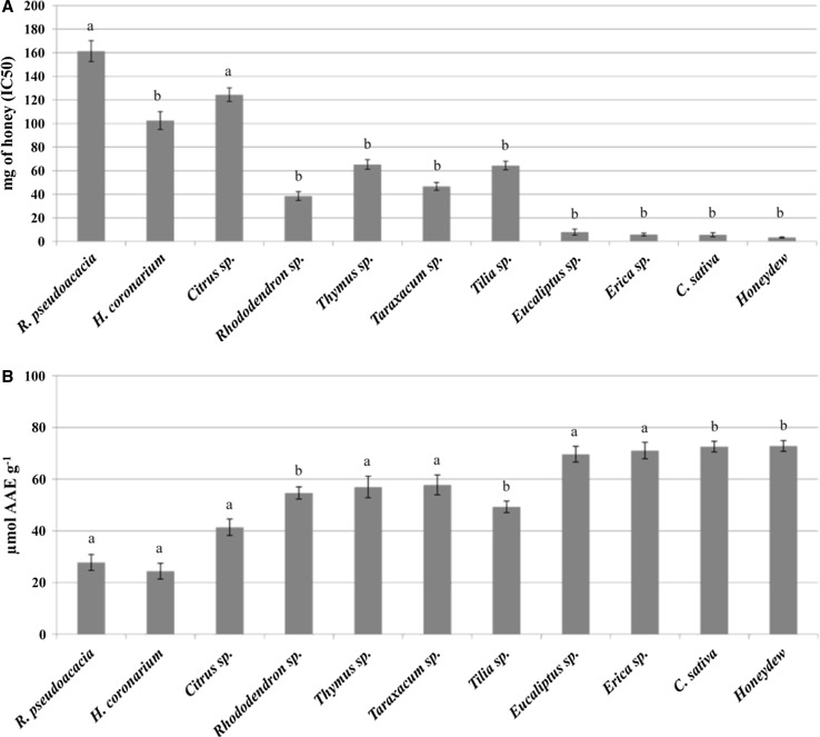
The antioxidant properties of honeys was evaluated by DPPH and FRAP assays, as displayed in Fig. 2. DPPH, expressed by IC50 index, gives an indirect measure of honey antiradical effect (Fig. 2A). The highest value of IC50, corresponding to lowest antioxidant power, was found in R. pseudoacacia honeys (161.3 ± 8.8 mg). On the other hand, Eucalyptus sp. (8.0 ± 2.6 mg), Erica sp. (5.9 ± 1.1 mg), C. sativa (5.7 ± 1.76 mg) and honeydew (3.4 ± 0.5 mg) showed the strongest antiradical properties.
Fig. 2.
Antioxidant assays. The antioxidant power of different monofloral honeys was evaluated by DPPH (A) and FRAP (B) assays. Results were expressed, respectively, by IC50 (mg of honey) and micromoles of ascorbic acid equivalent per g of sample (µmol AAE g−1) and represented the mean ± SD of three independent measurements
FRAP test offers a direct measurement of the antioxidant power of the samples, through comparison with a calibration curve obtained with ascorbic acid, a well-known antioxidant (Fig. 2B). Coherently with DPPH, the highest antiradical activity was registered in honeydew (72.8 ± 2.1 µmol L−1 AAE g−1), C. sativa (72.6 ± 2.1 µmol L−1 AAE g−1), Erica sp. (71.1 ± 3.2 µmol L−1 AAE g−1), and Eucalyptus sp. (69.7 ± 3.0 µmol L−1 AAE g−1) honeys. R. pseudoacacia (27.8 ± 3.1 µmol L−1 AAE g−1) and H. coronarium (24.4 ± 3.1 µmol L−1 AAE g−1) were the less antioxidant.
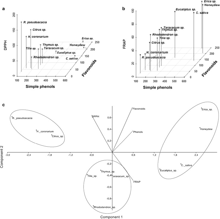
To put in prominence all possible correlations existing between antiradical activity and concentration of plant metabolites in honey, 3D-diagrams, reporting the data of simple phenols and flavonoids with respect to DPPH (Fig. 3a) or FRAP (Fig. 3b) results, were produced by R statistics software. As evidenced by these analyses, the high content of simple phenols and flavonoids in Erica sp. and honeydew samples clearly justified the strong antioxidant power of these honeys, revealed both by DPPH and FRAP assay. Since C. sativa and Eucalyptus sp. samples had similar antiradical effect although the first ones were richer in simple phenols but not in flavonoids than second ones, it is clear that the antioxidant tests performed in the present study are probably more sensible to flavonoid concentration in honey than simple phenol levels. However, on the contrary, Rhododendron sp. honeys, generally poorer of secondary metabolites compared to Tilia sp., Thymus sp. and Taraxacum sp. ones, appeared to be equally or even more reducing that these last samples. This phenomenon could be easily justified by the presence in Rhododendron sp. samples of other classes of plant compounds (i.e. terpens) which were not considered in this research but whose free radical scavenging activity in honey was widely documented in literature (Genovese et al. 2016). Coherently, H. coronarium and R. pseudoacacia honeys, showing the lowest amounts of plant antioxidants, were the less antiradical samples. The case of Citrus sp. honeys needs a little comment. In fact, the reducing property of these honeys was strongly lower (especially according to DPPH assay) than Rhododendron sp. honeys, although both these samples presented a very similar concentration of secondary metabolites. However, it is well known that plant compounds, on the basis of their chemical structure, are able to carry out diverse antioxidant activities, as reported in Gismondi et al. (2017a, b). Therefore, the observation previously raised on Citrus sp. and Rhododendron sp. honeys would find an explanation in the different metabolic profiles of these samples, as also revealed by LC–MS analysis (Table 1). Finally, a Multivariate Analysis of principal components (Component-1, phenols/flavonoids/DPPH/FRAP = 0.9487/0.9178/− 0.9395/0.9568; Component 2: phenols/flavonoids/DPPH/FRAP = 0.1675/0.3452/0.2861/− 0.2164) was carried out by PAST software (Fig. 3c). The two axes of the graph explained more than 94% of the whole variance, suggesting a high significance of the analysis. According to it, three main groups of samples were proposed. Erica sp., honeydew, C. sativa and Eucalyptus sp. honeys, being the most antiradical and the richest in secondary metabolites among all samples, were clustered together. Rhododendron sp., Tilia sp., Thymus sp. and Taraxacum sp. made up an intermediate group, while H. coronarium, Citrus sp. and R. pseudoacacia composed the less antioxidant cluster, confirming the results reported by 3D-diagrams (Fig. 3a, b).
Fig. 3.
Statistical inferences of monofloral honey parameters. 3D-diagrams, produced by R statistics software and reporting the quantitation data of simple phenols and flavonoids with respect to DPPH (a) or FRAP (b) results, were reported. The analysis of the principal components (c) was carried out by PAST software on the results obtained in the present work (simple phenols, flavonoids, DPPH and FRAP). The axes of the graph (component 1 and 2) explained, respectively, 88.9 and 6.9% of the whole variance
The present work documents that honey is a good antiradical food. Indeed, according to our data, 100 g of honey (about 5 teaspoons) contain, on average, the same amount of antioxidants detectable in 100 g of fresh fruit (i.e. pineapple, raspberry, cherry, peach, strawberry, banana) or vegetables (i.e. broccoli, cabbage, spinach, tomato, onion) (Balasundram et al. 2006; Klimczak et al. 2007).
Conclusion
In the present work, 460 honey samples were studied, in order to correlate the amount of secondary metabolites present in these matrixes with their antioxidant power. Spectrophotometric and LC–MS analyses revealed that each monofloral honey was characterized by a specific metabolic profile. In detail, we deduced that peculiar chemical compounds could be typically linked to certain flowerings and, consequently, to their deriving monofloral samples (i.e. myricetin and chrysin for R. pseudoacacia honey; apigenin and quercetin for Thymus sp. honey). Although these metabolites may be used as potential markers for the identification of honey botanic origin, the bioactivity of each sample is surely due to the synergy of all its molecular components. Dark honeys were demonstrated to be more antiradical than light ones, as consequence of their higher concentration of phenolic compounds. This evidence indicates that honeys are certainly good sources of natural antioxidants.
Acknowledgements
The present research was funded by Regione Lazio through FILAS-RU-2014-1122 project (SMART CAMPUS PROGRAM, “Analisi qualità delle materie prime, origine e verifica di contaminazione di alimenti vegetali”, code F1-2016-0069, CUP: E82I15000980002), and by Italian Beekeepers’ Federation (FAI) through MIPAAF FAI-LIGUSTICA program (AZIONE MELITAPIS). The authors also thank Dr. Simona Iacobelli and Dr. Giulia Sbianchi for their assistance in statistical analysis. The authors state no conflict of interest about the present work.
References
- Balasundram N, Sundram K, Samman S. Phenolic compounds in plants and agri-industrial by-products: antioxidant activity, occurrence, and potential uses. Food Chem. 2006;99(1):191–203. doi: 10.1016/j.foodchem.2005.07.042. [DOI] [Google Scholar]
- Benzie IF, Strain JJ. The ferric reducing ability of plasma (FRAP) as a measure of “antioxidant power”: the FRAP assay. Anal Biochem. 1996;239(1):70–76. doi: 10.1006/abio.1996.0292. [DOI] [PubMed] [Google Scholar]
- Beretta G, Granata P, Ferrero M, Orioli M, Facino RM. Standardization of antioxidant properties of honey by a combination of spectrophotometric/fluorimetric assays and chemometrics. Anal Chim Acta. 2005;533(2):185–191. doi: 10.1016/j.aca.2004.11.010. [DOI] [Google Scholar]
- Bertoncelj J, Doberšek U, Jamnik M, Golob T. Evaluation of the phenolic content, antioxidant activity and colour of Slovenian honey. Food Chem. 2007;105(2):822–828. doi: 10.1016/j.foodchem.2007.01.060. [DOI] [Google Scholar]
- Bertoncelj J, Polak T, Kropf U, Korošec M, Golob T. LC-DAD-ESI/MS analysis of flavonoids and abscisic acid with chemometric approach for the classification of Slovenian honey. Food Chem. 2011;127(1):296–302. doi: 10.1016/j.foodchem.2011.01.003. [DOI] [Google Scholar]
- Bogdanov S, Jurendic T, Sieber R, Gallmann P. Honey for nutrition and health: a review. J Am Coll Nutr. 2008;27(6):677–689. doi: 10.1080/07315724.2008.10719745. [DOI] [PubMed] [Google Scholar]
- Cai YZ, Sun M, Xing J, Luo Q, Corke H. Structure–radical scavenging activity relationships of phenolic compounds from traditional Chinese medicinal plants. Life Sci. 2006;78(25):2872–2888. doi: 10.1016/j.lfs.2005.11.004. [DOI] [PubMed] [Google Scholar]
- Can Z, Yildiz O, Sahin H, Turumtay EA, Silici S, Kolayli S. An investigation of Turkish honeys: their physico-chemical properties, antioxidant capacities and phenolic profiles. Food Chem. 2015;180:133–141. doi: 10.1016/j.foodchem.2015.02.024. [DOI] [PubMed] [Google Scholar]
- Crozier A, Clifford MN, Ashihara H. Plant secondary metabolites: occurrence, structure and role in the human diet. In: Crozier A, Clifford MN, Ashihara H, editors. Plant secondary metabolites: occurrence, structure and role in the human diet. Blackwell. New York: Wiley; 2008. [Google Scholar]
- Di Marco G, Canuti L, Impei S, Leonardi D, Canini A. Nutraceutical properties of honey and pollen produced in a natural park. Agric Sci. 2012;3(2):187. [Google Scholar]
- Di Marco G, Gismondi A, Canuti L, Scimeca M, Volpe A, Canini A. Tetracycline accumulates in Iberis sempervirens L. through apoplastic transport inducing oxidative stress and growth inhibition. Plant Biol. 2014;16(4):792–800. doi: 10.1111/plb.12102. [DOI] [PubMed] [Google Scholar]
- Di Marco G, Manfredini A, Leonardi D, Canuti L, Impei S, Gismondi A, Canini A. Geographical, botanical and chemical profile of monofloral Italian honeys as food quality guarantee and territory brand. Plant Biosyst. 2016;151(3):450–463. doi: 10.1080/11263504.2016.1179696. [DOI] [Google Scholar]
- Dudareva N, Pichersky E. Metabolic engineering of plant volatiles. Curr Opin Biotechnol. 2008;19(2):181–189. doi: 10.1016/j.copbio.2008.02.011. [DOI] [PubMed] [Google Scholar]
- Ferreres F, Pereira DM, Valentao P, Andrade PB, Seabra RM, Sottomayor M. New phenolic compounds and antioxidant potential of Catharanthus roseus. J Agric Food Chem. 2008;56(21):9967–9974. doi: 10.1021/jf8022723. [DOI] [PubMed] [Google Scholar]
- Fiehn O. Metabolomics—the link between genotypes and phenotypes. Plant Mol Biol. 2002;48(1–2):155–171. doi: 10.1023/A:1013713905833. [DOI] [PubMed] [Google Scholar]
- Genovese S, Taddeo VA, Fiorito S, Epifano F. Quantification of 4′-geranyloxyferulic acid (GOFA) in honey samples of different origin by validated RP-HPLC-UV method. J Pharm Biomed Anal. 2016;117:577–580. doi: 10.1016/j.jpba.2015.09.018. [DOI] [PubMed] [Google Scholar]
- Gismondi A, Canuti L, Impei S, Di Marco G, Kenzo M, Colizzi V, Canini A. Antioxidant extracts of African medicinal plants induce cell cycle arrest and differentiation in B16F10 melanoma cells. Int J Oncol. 2013;43(3):956–964. doi: 10.3892/ijo.2013.2001. [DOI] [PubMed] [Google Scholar]
- Gismondi A, Canuti L, Grispo M, Canini A (2014) Biochemical composition and antioxidant properties of Lavandula angustifolia Miller essential oil are shielded by propolis against UV radiations. Photochem Photobiol 90(3):702–708 [Erratum Photochem Photobiol 90(5):1214] [DOI] [PubMed]
- Gismondi A, Di Marco G, Canuti L, Canini A. Antiradical activity of phenolic metabolites extracted from grapes of white and red Vitis vinifera L. cultivars. Vitis. 2017;56:19–26. [Google Scholar]
- Gismondi A, Di Marco G, Canini A. Detection of plant microRNAs in honey. PLoS ONE. 2017;12(2):e0172981. doi: 10.1371/journal.pone.0172981. [DOI] [PMC free article] [PubMed] [Google Scholar]
- Impei S, Gismondi A, Canuti L, Canini A. Metabolic and biological profile of autochthonous Vitis vinifera L. ecotypes. Food Funct. 2015;6(5):1526–1538. doi: 10.1039/C5FO00110B. [DOI] [PubMed] [Google Scholar]
- Jaganathan SK, Mandal M. Antiproliferative effects of honey and of its polyphenols: a review. Biomed Res Int. 2009;2009:1–13. doi: 10.1155/2009/830616. [DOI] [PMC free article] [PubMed] [Google Scholar]
- Kaufman PB, Cseke LJ, Warber S, Duke JA, Brielmann HL. Natural products from plants. In: Cseke LJ, Kirakosyan A, Kaufman PB, Warber S, Duke JA, Brielmann HL, editors. Natural products from plants. 2. Boca Raton: CRC Press; 1999. pp. 183–205. [Google Scholar]
- Klimczak I, Małecka M, Szlachta M, Gliszczyńska-Świgło A. Effect of storage on the content of polyphenols, vitamin C and the antioxidant activity of orange juices. J Food Compos Anal. 2007;20(3):313–322. doi: 10.1016/j.jfca.2006.02.012. [DOI] [Google Scholar]
- Meda A, Lamien CE, Romito M, Millogo J, Nacoulma OG. Determination of the total phenolic, flavonoid and proline contents in Burkina Fasan honey, as well as their radical scavenging activity. Food Chem. 2005;91(3):571–577. doi: 10.1016/j.foodchem.2004.10.006. [DOI] [Google Scholar]
- Muhammad A, Odunola OA, Ibrahim MA, Sallau AB, Erukainure OL, Aimola IA, Malami I. Potential biological activity of acacia honey. Front Biosci (Elite Ed) 2016;8:351–357. doi: 10.2741/e771. [DOI] [PubMed] [Google Scholar]
- Pichichero E, Canuti L, Canini A. Characterisation of the phenolic and flavonoid fractions and antioxidant power of Italian honeys of different botanical origin. J Sci Food Agric. 2009;89(4):609–616. doi: 10.1002/jsfa.3484. [DOI] [Google Scholar]
- Pichichero E, Cicconi R, Mattei M, Muzi MG, Canini A. Acacia honey and chrysin reduce proliferation of melanoma cells through alterations in cell cycle progression. Int J Oncol. 2010;37(4):973. doi: 10.3892/ijo_00000748. [DOI] [PubMed] [Google Scholar]
- Pichichero E, Cicconi R, Mattei M, Canini A. Chrysin-induced apoptosis is mediated through p38 and Bax activation in B16-F1 and A375 melanoma cells. Int J Oncol. 2011;38(2):473. doi: 10.3892/ijo.2010.876. [DOI] [PubMed] [Google Scholar]
- Raskin I, Ribnicky DM, Komarnytsky S, Ilic N, Poulev A, Borisjuk N, Brinker A, Moreno DA, Ripoll C, Yakobv N, O’Neal JM, Cornwell T, Pastor I, Fridlender B. Plants and human health in the twenty-first century. Trends Biotechnol. 2002;20(12):522–531. doi: 10.1016/S0167-7799(02)02080-2. [DOI] [PubMed] [Google Scholar]
- Tahir HE, Xiaobo Z, Zhihua L, Jiyong S, Zhai X, Wang S, Mariod AA. Rapid prediction of phenolic compounds and antioxidant activity of Sudanese honey using Raman and Fourier transform infrared (FT-IR) spectroscopy. Food Chem. 2017;226:202–211. doi: 10.1016/j.foodchem.2017.01.024. [DOI] [PubMed] [Google Scholar]
- Vela L, de Lorenzo C, Perez RA. Antioxidant capacity of Spanish honeys and its correlation with polyphenol content and other physicochemical properties. J Sci Food Agric. 2007;87(6):1069–1075. doi: 10.1002/jsfa.2813. [DOI] [Google Scholar]
- Yaniv Z, Rudich M. Medicinal herbs as a potential source of high-quality honeys. In: Mizrahi A, Lensky Y, editors. Bee products. 1. US: Springer; 1997. pp. 77–81. [Google Scholar]


