“Sulla struttura dei canalicoli seminiferi dei testicoli studiata in rapporto allo sviluppo dei nemaspermi (The structure of seminiferous tubules and the development of spermatids in rats)”
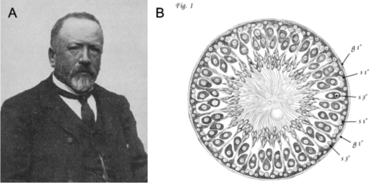
Enrico Sertoli (1842–1910) was an Italian histologist and physician (Figure 1A, [1–3]) who studied a wide variety of subjects during his 40+ year academic career including spermatogenesis, blood proteins, smooth muscle function, the contents of saliva and urine, acinous glands in the renal pelvis, and the histology of the tongue [4–13]. However, he is remembered best for the contents of his first manuscript entitled “About the existence of special branched cells in the seminiferous tubules of the human testis” [4]. This was published the year he graduated from medical school at the ripe age of 23, and described his discovery of the eponymous somatic cell of the seminiferous epithelium.
Figure 1.
(A) Photograph of Sertoli, circa. 1900 [17]. (B) A Camera lucida drawing of the rat seminiferous epithelium by Sertoli [14], which depicts a stage XI-XIV tubule.
Sertoli's legacy as one of the most famous figures in the field of male reproductive biology was ensured based solely on the findings in that first manuscript. However, he published several other reports on the topic of spermatogenesis that, to the best of my knowledge, have not been translated to English and remained largely unexamined by his contemporaries or by scientists of the modern era [6, 9, 10, 13]. Therefore, it is entirely possible that Sertoli made other important discoveries that have not been appropriately attributed to him. My laboratory group recently became interested in one of these works, a book chapter published in 1878 entitled “The structure of seminiferous tubules and the development of spermatids in rats” [14]. While working on a project to characterize cytoplasmic processes in spermatogonia, we realized that Sertoli may have described them in one of the figures in this chapter. Its title resembles those of two of his previously published (and also untranslated) manuscripts on spermatogenesis, which were much shorter [6, 9]; therefore, we suspected that this chapter included results from his work from medical school through 1878. In addition, its length suggested that it contained additional thoughts and interpretations. Therefore, I commissioned the translation of this work as well as faithful reproductions of the artwork (an example is shown in Figure 1B), since the pages of the original were yellowed, faded, and water-stained. The translation was done by a native Italian speaker who is currently a doctoral student in the Department of Romance Studies at the University of North Carolina at Chapel Hill, and the artwork was done by a Master's student in the School of Art and Design at East Carolina University. Afterwards, I edited the manuscript to modernize the terminology and provide context in the form of comments and footnotes, as I felt that would enhance its “readability” and make it as accessible as possible for today's readers.
The major debate among scientists studying spermatogenesis in the late 19th century appears to have been whether male gametes (spermatozoa or sperm) originated from germ cells or Sertoli cells. Sertoli led a group of scientists who shared the former (and we now know, correct) view, and in this chapter, he presented compelling evidence that male germ cells progress through a stepwise developmental program leading to the production of sperm, and he proposed that this process was supported by Sertoli cells. A contemporary, Victor von Ebner (1842–1925), championed the opposing view that Sertoli cells gave rise to sperm, while germ cells disintegrated to produce seminal fluid [15]. I was particularly intrigued by the extent to which Sertoli criticized the approaches and conclusions of von Ebner. Admittedly, we know little of either's temperament or shared personal history. However, in addition to being a scientific rival, von Ebner was also Austrian, and Italy and Austria had recently fought the Three Wars of Italian Independence (1848–1849, 1859–1861, and 1866). Sertoli served in the Italian military in the third war, and so his opinions of Austria and its citizens were likely to have been shaped a great deal by those wartime experiences. Whatever the reasoning behind the acrimony, this manuscript provides a clear depiction of how strongly Sertoli felt about von Ebner's work at that time.
As I read through the manuscript for the first time I was amazed at its sophistication and by Sertoli's clear and prescient observations. It is worth remembering that all his work was done using a crude light microscope (at least by today's standards) to view tissue samples illuminated by sunlight and without the benefits of currently used stains such as hematoxylin or fixatives such as Bouin's solution. It is difficult to know with surety whether all of the observations presented in this chapter were entirely novel, as several important manuscripts of that era have not yet been translated to English. With that caveat, here are the clear advances as I perceive them:
Clear identification of three stages of male germ cell development (mitotic, meiotic, and post-meiotic)
Accurate description and depiction of the stages of the adult seminiferous epithelium, which were not codified until almost 100 years later [16]; the stages are easily interpretable based on his precisely drawn figures and their accompanying figure legends
First description of mammalian spermatogonia
First description of spermatogonial intercellular bridges as well as cytoplasmic projections
Identification of the blood–testis barrier (although he was obviously unaware of its function at the time), and the correct observation that mitotic spermatogonia reside on the basal side, while meiotic spermatocytes and post-meiotic spermatids reside on the adluminal side
It is unfortunate that this work has only been cited a handful of times and was largely overlooked by generations of scientists. It is my hope that re-publication of this translated chapter will reveal the full scope of Sertoli's studies on spermatogenesis and provide a historical perspective of the state of research on male reproduction at the end of the 19th century.
Acknowledgments
I would like to thank Ms. Elisa Salemi (UNC-CH) for her efforts in translating the bulk of the manuscript, Dr. Susanne Jones (ECU) for her assistance with the German passages, and Ms. Katya Harris (ECU) for faithfully reproducing the illustrations. I am grateful to Drs. Rex Hess and Michael Griswold for providing helpful suggestions.
Grant Support
This project was funded by grants from the Eunice Kennedy Shriver National Institute of Child Health and Human Development (2R15HD072552 and 1R01HD090083 to CBG).
References
- 1. Baratelli GM, Lanzani A, Sacco RN. Biography of Enrico Sertoli. Urology 2002; 60:196–198. [DOI] [PubMed] [Google Scholar]
- 2. Franca LR, Hess RA, Dufour JM, Hofmann MC, Griswold MD. The Sertoli cell: one hundred fifty years of beauty and plasticity. Andrology 2016; 4:189–212. [DOI] [PMC free article] [PubMed] [Google Scholar]
- 3. Hess RA, Franca LR. History of the Sertoli Cell Discovery. In: Skinner MK, Griswold MD (eds.), Sertoli Cell Biology. San Diego, CA: Elsevier Academic Press; 2005. [Google Scholar]
- 4. Sertoli E. Dell’esistenza di particolari cellule ramificatenei canalicoli seminiferi del testicolo umano (About the existence of special branched cells in the seminiferous tubules of the human testis). Morgagni 1865; 7:31–33. [Google Scholar]
- 5. Sertoli E. Uber die Bindung der Kohlensaure im Bluteund ihre Ausscheidung in der Lunge (On the binding of carbonic acid in the blood and its excretion in the lungs). Medizinisch-chemische Untersuchungen 1867; 2:350–353. [Google Scholar]
- 6. Sertoli E. Osservazioni sulla struttura dei canalicoli seminiferi del testicolo (Observations on the structure of seminiferous tubules in the testis). Gazzetta Medica Lombarda 1871; 4:413–419. [Google Scholar]
- 7. Sertoli E. Inaugurale di apertura dell’anno scolastico 1872–73 della R. Scuola Superiore di Veterinaria di Milano (Inaugural address of the school year of the Superior Veterinary School of Milan, 1872–73). Milano, Archivio della Regia Scuola Superiore di Veterinaria di Milano 1872. [Google Scholar]
- 8. Sertoli E. Compendio di anatomia speciale degli animali domestici (Compendium of special anatomy of domestic animals). Milano, Tipografi Ronchi 1874. [Google Scholar]
- 9. Sertoli E. Sulla struttura dei canalicoli seminiferi dei testicoli studiata in rapporto allo sviluppo dei nemaspermi (On the structure of seminiferous tubules studied in relation to the development of round spermatids). Gazzetta Medica Lombarda 1875; 2:401–413. [Google Scholar]
- 10. Sertoli E. Di uno pseudoermafroditismo in una capra (On the pseudohermaphroditism in a goat). Arch Medicina Veterinaria 1876; 1:22–31. [Google Scholar]
- 11. Sertoli E. Contribuzioni alla fısiologia generale deimuscoli lisci (Contributions to the general physiology of smooth muscles). Rendiconti dell’Istituto Lombardo di Scienze e Lettere 1882; 15:567–579. [Google Scholar]
- 12. Sertoli E. Contribution a` la physiologie general des muscles lisses (Contribution to the general physiology of smooth muscles). Arch Ital Biol 1883; 3:78–83. [Google Scholar]
- 13. Sertoli E. Sur la caryokinese dans la spermatogenese (On cell division in spermatogenesis). Arch Italiennes Biologie 1886; 7:369–375. [Google Scholar]
- 14. Sertoli E. The structure of seminiferous tubules and the development of spermatids in rats. In: Archivio per le scienze mediche. Turin: Vincenzo Bona; 1878: 1–76. [Google Scholar]
- 15. von Ebner V. Untersuchungen über den Bau der Samencanälchen und die Entwicklung der Spermatozoiden (Studies on the construction of the seminal tubules, and the development of sperm). 1871. [Google Scholar]
- 16. Perey B, Clermont Y, Leblond CP. The wave of the seminiferous epithelium in the rat. Am J Anat 1961; 108:47–77. [Google Scholar]
- 17. Negrini F. Onoranze al Prof. Enrico Sertoli (Honoring professor Enrico Sertoli). Clinica Veterinaria 1908; 31:49–62. [Google Scholar]


