Summary
Chagas disease, also known as American trypanosomiasis, is a tropical parasitic disease caused by the protozoan Trypanosoma cruzi. T. cruzi targets adipose tissue, which serves as a reservoir of this parasite. T. cruzi infection of adipose tissue is characterized by increased lipolysis, oxidative stress, and parasitemia. High fat diet (HFD) decreases lipolysis and increases the survival rate in the mice infected with T. cruzi during acute infection. However, the effect of HFD on oxidative stress in adipose tissue has not been examined in detail. In the present study we evaluated the effect of HFD on oxidative stress markers in both white and brown adipose tissues (WAT and BAT) during acute infection. We used qPCR to examine the mRNA expression levels of genes involved in several antioxidant defence systems, such as those acting in ROS metabolism, peroxidases, and relevant oxygen transporter genes. The result of our study showed that HFD regulates the expression levels of oxidative stress genes in adipose tissues and that these effects are often different in WAT and BAT. For instance, while HFD down-regulated the levels of most antioxidant genes in both WAT and BAT, it differentially affected the expression pattern of genes involved in ROS metabolism (e.g. peroxidases) in WAT and BAT tissues of infected mice. Together with our previous studies, these findings show that infection and diet both regulate antioxidant enzymes and other oxidative stress defenses in mouse adipose tissues during acute T. cruzi infection.
Keywords: Adipose tissue, Oxidative stress, Antioxidant system, High fat diet
INTRODUCTION
Trypanosoma cruzi is an intracellular parasite, which causes debilitating Chagasic cardiomyopathy. Currently nearly 100 million people are at risk of being infected with T. cruzi in Latin America [1]; furthermore, the risk of this disease spreading to United States, Canada, Europe, and the Western Pacific region is increasing due to migration from the T. cruzi endemic regions [2–5].
The transition of traditional Latin American diet to western-style diet increases the risk of diabetes and obesity in the Chagas disease endemic region. This change in lifestyle may influence the pathogenesis and outcome of Chagas disease, especially as we have demonstrated previously that T. cruzi targets adipose tissue during infection and that adipose tissue plays a major role in the pathogenesis of Chagas disease [6]. We have also demonstrated that fat loss and lipolysis are key factors involved in the pathogenesis of heart in Chagas Disease [7]. Interestingly, we found that fat content of the diet influences disease progression and outcome. For instance, we showed that high fat diet (HFD) ameliorated cardiac pathology and increased the survival rate of the T. cruzi infected CD1 mice compared to the infected mice fed on a regular diet (RD) [7]. These outcomes were attributed to reduced fat cell necrosis and lipolysis in HFD-fed infected mice compared to RD-fed infected mice [7]. However, why lipolysis affects cardiac pathology and survival in T. cruzi infected mice is still not clear.
One well established consequence of lipolysis is increased fatty acid oxidation and ROS generation [8], suggesting that lipolysis-induced oxidative damage may promote Chagas disease pathology. Indeed, it has been shown that T. Cruzi infection causes oxidative stress in both the white and brown adipose tissues (WAT and BAT) [9]. Furthermore, it has been shown that the levels of several antioxidants, including glutathione peroxidase, superoxide dismutase and nitric oxide synthase, were reduced in the adipose tissues of a murine Chagas model relative to uninfected mice, suggesting that higher levels of free radicals may be present during acute infection and contribute to disease progression [9]. Indeed, we showed that antioxidant depletion by T. cruzi infection in adipose tissue increases oxidative stress and contributes in the pathogenesis of Chagas disease [9].
In the present study we evaluated the effect of different fat content in the diet on oxidative stress and antioxidant defence genes in different adipose depots (WAT and BAT) during acute T. cruzi infection. In particular, we measured glutathione, catalase, peroxidase and nitric oxide synthase systems in WAT and BAT from mice fed on either a HFD or RD.
MATERIALS AND METHODS
Ethical approval
All animal experimental protocols were approved by the Institutional Animal Care and Use Committee (IACUC) of the Albert Einstein College of Medicine (No. 20130202) and the Rutgers Biomedical and Health Sciences (No. 15107), and adhere to the National Research Council guidelines (Guide for the Care and Use of Laboratory Animals: Eighth Edition, Washington, DC: The National Academies Press, 2011).
Experimental animal model
The Brazil strain of T. cruzi was maintained by passage in C3H/Hej mice (Jackson Laboratories, Bar Harbor, ME). Male CD-1 mice (Jackson Laboratories) were infected intraperitoneally (i.p.) at 8–10 weeks of age with 5 × 10⁴ trypomastigotes of the Brazil strain (Combs et al, 2005). Mice were maintained on a 12-h light/dark cycle. Mice, starting at the day of infection, were randomly divided into 2 groups (15 per group) and fed on either high fat diet (HFD; 60% fat D12492 Research Diets, Inc., New Brunswick, NJ) or regular diet (RD, 10% fatD12450 Research Diets, Inc., New Brunswick, NJ) [7].
Polymerase chain reaction array
An RT2 Profiler (SA Biosciences, Valencia, CA) custom designed PCR array for mouse genes in Oxidative Stress PCR Array Data analysis was performed normalized to the expression of 18sRNA using the DDCT method according to the manufacturer’s protocol (SABiosciences) and as previously mentioned [7].
Gene list with NCBI accession number
Gpx1 NM_008160, Gpx2 NM_030677, Gpx3 NM_008161, Gpx4 NM_008162, Gpx5 NM_010343, Gpx6 NM_145451, Gpx7 NM_024198, Gsr NM_010344, Gss NM_008180, Cat NM_009804, Alb NM_009654, Prdx1 NM_011034, Prdx2 NM_011563, Prdx3 NM_007452, Prdx4 NM_016764, Prdx5 NM_012021, Prdx6 NM_007453, CygbNM_030206, Mpo NM_010824, Lpo NM_080420, Epx NM_007946, Sod1 NM_011434, Sod2 NM_013671, Sod3 NM_011435, Txnrd1 NM_015762, Txnrd2 NM_013711, Txnrd3 NM_153162, Ccl5 NM_013653, Il19 NM_001009940, Il22 NM_016971, Nos2 NM_010927, Nox1 NM_172203, Nox4 NM_015760, Scd1 NM_009127,
STATISTICAL ANALYSIS
Data were pooled and statistical analysis was performed using a Student’s t-test (Microsoft Excel) as appropriate and significance differences were determined as p values between < 0.05 and < 0.005 as appropriate. Gene array analyses were done in duplicates as described earlier.
RESULTS AND DISCUSSION
T. cruzi infection and HFD alters mRNA levels of glutathione system enzymes in the adipose tissue
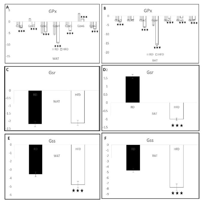
It has been shown that in response to acute T. cruzi infection, the host up-regulates glutathione antioxidant defence by overexpressing glutathione peroxidase (GPx), glutathione reductase (GSR), and glutathione synthetase (GSS) [10]. GPx protects cells from the H2O2 induced oxidative stress and limits free radical formation. The GPx reaction is coupled to GSR, which maintains reduced glutathione (GSH) levels [11]. Earlier we have demonstrated that Gpx mRNA levels decrease in BAT and increase in WAT during acute T. cruzi infection in CD1 mice fed on a chow diet [9]. Here we analyzed the effect of a HFD on the mRNA levels of GPx, GSR, and GSS in WAT and BAT of T. cruzi infected mice by qPCR compared to feeding a RD. In WAT, HFD drastically decreased the mRNA levels of all examined isoforms of GPx, except GPx3 (Figure 1A). In BAT, only GPx 1, 3 and 4 isoforms were significantly decreased by HFD, while other isoforms, like GPx 2, 5, 6 and 7, were significantly increased compared to RD infected mice (Figure 1B). We found no significant difference in the mRNA levels of GSR in WAT samples of infected mice fed on HFD (Figure 1C). However, the GSR levels significantly decreased in BAT in infected mice fed on HFD compared to RD (Figure 1D). Finally, the mRNA levels of GSS significantly decreased in both the WAT and BAT of infected mice fed on a HFD compared to RD fed infected mice (Figure 1E,1F). The qPCR analysis data suggest that T. cruzi infection reduces, and HFD further decreases, the activation of glutathione antioxidant system in white and brown adipose tissues during acute T. cruzi infection. Earlier we have shown that GPX5 is significantly reduced in the adipose tissues of T. cruzi infected mice fed on a chow diet [9]. However, the above data demonstrate that diet influences the glutathione oxido-reductive system during infection. HFD regulates glutathione peroxidase expression in mice as reported by Sekine et al., which has a synergistic effect on GPx expression during T. cruzi infection [12].
Figure 1. T. cruzi infection and HFD synergistically alter glutathione signalling system in different adipose tissues.
The qPCR analysis data suggest that infection reduces mRNA level of genes involved in glutathione system and HFD further decreases the activation of glutathione antioxidant system in the adipose tissues during acute T. cruzi infection. P ≤ 0.005, represented by black stars. Bars represent mean values of the data with Standard Error of the mean (SEM) as vertical lines. RD - regular diet; HFD-high fat diet.
T. cruzi infection and HFD regulates mRNA levels of catalase and albumin in the adipose tissue
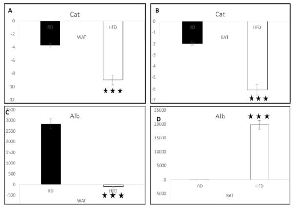
We have previously shown that T. cruzi infection causes oxidative stress and results in the accumulation of intracellular free radicals in adipose tissue in a murine Chagas model [9]. Here we analyzed the effect of HFD on the expression levels of antioxidant enzyme catalase (Cat), which protects cells against free radical induced toxicity and whose deficiency leads to the accumulation of H2O2 and other highly reactive hydroxyl radicals [13]. qPCR analysis demonstrated that HFD significantly reduced the mRNA levels of catalase in both the WAT (Figure 2A) and BAT (Figure 2B) compared to RD during acute T. cruzi infection, suggesting that HFD further reduces catalase levels during infection. Catalase expression in adipose tissue is mainly regulated by PPAR-g [14]. We have earlier demonstrated that PPAR-g decreases in the adipose tissues of T. cruzi infected mice [7]. It has been shown that antioxidant catalase rescues against HFD-induced cardiac dysfunction in mice [15]. This suggests that reduced catalase levels during infection may responsible for the cardiac pathogenesis during acute T. cruzi infection. We also analyzed the effect of HFD during infection on albumin (Alb), which is the most abundant circulating protein in the plasma and has important antioxidant properties [16]. Pereira et al, showed that T. cruzi infection alters Alb level [17]. We found that HFD drastically decreases the mRNA levels of Alb in WAT (Figure 2C) but, surprisingly, increases Alb mRNA in BAT (Figure 2D) compared to RD fed infected mice. This suggests that HFD has a differential effect on Albumin in WAT and BAT. Adipose tissue expresses Albumin and hypoalbumin is associated with inflammation and oxidative stress [18,19].
Figure 2. Diet regulates mRNA levels of catalase and albumin in the adipose tissues of T. cruzi infected CD1 mice.
qPCR analysis demonstrated that HFD significantly reduces the mRNA levels of catalase in both the WAT (Figure 2A) and BAT (Figure 2B) compared to RD in acute T. cruzi infection. HFD drastically decreases the mRNA levels of Alb in WAT (Figure 2C) and increases in BAT (Figure 2D) compared to RD fed infected mice. P ≤ 0.005, represented by black stars. Bars represent mean values of the data with Standard Error of the mean (SEM) as vertical lines. RD - regular diet; HFD-high fat diet.
Alteration in peroxiredoxins and cytoglobin signalling by HFD in T. cruzi infected mice
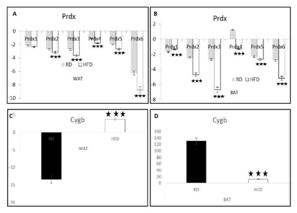
Peroxiredoxins (Prdx) are members of a ubiquitous family of cysteine-dependent peroxidase enzymes that play important roles in regulating peroxide levels within cells. They have cytoprotective functions against intracellular and extracellular reactive oxygen species (ROS) and reactive nitrogen species (RNS) [20]. Our qPCR analysis demonstrated a significant down regulation of various isoforms of Prdx (Prdx 2, 3, 4, 5, & 6) mRNA levels in both WAT (Figure 3A) and BAT (Figure 3B) of infected mice fed on a HFD compared to RD, except Prdx1. It has been demonstrated that Peroxiredoxin 3 regulates adipocyte oxidative stress, mitochondrial biogenesis, and adipokine expression [21]. We also measured the mRNA levels of cytoglobin (CygB), an antioxidant protein expressed in tissues as part of the defense against oxidative stress [22]. Interestingly, HFD increased CygB mRNA levels 13-fold in WAT and decreased them 13-fold in BAT compared to RD fed infected mice (Figure 3C,3D). Thus far, the exact mechanism of CygB function in WAT and BAT is not known. However, HFD has shown to reduce the levels of CygB in mice with time [23].
Figure 3. Alteration in the mRNA levels of genes involved in peroxiredoxins and cytoglobin levels in the adipose tissues during acute T. cruzi infection.
Study at mRNA level by qPCR analysis demonstrated a significant down regulation of various isoforms of Prdx (Prdx 2, 3, 4, 5, & 6) mRNA levels in both the WAT (Figure 3A) and BAT (Figure 3B) of infected mice fed on a HFD compared to RD, except Prdx1. HFD increases CygB mRNA levels to 10 fold in the WAT and decreases to 100 fold in the BAT compared to RD fed infected mice (Figure 3C and 3D). Bars represent mean values of the data with Standard Error of the mean (SEM) as vertical lines. RD - regular diet; HFD-high fat diet.
HFD changes peroxidase signalling in the adipose tissue of T. cruzi infected mice
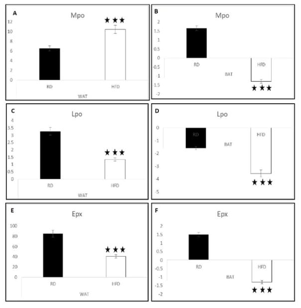
Previously, Dhiman et al., demonstrated that T. cruzi infection leads to myeloperoxidase (MPO) mediated activation of neutrophils and causes oxidative and nitrosative stress in Chagas patients [24]. Treatments that down-regulate the activity of MPO have been suggested as therapeutic strategies for chagasic cardiomyopathic patients [24]. Here we measured the mRNA levels of various peroxidases, including MPO, lactoperoxidase and eosinophil peroxidase in the different adipose tissue depots. MPO levels significantly increases (4-fold) in the WAT and significantly decreased (2.5-fold) in the BAT of infected mice fed on a HFD compared to RD (Figure 4A,4B). Lactoperoxidase (Lpo) is one of the family members of heme peroxidases - a family of peroxidases involved in the production of strong oxidants which act as a shield against pathogenic bacteria, fungi or parasites [25]. HFD significantly down-regulated the expression of Lpo in both WAT (Figure 4C) and BAT (Figure 4D) compared to RD. Another peroxidase, Eosinophil peroxidase (Epx) was significantly decreased in both WAT (Figure 4E) and BAT (Figure 4F) (40- and 3-fold, respectively) of the T. cruzi infected mice fed on a HFD compared to RD. Epx is usually released to the site of parasitic infection for active killing of protozoans and has been shown to be involved in the formation of cytotoxic oxidants in various disease models [26]. Earlier we have shown an increased Epx levels in the WAT of T. cruzi infected mice fed on a chow diet compared to the uninfected mice [9]. Even though, the levels of Lpo and dEpx are significantly greater in the infected mice compared to the uninfected mice fed on a HFD, their levels were significantly lower compared to the infected mice fed on a RD in both the WAT and BAT. Together, these results indicate that HFD regulates the levels of several peroxidases involved in host defence against T. cruzi.
Figure 4. HFD changes peroxidase levels in the adipose tissue of T. cruzi infected mice.
qPCR analysis of MPO gene showed that it was significantly increased (4-fold) in the WAT and significantly decreased (2.5-fold) in the BAT of infected mice fed on a HFD compared to RD (Figure 4A,4B). HFD significantly down regulated the expression of Lpo in both the WAT (Figure 4C) and BAT (Figure 4D) during acute infection in mice compared to feeding with RD. Epx significantly decreased in both the WAT (Figure 4E) and BAT (Figure 4F) (40- and 3-fold, respectively) of the T. cruzi infected mice fed on a HFD compared to RD. Bars represent mean values of the data with Standard Error of the mean (SEM) as vertical lines. RD - regular diet; HFD-high fat diet.
Modulation of superoxide and thioredoxin signalling by HFD during acute T. cruzi infection
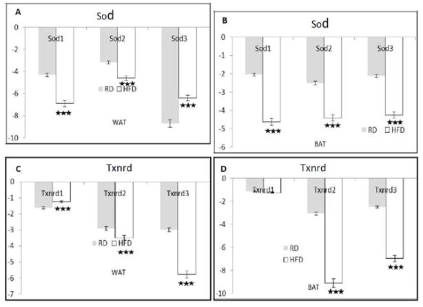
Superoxide dismutase (Sod) is an antioxidant enzyme that plays an important role in protecting cells from oxidative insults by reducing superoxide anions to hydrogen peroxide. T. cruzi infection causes oxidative stress, which acts as an inflammatory mediator, and Sod is induced concomitantly with pro inflammatory cytokines to rescue infected cells/tissues from oxidative damage [27,28]. We analyzed various isoforms of Sod (Sod 1, 2 and 3) by qPCR during acute T. cruzi infection in a murine Chagas disease model. HFD significantly decreased the mRNA levels of Sod in both WAT (Figure 5A) and BAT (Figure 5B) compared to infected RD fed mice (except Sod3 in the WAT). Decreased Sod in adipose tissue is associated with an increased perivascular adipose tissue dysfunction in HFD fed mice [29]. HFD has shown to decrease the levels of Sod and increase oxidative stress in mice [30] which backs our observation. Thioredoxin system (Txn) is one of the oxidative stress scavenging systems that protect cells by reducing ROS and maintaining the intracellular redox state [31]. Thioredoxin reductase (Txnrd) is an important part of the Txn system [32], which plays a major role in maintaining the cellular redox state. We assessed the levels of all three isoforms of Txnrd: cytosolic (Txnrd 1), mitochondrial (Txnrd 2) and testis-specific thioredoxin reductase (Txnrd3) in the adipose tissues of the infected mice fed on either a HFD or RD. The levels of Txnrd 2 and 3 significantly decreased in both WAT and BAT of the infected mice fed on a HFD compared to RD (Figure 5C,5D). Our qPCR data provides the evidence that T. cruzi infection and HFD synergistically alter the Txn system in the different depots of adipose tissues. Txnrd 1 suppresses adipocyte differentiation and insulin responsiveness, which supports our observation that acute T. cruzi infection reduces fat anabolism [10,33].
Figure 5. Modulation of superoxide and thioredoxin system signalling in the adipose tissue with diet during acute T. cruzi infection.
HFD significantly decreased the mRNA levels of Sod in both the WAT (Figure 5A) and BAT (Figure 5B) when compared to infected RD fed mice (except Sod3 in the WAT). The levels of Txnrd 2 and 3 significantly decreases in both the WAT and BAT of the infected mice fed on a HFD compared to RD (Figure 5C,5D). Bars represent mean values of the data with Standard Error of the mean (SEM) as vertical lines. RD - regular diet; HFD-high fat diet.
Diet regulates NO and NOX signalling in T. cruzi infected adipose tissues
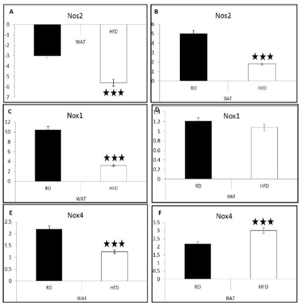
Inducible nitric oxide synthase 2 (iNOS2) produces nitric oxide (NO), which plays a prominent role in controlling early murine T. cruzi infection [34–36]. Our qPCR analysis demonstrated a significant down-regulation of NOS2 in both types of adipose tissue (WAT and BAT) of the infected mice fed on a HFD compared to a RD (Figure 6A,6B). Also, T. cruzi infection activates NADPH oxidase (Nox) in mice and causes an oxidative burst, inflammatory cell proliferation, and cytokine production [37]. Nox activation results in the production of high amount of ROS, which act as primary defensive barrier against infection [38]. We found that HFD caused a significant down-regulation of Nox1 and Nox4 (approximately 3.5- and 2-fold, respectively) in the WAT of the infected mice compared to RD (Figure 6C). mRNA levels of Nox1 were not significantly altered in the BAT (Figure 6D) of the infected mice fed on either a HFD or RD. However, Nox4 levels significantly decreased in WAT (Figure 6E) and increased in the BAT (Figure 6F) in the infected mice fed on a HFD compared to RD. Earlier we have demonstrated that T. cruzi acute infection in WAT of CD1 mice fed on a chow diet increases Nox and decreases Nos2. Here also, we observed a similar result that HFD significantly reduces Nos2 mRNA levels compared to RD during acute infection in WAT as well as in BAT. Together, these results indicate that HFD prevents the formation of Nos2 which could control early murine T. cruzi infection [34–36].
Figure 6. Diets differentially regulate T. cruzi infection induced nitrosative stress in different adipose depots during T. cruzi infection.
qPCR study showed down regulation expression of Nox1 and Nox4 approximately 3.5 and 2 folds respectively, in the WAT of the infected mice (Figure 6C) on HFD compared to RD. mRNA levels of Nox1 significantly does not alter in the BAT (Figure 6D) of the infected mice fed on either an HFD or RD. However, Nox4 levels significantly decreases on HFD in WAT (Figure 6E) but it is reverse in the case of BAT (Figure 6F), as it is increased on HFD when compared to RD infected mice. Bars represent mean values of the data with Standard Error of the mean (SEM) as vertical lines. RD - regular diet; HFD-high fat diet.
HFD alters MUFA signalling in T. cruzi infected adipose tissue
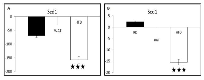
Stearoyl coenzyme A desaturase (Scd1) is an iron-containing enzyme that catalyzes a rate-limiting step in the synthesis of unsaturated fatty acids, including monounsaturated fatty acids (MUFA), which are the major lipids in tissues. Any alterations in Scd1 levels significantly alter the steady lipid profile and have a drastic impact on cellular functions by mediating diverse inflammatory stresses [39]. Our qPCR analysis showed that HFD significantly decreased the level of Scd1 in both WAT (3-fold) (Figure 7A) and BAT (17-fold) (Figure 7B) compared to RD fed infected mice. It has been previously shown that Scd1 deficiency has beneficial effect in WAT from inflammatory insult caused by HFD [40], in our study the highly decreased levels of Scd1 may have protective role in the adipose tissues of the T. cruzi infected mice from inflammatory stress. Future studies are required to clarify the exact mechanism and role of Scd1 in adipose tissues during infection.
Figure 7. High fat diet alters the level of mono unsaturated fatty acids by limiting the expression of a key fatty acid synthesizing protein Scd1 in the adipose tissues during acute T. cruzi infection.
qPCR analysis showed that HFD highly down regulated the level of Scd1 in both WAT (Figure 7A) and BAT (Figure 7B) when compared to RD infected mice. Bars represent mean values of the data with Standard Error of the mean (SEM) as vertical lines. RD - regular diet; HFD-high fat diet.
We previously showed that acute T. cruzi infection significantly increases oxidative stress and inflammation in the adipose tissues of the infected mice [10]. In this manuscript we report the effect of HFD on oxidative and nitrosative stresses and inflammatory markers in different depots of the adipose tissues during acute T. cruzi infection. Our results indicate that HFD further worsens the oxidative stress induced pathogenesis in adipose tissue by significantly reducing the expression levels of anti-oxidant genes. Suppressing inflammation and anti-oxidant levels may help the parasites stay within the adipose tissue during acute infection and escape the host immune attack. The differences in the mRNA levels of the oxidant and anti-oxidant genes between the WAT and BAT during infection in a murine Chagas model fed on either a HFD or RD may be due to the variations in the mitochondrial oxidation levels in these tissues.
CONCLUSION
Adipose tissue undergoes oxidative and nitrosative stress during acute T. cruzi infection. HFD increases parasite load in the adipose tissues which could alter the levels of oxidative and nitrosative stresses during acute infection. HFD further amplifies the pathological state of adipose tissue by expressing lower levels of many stress and antioxidant defence genes (ROS metabolism, peroxidases genes and relevant oxygen transporter genes) in both the WAT and BAT. Our study demonstrated the redox condition of the different adipose depots when fed on a HFD or RD during acute T. cruzi infection, which helps understand the pathological status of these tissues during infection. In conclusion, we showed that HFD reduces oxidative and nitrosative stresses during acute infection and thus may facilitate the parasite survival in adipose tissues during acute infection. Further studies are required to understand the specific mechanisms involved in the role of diet in regulating antioxidant and oxidative stress in acute T. cruzi infected adipose tissue.
Acknowledgments
We acknowledge Erika Shor at the Public Health Research Institute for a critical reading of the manuscript. This study was supported by grants from the National Heart, Lung, and Blood Institute (National Institutes of Health HL-112099 and HL-122866) to Jyothi Nagajyothi.
References
- 1.Montgomery SP, Starr MC, Cantey PT, Edwards MS, Meymandi SK. Neglected parasitic infections in the United States: Chagas disease. Am J Trop Med Hyg. 2014;90:814–818. doi: 10.4269/ajtmh.13-0726. [DOI] [PMC free article] [PubMed] [Google Scholar]
- 2.Bern C, Kjos S, Yabsley MJ, Montgomery SP. Trypanosoma cruzi and Chagas’ Disease in the United States. Clin Microbiol Rev. 2011;24:655–681. doi: 10.1128/CMR.00005-11. [DOI] [PMC free article] [PubMed] [Google Scholar]
- 3.Gascon J, Bern, Pinazo MJ. Chagas Disease in Spain, the United States and Other Non-Endemic Countries. Acta Trop. 2010;115:22–27. doi: 10.1016/j.actatropica.2009.07.019. [DOI] [PubMed] [Google Scholar]
- 4.Jackson Y, Pinto A, Pettt S. Chagas Disease in Australia and New Zealand: Risks and Needs for Public Health Interventions. Trop Med Int Health. 2014;19:212–218. doi: 10.1111/tmi.12235. [DOI] [PubMed] [Google Scholar]
- 5.Navarro M, Navaza B, Guionnet A, López-Vélez R. Chagas Disease in Spain: Need for Further Public Health Measures. PLoS Negl Trop Dis. 2012;6:e1962. doi: 10.1371/journal.pntd.0001962. [DOI] [PMC free article] [PubMed] [Google Scholar]
- 6.Nagajyothi F, Desruisseaux MS, Weiss LM, Chua S, Albanese C, Machado FS, et al. Chagas disease, adipose tissue and the metabolic syndrome. Mem Inst Oswaldo Cruz. 2009;104:219–225. doi: 10.1590/s0074-02762009000900028. [DOI] [PMC free article] [PubMed] [Google Scholar]
- 7.Nagajyothi F, Weiss LM, Zhao D, Koba W, Jelicks LA, Cui MH, et al. High fat diet modulates Trypanosoma cruzi infection associated myocarditis. PLoS Negl Trop Dis. 2014;8:e3118. doi: 10.1371/journal.pntd.0003118. [DOI] [PMC free article] [PubMed] [Google Scholar]
- 8.den Hartigh LJ, Han CY, Wang S, Omer M, Chait A. 10E,12Z-conjugated linoleic acid impairs adipocyte triglyceride storage by enhancing fatty acid oxidation, lipolysis, and mitochondrial reactive oxygen species. J Lipid Res. 2013;54:2964–2978. doi: 10.1194/jlr.M035188. [DOI] [PMC free article] [PubMed] [Google Scholar]
- 9.Wen JJ, Nagajyothi F, Machado FS, Weiss LM, Scherer PE, Tanowitz HB. Markers of oxidative stress in adipose tissue during Trypanosoma cruzi infection. Parasitol Res. 2014;113:3159–3165. doi: 10.1007/s00436-014-3977-7. [DOI] [PMC free article] [PubMed] [Google Scholar]
- 10.Gupta S, Wen JJ, Garg NJ. Oxidative Stress in Chagas Disease. Interdiscip Perspect Infect Dis. 2009;2009:1–8. doi: 10.1155/2009/190354. [DOI] [PMC free article] [PubMed] [Google Scholar]
- 11.Bhattacharyya A, Chattopadhyay R, Mitra S, Crowe SE. Oxidative stress: an essential factor in the pathogenesis of gastrointestinal mucosal diseases. Physiol Rev. 2014;94:329–354. doi: 10.1152/physrev.00040.2012. [DOI] [PMC free article] [PubMed] [Google Scholar]
- 12.Sekine Y, Osei-Hwedieh D, Matsuda K, Raghavachari N, Liu D, Furuya Y, et al. High fat diet reduces the expression of glutathione peroxidase 3 in mouse prostate. Prostate. 2011;71:1499–1509. doi: 10.1002/pros.21365. [DOI] [PMC free article] [PubMed] [Google Scholar]
- 13.Al-Abrash AS, Al-Quobaili FA, Al-Akhras GN. Catalase evaluation in different human diseases associated with oxidative stress. Saudi Med J. 2000;21:826–830. [PubMed] [Google Scholar]
- 14.Okuno Y, Matsuda M, Kobayashi H, Morita K, Suzuki E, Fukuhara A, et al. Adipose expression of catalase is regulated via a novel remote PPARgamma-responsive region. Biochem Biophys Res Commun. 2008;366:698–704. doi: 10.1016/j.bbrc.2007.12.001. [DOI] [PubMed] [Google Scholar]
- 15.Liang L, Shou XL, Zhao HK, Ren GQ, Wang JB, Wang XH, et al. Antioxidant catalase rescues against high fat diet-induced cardiac dysfunction via an IKKβ-AMPK-dependent regulation of autophagy. Biochim Biophys Acta. 2015;1852:343–352. doi: 10.1016/j.bbadis.2014.06.027. [DOI] [PubMed] [Google Scholar]
- 16.Roche M, Rondeau P, Singh NR, Tarnus E, Bourdon E. The antioxidant properties of serum albumin. FEBS Lett. 2008;582:1783–1787. doi: 10.1016/j.febslet.2008.04.057. [DOI] [PubMed] [Google Scholar]
- 17.Pereira MG, Dorea JG, Johnson NE, Castro CN, Macêdo V. Serum albumin and gamma globulin in Trypanosoma cruzi infections. Trans R Soc Trop Med Hyg. 1983;77:32–34. doi: 10.1016/0035-9203(83)90006-8. [DOI] [PubMed] [Google Scholar]
- 18.Ellmerer M, Schaupp L, Brunner GA, Sendlhofer G, Wutte A, Wach P, et al. Measurement of interstitial albumin in human skeletal muscle and adipose tissue by open-flow microperfusion. Am J Physiol Endocrinol Metab. 2000;278:E352–E356. doi: 10.1152/ajpendo.2000.278.2.E352. [DOI] [PubMed] [Google Scholar]
- 19.Danielski M, Ikizler TA, McMonagle E, Kane JC, Pupim L, Morrow J, et al. Linkage of hypoalbuminemia, inflammation, and oxidative stress in patients receiving maintenance hemodialysis therapy. Am J Kidney Dis. 2003;42:286–294. doi: 10.1016/s0272-6386(03)00653-x. [DOI] [PubMed] [Google Scholar]
- 20.Perkins A, Nelson KJ, Parsonage D, Poole LB, Karplus PA. Peroxiredoxins: guardians against oxidative stress and modulators of peroxide signaling. Trends Biochem Sci. 2015;40:435–445. doi: 10.1016/j.tibs.2015.05.001. [DOI] [PMC free article] [PubMed] [Google Scholar]
- 21.Huh JY, Kim Y, Jeong J, Park J, Kim I, Huh KH, et al. Peroxiredoxin 3 is a key molecule regulating adipocyte oxidative stress, mitochondrial biogenesis, and adipokine expression. Antioxid Redox Signal. 2012;16:229–243. doi: 10.1089/ars.2010.3766. [DOI] [PMC free article] [PubMed] [Google Scholar]
- 22.Li D, Chen XQ, Li WJ, Yang YH, Wang JZ, Yu AC. Cytoglobin up-regulated by hydrogen peroxide plays a protective role in oxidative stress. Neurochem Res. 2007;32:1375–1380. doi: 10.1007/s11064-007-9317-x. [DOI] [PubMed] [Google Scholar]
- 23.Bae YJ, Kim SE, Seong YH, Park T, Lee SG, Cho MS, et al. Time-course microarray analysis for identifying candidate genes involved in obesity-associated pathological changes in the mouse colon. Genes Nutr. 2016;11:30. doi: 10.1186/s12263-016-0547-x. [DOI] [PMC free article] [PubMed] [Google Scholar]
- 24.Dhiman M, Estrada-Franco JG, Pando JM, Ramirez-Aguilar FJ, Spratt H, Vazquez-Corzo S, et al. Increased myeloperoxidase activity and protein nitration are indicators of inflammation in patients with Chagas’ disease. Clin Vaccine Immunol. 2009;16:660–666. doi: 10.1128/CVI.00019-09. [DOI] [PMC free article] [PubMed] [Google Scholar]
- 25.Bafort F, Parisi O, Perraudin JP, Jijakli MH. Mode of action of lactoperoxidase as related to its antimicrobial activity: a review. Enzyme Res. 2014;2014:1–13. doi: 10.1155/2014/517164. [DOI] [PMC free article] [PubMed] [Google Scholar]
- 26.Ranguelova K, Chatterjee S, Ehrenshaft M, Ramirez DC, Summers FA, Kadiiska MB, et al. Protein Radical Formation Resulting from Eosinophil Peroxidase-catalyzed Oxidation of Sulfite. J Biol Chem. 2010;285:24195–24205. doi: 10.1074/jbc.M109.069054. [DOI] [PMC free article] [PubMed] [Google Scholar]
- 27.Cardoni RL, Rottenberg ME, Segura EL. Increased production of reactive oxygen species by cells from mice acutely infected with Trypanosoma cruzi. Cell Immunol. 1990;128:11–21. doi: 10.1016/0008-8749(90)90002-9. [DOI] [PubMed] [Google Scholar]
- 28.Hirose K, Longo DL, Oppenheim JJ, Matsushima K. Overexpression of mitochondrial manganese superoxide dismutase promotes the survival of tumor cells exposed to interleukin-1, tumor necrosis factor, selected anticancer drugs, and ionizing radiation. FASEB J. 1993;7:361–368. doi: 10.1096/fasebj.7.2.8440412. [DOI] [PubMed] [Google Scholar]
- 29.Fernández-Alfonso MS, Gil-Ortega M, García-Prieto CF, Aranguez I, Ruiz-Gayo M, Somoza B. Mechanisms of perivascular adipose tissue dysfunction in obesity. Int J Endocrinol. 2013;2013:402053. doi: 10.1155/2013/402053. [DOI] [PMC free article] [PubMed] [Google Scholar]
- 30.Meng R, Zhu DL, Bi Y, Yang DH, Wang YP. Anti-oxidative effect of apocynin on insulin resistance in high-fat diet mice. Ann Clin Lab Sci. 2011;41:236–243. [PubMed] [Google Scholar]
- 31.Koháryová M, Kolárová M. Oxidative stress and thioredoxin system. Gen Physiol Biophys. 2008;27:71–84. [PubMed] [Google Scholar]
- 32.Arner ES, Holmgren A. Physiological functions of thioredoxin and thioredoxin reductase. Eur J Biochem. 2000;267:6102–6109. doi: 10.1046/j.1432-1327.2000.01701.x. [DOI] [PubMed] [Google Scholar]
- 33.Peng X, Giménez-Cassina A, Petrus P, Conrad M, Rydén M, Arnér ES. Thioredoxin reductase 1 suppresses adipocyte differentiation and insulin responsiveness. Sci Rep. 2016;6:28080. doi: 10.1038/srep28080. [DOI] [PMC free article] [PubMed] [Google Scholar]
- 34.Gazzinelli RT, Oswald IP, Hienry S, James SL, Sher A. The microbicidal activity of interferon-gamma treated macrophages against Trypanosoma cruzi involves an l-arginine-dependent, nitrogen oxide-mediated mechanism inhibitable by IL-10 and transforming growth factor-beta. Eur J Immunol. 1992;22:2501–2506. doi: 10.1002/eji.1830221006. [DOI] [PubMed] [Google Scholar]
- 35.Vespa GN, Cunha FQ, Silva JS. Nitric oxide is involved in control of Trypanosoma cruzi-induced parasitemia and directly kills the parasite in vitro. Infect Immun. 1994;62:5177–5182. doi: 10.1128/iai.62.11.5177-5182.1994. [DOI] [PMC free article] [PubMed] [Google Scholar]
- 36.Dijkstra G, Blokzij H, Bok L, Homan M, van Goor H, Faber KN, et al. Opposite effect of oxidative stress on inducible nitric oxide synthase and haem oxygenase-1 expression in intestinal inflammation: anti-inflammatory effect of carbon monoxide. J Pathol. 2004;204:296–303. doi: 10.1002/path.1656. [DOI] [PubMed] [Google Scholar]
- 37.Dhiman M, Garg NJ. NADPH oxidase inhibition ameliorates Trypanosoma cruzi-induced myocarditis during Chagas disease. J Pathol. 2011;225:583–596. doi: 10.1002/path.2975. [DOI] [PMC free article] [PubMed] [Google Scholar]
- 38.Cardoni RL, Antunez MI, Morales C, Nantes IR. Release of reactive oxygen species by phagocytic cells in response to live parasites in mice infected with Trypanosoma cruzi. Am J Trop Med Hyg. 1997;56:329–334. doi: 10.4269/ajtmh.1997.56.329. [DOI] [PubMed] [Google Scholar]
- 39.Liu X, Strable MS, Ntambi JM. Stearoyl CoA desaturase 1: role in cellular inflammation and stress. Adv Nutr. 2011;2:15–22. doi: 10.3945/an.110.000125. [DOI] [PMC free article] [PubMed] [Google Scholar]
- 40.Liu X, Miyazaki M, Flowers MT, Sampath H, Zhao M, Chu K, et al. Loss of stearoyl-CoA desaturase-1 attenuates adipocyte inflammation: effects of adipocyte-derived oleate. Arterioscler Thromb Vasc Biol. 2010;30:31–38. doi: 10.1161/ATVBAHA.109.195636. [DOI] [PMC free article] [PubMed] [Google Scholar]


