Figure 3.

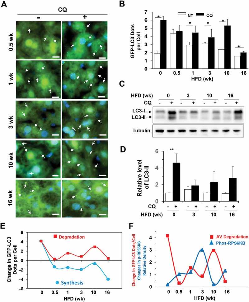
Autophagy degradation was dynamically regulated in HFD-fed mice. (a-b) GFP-LC3 transgenic mice at the age of 8 weeks were fed with HFD for the indicated time, and then treated with (CQ) or without (NT) CQ (60 mg/kg, i.p.) 16 h before sacrifice. Cryosections of the livers were stained with Hoechst 33328. GFP-LC3 puncta represented autophagosomes (a) and were quantified (b). The values at time zero were obtained from 8-week-old mice fed with regular diet. (c-d) Mice were fed with HFD for the indicated duration and then treated with or without CQ (60 mg/kg, i.p.) 16 h before sacrifice. Liver lysates were analyzed by immunoblotting for LC3. The level of LC3-II was normalized to that of tubulin and expressed as the fold of change over the control diet with no CQ treatment (0 week). (e) Autophagosome synthesis and degradation were calculated using the GFP-LC3 puncta data above and plotted against the duration of HFD feeding. (f) The changes in degradation of the GFP-LC3 puncta (autophagosomes or AV) and phosphorylated RPS6KB levels (from Figure 1F) were compared on the same time scale. The changes of the 2 parameters were in opposite directions at each time point. *, p < 0.05; **, p < 0.01; n = 3–6 per group. Scale bar: 10 μm. Arrows indicate GFP-LC3 puncta. For clarity and for the illustrative purpose, not all puncta are indicated.
