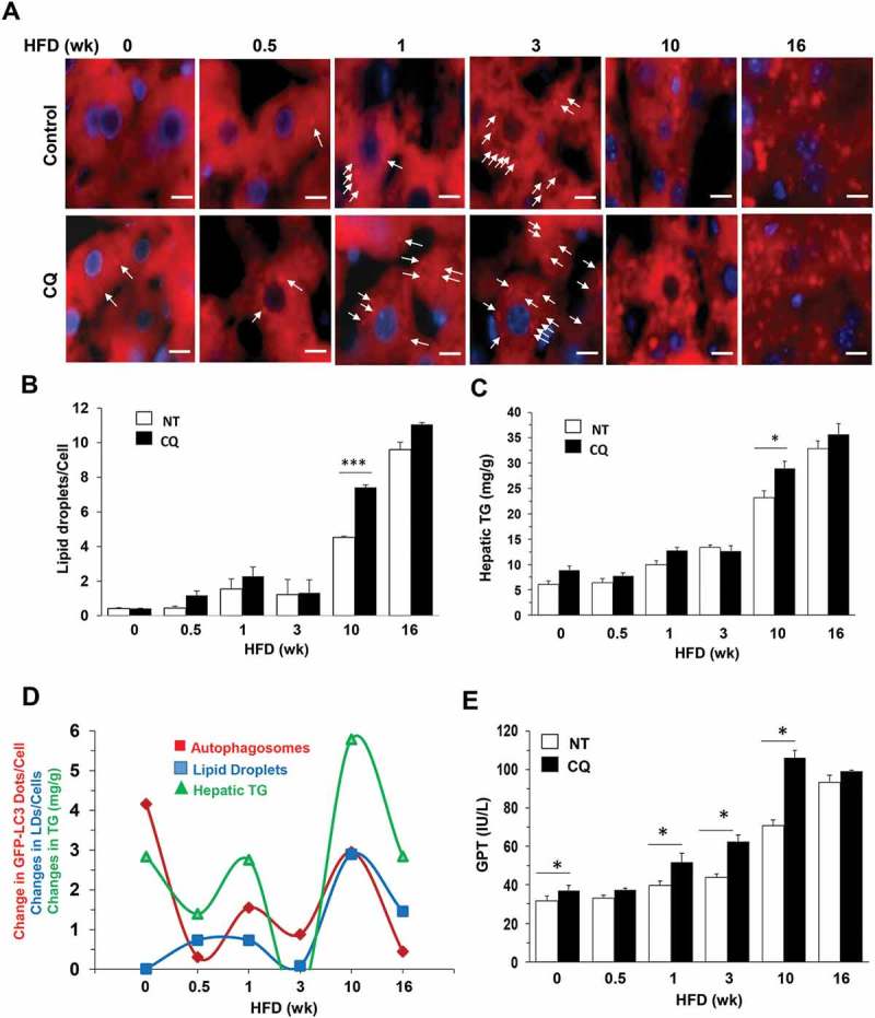
Figure 4.

Alterations of lipophagy in the liver of HFD-fed mice. (a-b) Mice were fed with HFD for different times and given CQ or not treated (NT) 16 h before sacrifice. Cryosections of the liver were stained with BODIPY-581/591 for lipids droplets (a), which were quantified as the number per cell (b). (c) Hepatic triglycerides (TG) were measured in the same group of mice. (d) The difference between the CQ plus group and control group was what was degraded in the lysosome. The difference was noted as the ‘changes’ in the degradation of autophagosomes, the level of lipid droplets or the level of hepatic TG, which were plotted against the duration of HFD feeding. Data of the degraded autophagosomes (GFP-LC3 puncta) were from Figure 3D. The changes of these 3 parameters were in the same directions at almost all time points, implying that they are associated with the same process related to lipophagy. (e) GPT/ALT levels were measured in the same group of mice as in panel C. *: p < 0.05, n = 3–6 per group. Scale bar: 10 μm. Lipid droplets were small in both size and number in samples with shorter HFD duration (0–3 weeks) and are indicated by arrows. Lipid droplets in samples with longer HFD duration (10–16 wk) were large in size and number and are not indicated by arrows.
