Figure 1.
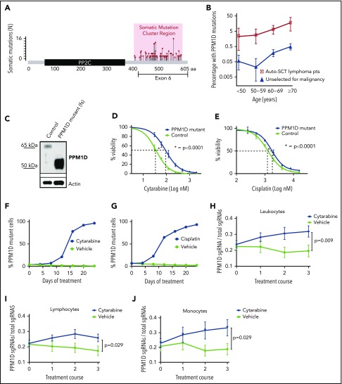
Truncating PPM1D mutations lead to the selective outgrowth of PPM1D-mutant hematopoietic cells during chemotherapy treatment in vitro and in vivo. (A) The location of the absolute number of somatic frameshift and nonsense mutations in PPM1D identified in the blood cells of a cohort of 28 418 persons is shown.24 (B) The prevalence of PPM1D mutations in 28 418 people unselected for malignancy was compared with 401 lymphoma patients who had received chemotherapy and were undergoing stem cell collection for autologous transplantation (Auto-SCT).14,24 Only PPM1D mutations with a variant allele frequency (VAF) > 0.05 are included. Error bars represent the 95% confidence intervals in each age bin. (C) Whole-cell lysates from Molm13 isogenic control cells (control) and PPM1D frameshift (fs) mutant (mut) single-cell clones probed with anti-PPM1D and anti-Actin. (D-E) Viability assays in Molm13 PPM1D-mutant or control single-cell clones that were treated with increasing concentrations of cytarabine (D) or cisplatin (E) for 72 hours. Experiments were performed in triplicate and data are shown as the mean ± standard deviation (SD). Nonlinear logistic regression analyses and a sum of squares F test was performed to compare inhibitory responses. (F-G) Flow cytometric readout of competition experiment with 5% Molm13 PPM1D-mutant cells (tdTomato+) and 95% Molm13 control cells (tdTomato−), exposed to 150 nM cytarabine (F) and vehicle or 1 μM cisplatin (G) and vehicle for 24 days. Experiments were performed with biological triplicates and data are shown as the means ± SD. (H-J) Flow cytometric analysis of the peripheral blood of chimeric mice transduced with a gRNA-targeting PPM1D (BFP+) or a control locus (tRFP+). Mice were exposed to 3 rounds of cytarabine or vehicle treatment. Data represent the mean ± standard error of the mean (SEM) of 18 mice in the vehicle and 18 mice in the treatment arm, and are shown for the leukocyte (H), lymphocyte (I), and monocyte (J) cell compartments. Peripheral expansion of Ppm1d-mutant cells was analyzed using Wilcoxon rank sum tests.

