
Figure 1.
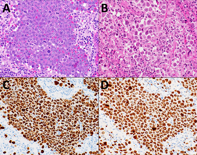
Cervical lymph node biopsy microphotographs from Case 1 and Case 2, A. Case 1 showing tumor cells with nest-like growth pattern of large pleomorphic cells, with prominent nucleoli and frequent mitotic figures (H&E, 40X) B. Case 2 showing more loosely arranged cells with an alveolar pattern and plasmacytoid appearance (H&E, 40X). C. Case 1 with HHV8 LANA IHC staining of the tumor cells showing characteristic punctuate nuclear staining pattern (40X). D. Case 1 with tumor cells showing with very high Ki67% (40X)