Abstract
Purpose
Regulatory T (Treg) cells are important suppressive cells among tumor infiltrating lymphocytes (TIL). Treg express the well-known immune checkpoint receptor PD-1, which is reported to mark “exhausted” Treg with lower suppressive function. T cell immunoglobulin mucin (Tim)-3, a negative regulator of Th1 immunity, is expressed by a sizeable fraction of TIL Tregs, but the functional status of Tim-3+ Tregs remains unclear.
Experimental design
CD4+CTLA-4+CD25high Treg were sorted from freshly excised head and neck squamous cell carcinoma (HNSCC) TIL based on Tim-3 expression. Functional and phenotypic features of these Tim-3+ and Tim-3− TIL Tregs were tested by in vitro suppression assays and multi-color flow cytometry. Gene expression profiling and NanoString analysis of Tim-3+ TIL Treg were performed. A murine HNSCC tumor model was used to test the effect of anti-PD-1 immunotherapy on Tim-3+ Treg.
Results
Despite high PD-1 expression, Tim-3+ TIL Treg displayed a greater capacity to inhibit naïve T cell proliferation than Tim-3− Treg. Tim-3+ Treg from human HNSCC TIL also displayed an effector-like phenotype, with more robust expression of CTLA-4, PD-1, CD39 and IFN-γ receptor. Exogenous IFN-γ treatment could partially reverse the suppressive function of Tim-3+ TIL Treg. Anti-PD-1 immunotherapy downregulated Tim-3 expression on Tregs isolated from murine HNSCC tumors, and this treatment reversed the suppressive function of HNSCC TIL Tregs.
Conclusion
Tim-3+ Treg are functionally and phenotypically distinct in HNSCC TIL, and are highly effective at inhibiting T cell proliferation despite high PD-1 expression. IFN-γ induced by anti-PD-1 immunotherapy may be beneficial by reversing Tim-3+ Treg suppression.
Keywords: regulatory T cells, Tim-3, suppressive function, immunotherapy, TIL, anti-PD-1 Ab
Introduction
Head and neck squamous cell carcinoma (HNSCC) is the sixth leading cause of cancer by incidence worldwide and is one of the most morbid and genetically diverse malignancies (1). Despite improvements in treatment, survival rates remain low. Co-inhibitory immune checkpoint receptors, such as CTLA-4 and PD-1, have a critical role in the regulation of T cell responses and have proven to be effective targets in the setting of cancer therapy (2, 3). However, the objective response to anti-PD-1 monotherapy is still only 15–20% (4) and some tumor types remain largely refractory to these therapies. In order to broaden therapeutic methods, intense investigation has focused on targeting of other co-inhibitory receptors, particularly those that are uniquely expressed in the tumor microenvironment.
T cell immunoglobulin and mucin domain (Tim-3) is often recognized as a checkpoint receptor due to its apparent inhibitory function on T cells and its association with activation-induced T cell exhaustion in tumors and chronic viral infection (5, 6). Initial examination of Tim-3 function suggested that Tim-3 is a negative regulator of type I immunity. Blockade of Tim-3 can increase IFN-γ secretion and the susceptibility of mice to develop multiple autoimmune diseases in which IFN-γ secreting cells play an important role and exacerbate the disease severity (5, 7–9). These data suggest that Tim-3 might function as an inhibitory molecule counteracting IFN-γ driven inflammation (10).
In addition to its expression on T cells, Tim-3 has also been identified on regulatory T cells (Treg) and on innate immune cells (DCs, NK cells, monocytes) (11–13), but its function on these cells is less clear. In addition to its role in regulating effector CD8+ T cell responses, Tim-3 might also have a role in regulating the function of Foxp3+ Treg cells (14–16). In human tumors, 25–50% of TIL Tregs express Tim-3 (13, 17) and Treg can be destabilized or differentiated in the tumor microenvironment (18, 19). However, the functional impact of a suppressive checkpoint receptor on Treg is controversial. Treg specialize in regulating immune responses to pathogens and in maintaining self-tolerance (20), however, they also restrain critical tumor-specific T cell responses. The frequency of Treg is increased at tumor sites and among the peripheral blood lymphocytes (PBMC) of patients with cancer (21, 22), including those with HNSCC (13), suggesting that Tregs suppress immune responses within the tumor environment (23–25). Several recent studies have led to the current concept that Foxp3+ Treg cells are heterogeneous, and contain multiple, functionally diverse populations with distinct phenotypes and specialized functions (26, 27). However, little is known about the mechanism of these specific suppressor functions or the checkpoint receptors that can identify Treg subsets.
Highly suppressive TIGIT+ Treg cells also express higher levels of Tim-3 than TIGIT− Treg cells (26) and Tim-3 and TIGIT cooperate to suppress antitumor responses (28). A recent report found that PD-1 marks dysfunctional Treg in malignant gliomas, and PD-1hi Tregs also upregulated Tim-3 expression (29). We investigated the function and phenotype of human Tim-3+ and Tim-3− TIL Treg cells freshly isolated from HNSCC patients, using a combination of ex vivo functional assays, phenotypic and gene expression profiling, and an in vivo HNSCC model. Tim-3+ TIL Treg displayed a greater capacity for inhibiting T cell proliferation than Tim-3− Treg. These Tim-3+ Treg also exhibited characteristics of highly suppressive Treg cell profile, including granzyme B and effector chemokines. In addition, suppression mediated by Tim-3+ Treg was partially reversed by exogenous IFN-γ treatment. Tim-3 expression on Treg was nearly abolished after PD-1 blockade in TIL from a murine HNSCC tumor, and anti-PD-1 Ab could reverse the suppressive function of HNSCC TIL Tregs, indicating the importance of overcoming this pathway to destabilize Treg activity in the tumor microenvironment.
Patients and Methods
Patients and specimens
Human tissue and peripheral blood were collected from patients in the Department of Otolaryngology at the University of Pittsburgh Medical Center (Pittsburgh, PA). All subjects gave written informed consent approved by the Institutional Review Board of the University of Pittsburgh (IRB #99-069) and followed the Declaration of Helsinki guidelines. The clinical characteristics of the patients are shown in Table 1.
Table 1.
Clinicopathologic features of the HNSCC patients in this study.
| Patient# | Gender | Age | Tumor site | T stage | N stage | M stage |
|---|---|---|---|---|---|---|
| 1 | F | 46 | Oral Cavity | T3 | N0 | M0 |
| 2 | M | 59 | Oral Cavity | T2 | N0 | M0 |
| 3 | M | 52 | Oropharynx | T1 | NX | M0 |
| 4 | M | 53 | Oropharynx | T3 | NX | M0 |
| 5 | M | 41 | Larynx | T3 | N2A | M0 |
| 6 | M | 63 | Oropharynx | T2 | N0 | M0 |
| 7 | M | 55 | Oropharynx | T2 | N1 | M0 |
| 8 | F | 79 | Oropharynx | T2 | N3 | M0 |
| 9 | M | 64 | Oral Cavity | T4 | N2B | M0 |
| 10 | M | 73 | Oral Cavity | T2 | N1B | M0 |
| 11 | F | 86 | Nasal Cavity | T2 | NX | M0 |
| 12 | M | 61 | Oral Cavity | T4 | N0 | M0 |
| 13 | M | 66 | Oral Cavity | T2 | N0 | M0 |
| 14 | M | 60 | Oropharynx | T3 | N1 | M0 |
| 15 | M | 90 | Oropharynx | T3 | NX | M0 |
| 16 | M | 59 | Oropharynx | T3 | N2 | M0 |
| 17 | F | 90 | Oral Cavity | T4 | N2B | M0 |
| 18 | M | 62 | Oral Cavity | T1 | N0 | M0 |
| 19 | M | 56 | Oral Cavity | T1 | N0 | M0 |
| 20 | M | 75 | Oral Cavity | T4 | N2B | M0 |
| 21 | M | 81 | Oral Cavity | T4 | N1 | M0 |
| 22 | M | 78 | Larynx | T4 | N0 | M0 |
| 23 | M | 79 | Oral Cavity | T2 | N0 | M0 |
Collection and processing of PBMC and TILs
Blood samples from patients with HNSCC and healthy donors were drawn into heparinized tubes and centrifuged on Ficoll-Paque™ PLUS gradients (GE Healthcare Bioscience). For extraction of TIL, freshly resected tumor specimens were manually minced into small pieces, then transferred to 70 µm cell strainers (Corning) and mechanically separated using the plunger of a 5 mL syringe. Tumor homogenates were passed through another 70 µm cell strainer prior to separation on Ficoll-Paque™ PLUS gradients. After centrifugation, mononuclear cells were recovered and restored at −80 °C until flow cytometry analysis or immediately used for experiments.
Antibodies and cytokine treatment
The following anti-human antibodies were used for staining: CD3-FITC, CD3- Alexa Fluor 700, CD8a-Alexa Fluor700, CD4-Percp-Cy5.5, CD4-Alexa Fluor 700, CD25-PE, CD25-PE-Cy7, Foxp3-FITC, Foxp3- Percp-Cy5.5, Foxp3-BV421, CD127-APC, CD39-APC, LAP-PE, IL-10-PE, TIM-3-PE, TIM-3-BV421, IFN-γ receptor-PE, IFN-γ- APC, Granzyme B-PE-dazzle E, PD-1-APC, PD-1- PE-Cy7, PD-1- Percp-Cy5.5, CTLA-4-PE, CTLA-4-FITC, ki67-Alexa Fluor 488 and their respective isotype controls were purchased from Biolegend. Recombinant human IFN-γ (R&D Systems) was used at 200 ng/ml. Anti-PD-1 Ab (Nivolumab from Bristol-Myers Squibb), anti-Tim-3 (clone 2E2 from Biolegend) and isotype were used at 10µg/ml.
Flow cytometry analysis
Single cell suspensions from peripheral blood and tumor tissue were surface labeled using Abs mentioned above for 15 mins at room temperature (RT). For intracellular cytokine production of IFN- γ, IL-10 and Granzyme B, TILs were stimulated in vitro with anti-CD3/CD28 beads and protein transport inhibitor (BD GolgiPlug™) for 6h. Intracellular staining for Foxp3, IL-10, granzyme B, ki67 and IFN- γ was performed as follows. After PBMC or TIL were stained with mAb for surface markers, they were fixed and permeabilized 30 mins (eBioscience, San Diego, USA). After washing, cells were subjected to intracellular staining for 30 mins at RT. Cell viability was determined by Zombie Aqua staining (Biolegend, San Diego, USA) or Fixable Viability Dye eFluor 780 (eBioscience, San Diego, USA). Samples were analyzed using an LSR Fortessa cytometer (BD Biosciences), and FlowJo version 10 software.
Cell sorting and Treg suppression assays
Tim-3+ or Tim-3− Tregs (defined by CD4+CD25highCD127−) were sorted from fresh isolated tumor-infiltrating lymphocytes (TIL). CD4+CD25highCD127− T cells showed >97% purity measured by Foxp3 staining after isolation (not shown). Naïve CD8 T cells were isolated from matched HNSCC or healthy donor PBMC using human naïve CD8 T cell isolating kit (STEMCELL Technologies, Vancouver, BC, Canada) and then were stained with CellTrace Violet (5µM, Life Technologies, CA, USA). APC was sorted by FSC vs. SSC in bulk. To assess the functional potential of Tim-3+ and Tim-3− Tregs, both were cultured alongside peripheral blood Naïve CD8 T cells at ratios ranging from 1:1 to 1:16 with 2,000 responder T cells in the round-bottom 96-well plates. To induce proliferation, responder T cells were stimulated with soluble OKT3 (anti-human CD3, 1µg/ml, Biolegend) antibody in the presence of autologous APC for 4 days. All CellTrace Violet data were collected by a Fortessa cytometry (Becton Dickinson) and analyzed using the Flowjo software (TreeStar, Inc.). For the IFN-γ treated experiment, Tim-3+ Tregs or Tim-3− Tregs were pretreated with IFN-γ (200ng/ml) or not for 48 hours with IL-2 (200 U/ml, R&D System). Treg suppressive capacity decline was defined by (Tim-3+ Treg suppression - IFN-γ treated Tim-3+Treg suppression)/Tim-3+ Treg suppression *100%. For the anti-PD-1 and anti-Tim-3 treated experiment, total Tregs were pretreated with nivolumab (10µg/ml) or anti-Tim-3 (clone 2E2 10µg/ml) or isotype (10µg/ml) for 48 hours with IL-2 (200 U/ml, R&D System) under TCR stimulation. Following incubation, Tregs were washed, counted, and plated in a suppression assay in T cell medium with freshly sorted naïve CD8 T cells stained with Celltrace Violet. Celltrace Violet was measured via flow cytometry after 4 days.
Gene expression profiling by Affymetrix and NanoString analysis
Total RNA were extracted from three sets of Tim-3+ and Tim-3− Treg. Total RNA from one set were amplified using Nugen Pico WTA System V2 kit to generate adequate amounts of cDNA for downstream Affymetrix genechip analysis using Human Transcriptome Array 2.0 by Genomics and Proteomics Core Laboratories of University of Pittsburgh. The differentially expressed genes and fold of changes were obtained using Affymetrix Transcriptome Analysis Console software. Total RNA from all three sets were subjected to pre-amplification steps to generate sufficient amounts of cDNA for downstream nanostring analysis using nCounter® Human Immunology v2 Gene Expression CodeSet profiling 594 immunology-related human genes and 15 internal reference genes by Genomics and Proteomics Core Laboratories of University of Pittsburgh. Differential expression levels was determined by Student’s t-test.
Murine ex vivo tumor studies
All murine experiments were approved and conducted according to the guidelines set forth by the Institutional Animal Care and Use Committee (IACUC) at the University of Pittsburgh. 6 to 8-week-old female C57BL/6 mice (Jackson Laboratory, Bar Harbor, Maine) were inoculated with 1×10⁶ murine tonsil epithelium E6/E7/H-ras transformed (MEER) cells (30), kindly provided by Dr John Lee (Sanford Institute, Sioux Falls, SD). Anti-PD-1 (clone 4H2) and isotype control (IgG1) antibodies were purchased from Bristol Myers-Squibb. Anti-PD-1 or isotype mAb was given at 3mg/kg every 2 days for 5 doses (day 12, day 14, day 16, day 18 and day 20). For in vivo IFN-γ neutralization experiments, 200 µg anti-IFN-γ (BioXCell, clone XMG1.2) or isotype control (rat IgG1) was administered 1 day before initiation of therapy and every 3 days thereafter. TILs were isolated when tumor size was larger than 20mm in its greatest dimension. The following anti-mouse antibodies were used for staining: CD4-APC-Cy7, CD25-Alexa Fluor 700, Foxp3-FITC, Tim-3-PE, Nrp1-PE and Zombie Aqua. Flow cytometry analysis is the same as human sample method.
Statistics
Statistical analysis was carried out using GraphPad Prism 7.0 (GraphPad Inc.). A two-way or one-way ANOVA, A two-tailed paired t-test or non-paired t-test was used to calculate whether observed differences were statistically significant, defined as p≤0.05*, p≤ 0.01**, p≤ 0.001***, p≤ 0.0001****.
Results
Tim-3 expression on Treg cells defines a functionally suppressive Treg cell subset
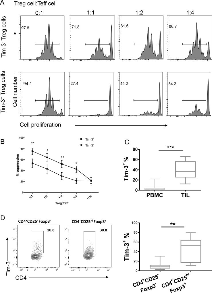
Functional properties of Tim-3+ TIL Treg were compared with Tim-3− TIL Treg, in terms of their ability to inhibit effector T cell proliferation after anti-CD3 stimulation using an in vitro suppression assay. Indeed, greater suppression by Tim-3+ Treg cells extracted from HNSCC patient tumors (n = 5) was observed compared to Tim-3− TIL Treg cells (p<0.05 at Treg/Teff cell ratio of 1:2 and 1:8, p<0.01 at Treg/Teff cell ratio of 1:1 and 1:4) (Figure 1A, B), indicating that Tim-3+ Treg cells are more suppressive. Previous work showed an increased frequency of CD4+CD25hi Foxp3+ Treg cells and immune checkpoint receptors on these Treg at tumor sites from HNSCC patients (13). In order to clarify functional differences based on Tim-3 expression, we then compared the expression of Tim-3 on CD4+CD25hiFoxp3+T cells from paired PBMC and TIL of HNSCC patients. As shown in Figure 1C, the proportion of Tim-3+ cells was significantly higher in CD4+CD25hi Foxp3+ cells in TIL compared with those in PBMC (p<0.01). We also found that Tim-3 was highly expressed on CD4+CD25hi Foxp3+ Tregs compared with CD4+CD25−Foxp3− effector T cells from HNSCC TILs (Figure 1D; p<0.01).
Figure 1. Tim-3+ Treg cells exhibit enhanced suppressive function in HNSCC patients.
(A) Naïve CD8+ T cells were sorted from HNSCC patient or healthy donor PBMC and labeled with Celltrace Violet. Tim-3+ CD4+ CD25hi CD127− and Tim-3− CD4+CD25hiCD127− cells were sorted from HNSCC TIL and titrated onto naïve CD8+ T cells stimulated with anti-CD3 (OKT3 1µg/ml) and APCs. Celltrace Violet dilution after proliferation was measured by flow cytometry after 4 days. Representative suppression assay is shown. (B) Line graph depicts a summary of experiments (n=5). (C) Gated CD4+CD25hiFoxp3+ Tregs freshly harvested from peripheral blood and tumor biopsies (n=10) were stained for Tim-3 expression by flow cytometry. CD4+CD25hiFoxp3+ Tim-3+ Tregs comprised about 40% +/− SEM of total TIL Tregs (n=10). (D) Left: Representative flow cytometric data showing Tim-3 expression on CD4+CD25−Foxp3− T cells and CD4+CD25hi Foxp3+ Tregs in HNSCC TILs. Right: Summarized frequency +/− SEM of Tim-3 expression on CD4+CD25−Foxp3− T cells and CD4+CD25hi Foxp3+ Tregs in HNSCC TILs.
Tim-3+ Treg cells express markers of reflecting a suppressive phenotype
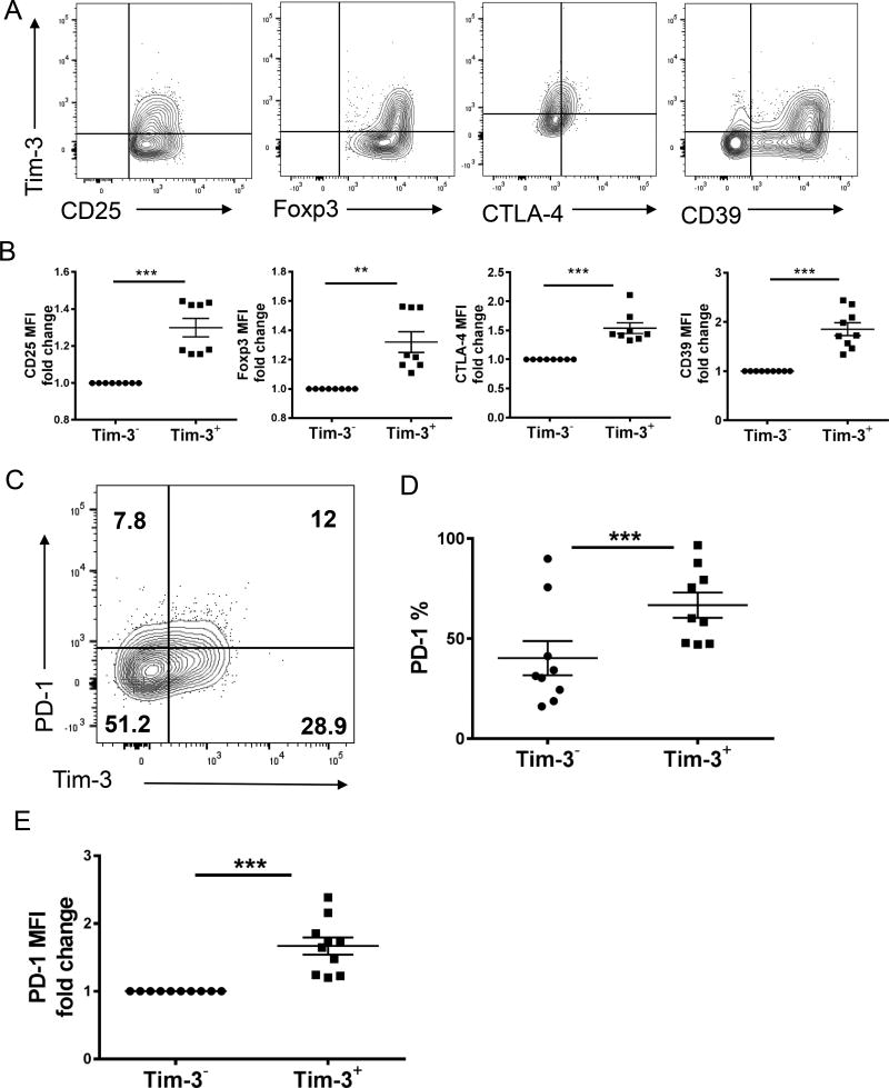
Effector and regulatory T cell function is shaped by the tumor microenvironment as well as engagement of co-signaling receptors or ligands. We therefore used flow cytometry to characterize the expression of membrane receptors associated with Treg activity in both the Tim-3+ and Tim-3− Treg fractions. A distinct pattern of phenotypic markers in Tim-3+ Treg cells was identified, including higher levels of the co-inhibitory molecules CTLA-4 and CD39 (Figure 2A, 2B). Tim-3+ Treg cells also expressed higher amounts of Foxp3 and CD25 (Figure 2A, 2B). Tim-3+ Treg cells therefore displayed features of an activated, more suppressive Treg cell subset. Expression of Tim-3 and PD-1 have been used to stratify populations of CD8+ TILs that exhibit different functional phenotypes (31). PD-1hi marks dysfunctional Treg in malignant gliomas, and PD-1hi Tregs also have upregulated Tim-3 genes (29). Thus, we compared PD-1 expression on Tim-3− or Tim-3+ Treg cells (Figure 2C). Over 70% of TIL Tim-3+ Treg cells expressed PD-1, compared with only 40% of Tim-3− Treg cells in the HNSCC microenvironment (Figure 2D). Surprisingly PD-1 intensity (MFI) was also higher on Tim-3+ Treg cells (Figure 2E), conflicting with prior suggestion that PD-1hi Tregs are exhausted and less suppressive (29).
Figure 2. Tim-3+ Treg cells phenotyping measured by flow cytometry.
(A) Gated CD4+CD25hiFoxp3+ cells freshly harvested from HNSCC tumor biopsies (n=8) were analyzed for Tim-3 expression and Treg suppressive markers by flow cytometry. Tim-3+ Treg cells expressed higher amounts of CD39 (p<0.001), Foxp3 (p<0.01), CD25 (p<0.001) and CTLA-4 (p<0.001). (B) Summary data of CD39, CTLA-4, Foxp3 and CD25 expression on gated Tim-3+ Treg and Tim-3− Treg cells from HNSCC patients’ tumors (n=8). (C) Representative PD-1 and Tim-3 flow cytometric dot plots gated on CD4+CD25hiFoxp3+ Treg cells from HNSCC TIL. (D–E) The proportion of PD-1-expressing Tim-3+ Tregs in comparison with Tim-3− Tregs in HNSCC TIL. Over 70% of Tim-3+ Treg cells from HNSCC co-expressed PD-1, compared with only 40% of Tim-3− Treg cells.
Tim-3+ Treg cells express effector cytokines and chemokines genes
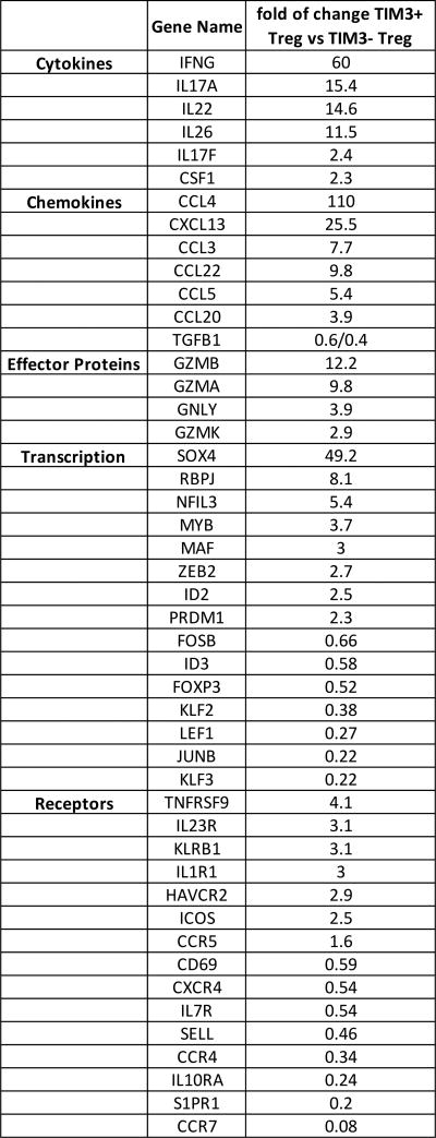
Chemokines play an important role in evading tumor immunity by recruiting CD4+ Treg cells into the tumor environment. To test the expression of relevant chemokines and cytokines that have been associated with Treg suppressive function, CTLA-4+CD25high Treg cells were sorted from HNSCC patients into Tim-3+ and Tim-3− subsets. Samples from different HNSCC patients (n=4) were pooled for RNA extraction and gene expression profiling and NanoString analysis. Tim-3+CTLA-4+CD25hi Treg cells expressed higher type I cytokines and chemokine genes than Tim-3−CTLA-4+CD25hi Treg cells (Figure 3). Our NanoString analysis also showed the same results as gene expression profiling (data not shown). Based on this data, we measured IL-10, granzyme B and IFN-γ secretion. TILs were stimulated in vitro with anti-CD3/CD28 coated beads in the presence of protein transport inhibitor (BD GolgiPlug™) for 6h. No significant difference was detected in IL-10, IFN-γ, LAP and Ki67 expression by flow cytometry between Tim-3+ and Tim-3− HNSCC patient-derived Treg. While Tim-3+ Treg have been shown to contain a higher frequency of IL-10-producing cells than Tim-3− Treg (16), we observed no significant difference in the frequency of these cells, although there was a trend towards an increase in the Tim-3+ Treg fraction (Supplemental Figure 1A). However, we did detect significantly more granzyme B expression by Tim-3+ Tregs (Supplemental Figure 1B).
Figure 3. Tim-3+ CTLA-4+CD25high Treg cells from HNSCC TIL secrete more cytokines.
Differentially expressed genes between TIM3+ and TIM3− Treg by Gene expression profiling. Tim-3+ Treg cells from HNSCC TIL secrete more cytokines.
Tim-3+ Treg suppressive activity is partially reversed by exogenous IFN-γ treatment
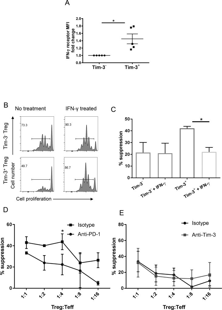
Previous reports suggested that IFN-γ could reverse the suppressive function of Treg (32), and we found a higher level of IFN-γ receptor expression on Tim-3+ Treg cells (Figure 4A). Thus, sorted Tregs were treated with IFN-γ (200ng/ml) for 48 hours, then tested functionally in a Treg suppression assay against labeled CD8+ T cells stimulated using anti-CD3 antibody. Proliferation using Celltrace Violet dye dilution was measured via flow cytometry after 4 days. Tim-3+ Tregs pretreated with IFN-γ partially lost their in vitro suppressor function (47.47% decline in suppressive capacity, p<0.05, n=3) at a Treg/Teff cell ratio of 1:4 (Figure 4B and C), however, no change in expression of Tim-3 on Treg was observed (data not shown). We also found similar results at a Treg/Teff cell ratio of 1:2 (data not shown). We excluded that a decrease in viability, proliferation (decreased ki-67 expression), or Foxp3 expression occurred after Treg were treated with IFN-γ for 48 hours (Supplemental Figure 2), as alternate explanations for our findings. In addition, anti-PD-1 Ab (Figure 4D), but not anti-Tim-3 (Figure 4E), overcame the suppressive function of HNSCC TIL Tregs. Indeed, total Tregs treated with anti-PD-1 48 hours partially lost their in vitro suppressive function (48.9 % decline in suppressive capacity, p<0.05, n=3). In an effort this further, total TIL was treated with anti-PD-1 for 48 hrs in a mixed-lymphocyte reaction with APCs. We found an increase in IFN-γ production by CD8+ T cells, and corresponding increase in Tim-3 expression on the surface of Treg (Figure 4F, p<0.05, n=3).
Figure 4. The suppressive function mediated by Tim-3+ Treg is significantly reversed by exogenous IFN-γ treatment.
(A) IFN-γ receptor expression on Tim-3− Tregs and Tim-3+ Tregs cells from HNSCC TIL (n=5). Summary data from HNSCC patients (n=5). (B) After sorting Tim-3+ CD4+CD25hiCD127− and Tim-3− CD4+CD25hiCD127− cells from HNSCC TIL, they were treated with IL-2 (200 IU/ml) with or without IFN- γ (200 ng/ml) for 48 hours. Then Tregs were washed, counted, and plated in a suppression assay in complete medium with sorted naïve healthy donor CD8+ cells labeled. Celltrace Violet dilution was measured via flow cytometry after 4 days. Representative data of a Celltrace Violet suppression assay at a Treg/naïve CD8 effector cell ratio of 1:4. (C) Column graph depicts a summary of experiments (n=3). (D–E) After sorting CD4+CD25hiCD127− cells from HNSCC TIL, Tregs were pretreated with nivolumab (10µg/ml) or isotype (10µg/ml) for 48 hours with IL-2 (200 U/ml, R&D System) with TCR stimulation (plate bound anti-CD3 2ng/ml and soluble anti-CD28 1µg/ml). For the anti-Tim-3 treated experiment, total Tregs were pretreated with anti-Tim-3 Ab (clone 2E2, 10µg/ml) or isotype (10µg/ml) for 48 hours with IL-2 (200 U/ml, R&D System) under TCR stimulation. Then Tregs were washed, counted, and plated in a suppression assay in complete medium with sorted naïve healthy donor CD8+ cells labeled. Celltrace Violet dilution was measured via flow cytometry after 4 days. Column graph depicts a summary of experiments (n=3 respectively).
Tim-3 is downregulated after PD-1 blockade ex vivo in mouse TIL
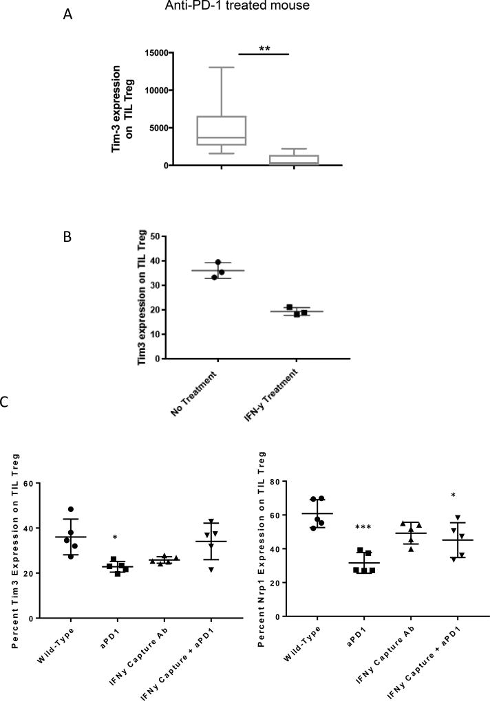
Next, we investigated the change in Tim-3 expression after PD-1 blockade in a murine HPV+ HNSCC model (33). Total TIL we extracted from the murine tumors, after treatment with anti-PD-1 (3mg/kg) or isotype Ab every other day (q.o.d) from days 12–20. Murine Tim-3 expression was evaluated by flow cytometry ex vivo. Strikingly, Tim-3 expression was decreased by approximately 90% in CD4+CD25hiFoxp3+ Treg (p<0.01) extracted from responding tumors after anti-PD-1 mAb immunotherapy (Figure 5A). Anti-PD-1 has been shown to stimulate CD8+ T cells to secrete more IFN-γ (34). Thus, we treated murine TIL Treg for 3 days ex vivo with IFN-γ and noted a decrease in Tim-3 expression following culture (Figure 5B). In an effort to investigate the role of IFN-γ further, we administered IFN-γ capture antibody to mice in combination with anti-PD-1 (Figure 5C). When mice were treated with anti-PD-1 or IFN- γ Ab alone, there was an expected decrease in Tim-3 expression on TIL Treg (p<0.05), however when mice were also given IFN-γ capture antibody, there was a slight change in Tim-3 expression compared to wild-type mice that was not statistically significant. It has recently been shown that the expression of neuropilin-1 (Nrp-1) is required to maintain the stability of intratumoral Treg (19). Administration of anti-PD-1 monotherapy decreased the expression of Nrp-1 on the surface of TIL Treg, however, the co-administration of IFN-γ capture antibody seemed to rescue this loss of Nrp-1 expression (Figure 5C, p<0.001).
Figure 5. Tim-3 expression is downregulated after PD-1 blockade ex vivo in mouse TIL.
(A) Mice bearing HNSCC tumors were treated with anti-PD-1 or isotype Ab (3mg/kg) for five doses on alternating days from days 12–20. TILs were extracted from anti-PD-1 or isotype mAb treated murine tumors, then Tim-3 expression was tested on TIL Treg by flow cytometry (n=7). (B) TIL Treg from mice bearing HNSCC tumors were treated with IFN-γ (200 ng/ml) for 3 days, prior to flow cytometry measurement of Tim-3 expression (n=3). (C–D) Mice bearing HNSCC tumors were treated with anti-PD-1 or isotype Ab (3mg/kg) for five doses on alternating days from days 12–20, alone, or in combination with 200 µg IFN-γ capture antibody or isotype control. TILs were extracted and Tim-3 and Nrp-1 expression determined by flow cytometry (n=5).
Discussion
Although Tim-3 has been shown to play an important role in effector T cell exhaustion, its role in Treg function is not as clear (6, 35). Several recent reports indicate that Treg cells in TIL are heterogeneous, and are composed of functionally diverse populations with different phenotypes and functions. In this study, we identified Tim-3+ as a marker of Treg cells that are highly effective at inhibiting T cell proliferation. These Tim-3+ TIL Treg were more suppressive despite higher PD-1 expression, conflicting with other data suggesting that PD-1hi Treg are exhausted and less suppressive (29). We did not observe significant differences in IL-10 or membrane bound TGF-β (LAP) expression in Tim-3+ Treg subsets. However, granzyme B expression was notably enriched on Tim-3+ TIL Treg, consistent with a more effector phenotype. Furthermore, gene expression profiling and NanoString analysis both support that Tim-3+ Treg cells express more effector cytokines and chemokines. Exogenous IFN-γ could partially reverse the suppressive activity of Tim-3+ Tregs. Notably, Tim-3 expression on Tregs was downregulated after anti-PD-1 Ab immunotherapy in a murine HNSCC model, and anti-PD-1 Ab could reverse the suppressive function of HNSCC TIL Tregs. In vtiro, we observed that IFN-γ was greeater in CD8+ T cells after treatment with anti-PD-1, and this correlated to an increase in Tim-3 expression on the surface of Treg.
In an effort to investigate the relationship between IFN-γ production and expression of Tim-3 in vivo, we employed the use of an IFN-γ capture antibody in combination with anti-PD-1. We observed a decrease in vivo of Tim-3 expression on TIL Treg after the asminitration of anti-PD-1, a result that was reversersed with the co-administration of anti-IFN- γ (Figure 5C). Furthermore, we observed a significant decrease in Nrp-1 when mice were treated with anti-PD-1 alone, suggesting that anti-PD-1 monotherapy increases the fragiligt of TIL Treg. This effect was also partially reversed by the co-administration of IFN-γ capture antibody.
While it remains clear that Treg fragility is required for response to PD-1 blockade and it has been reported that IFN-γ drives Treg fragility to promote anti-tumor immunity through regulation of the expression of Nrp-1 (19), it remains unclear the source of IFN-γ in vivo that regulates expression. We have presented in vitro evidence that the source of IFN-γ may come from CD8+ T cells following anti-PD-1 monotherapy (36), although a deeper analysis into this complex issue is ongoing. It is possible that Nrp-1 marks Treg that can be destabilized, whereas Tim-3 expression is unassociated with this phenotype. Ultimately, genetically engineered mice with selective deletion of Nrp-1, Tim-3 or IFN-γ, currently being generated, will be useful to definitively characterize the differential roles of Tim-3 vs Nrp-1 in TIL Treg.
Tim-3 was first identified as a cell surface molecule selectively expressed on IFN-γ-producing Th1 and Tc1 cells (7). Here, Tim-3 was shown to play an important role in the induction of autoimmune diseases by regulating macrophage activation and function and Tim-3 blockade enhanced the clinical and pathological severity of Th1-dependent autoimmune disease and increases the number of activated macrophages in mice. Furthermore, transgenic overexpression of Tim-3 on T cells resulted in an increased in granulocytic MDSC and inhibition of immune responses (37). In accordance with prior studies, we could only detect appreciable Tim-3 expression in HNSCC TIL Treg, with little expression on circulating Treg (14, 17). This localized expression within tumors makes it an attractive therapeutic target, directly or indirectly. Similarly, CD4+CD25hiFoxp3+ Tregs express more Tim-3 than CD4+CD25−Foxp3− T cells in HNSCC TIL. Taken together, Tim-3 is highly expressed in TIL Tregs, which appear to play an important role in antitumor immune responses. Interestingly, the suppressive effects of Tim-3+ TIL Treg appeared to be reversible during anti-PD-1 based immunotherapy in a murine model, and the suppressive function of HNSCC TIL Treg could also be reversed by anti-PD-1 Ab.
Increased suppression by Tim-3+ Treg cells compared to Tim-3− Treg cells indicates that Tim-3+ Treg cells from HNSCC patients are more potent in the microenvironment. Previous studies reported that PD-1+Tim-3+ CD8+ T cells are dysfunctional in melanoma patients (38). PD-1hi TIL Treg are dysfunctional also, losing their suppressive function in malignant gliomas as compared to PD-1low TIL Treg (29). We observed that Tim-3+ TIL Treg express greater PD-1 than Tim-3− Treg. Indeed, PD-1+ Treg cells, Tim-3+ Treg have higher PD-1 MFI than Tim-3− Treg. Since PD-1 is an emerging marker for Treg function, we explored other immune receptors that have been shown to be related to Treg suppressive function. Tim-3+ TIL Treg express more CD39 and CTLA-4, both of them have been reported to play an important role in Treg suppressive function (39–41). Besides those cell surface markers, Tim-3+ Treg cells have a higher MFI of Foxp3 and CD25 than Tim-3− Treg. As CD25 and Foxp3 are the key markers for CD4+ regulatory T cells. These phenotypic analysis further verified the functional assays comparing Tim-3+ and Tim-3− Treg. Although many reports have reported that IL-10 and LAP are the most functional factors of Tregs (42, 43), we did not identify any differences between Tim-3+ and Tim-3− Treg cells. This is consistent with previous research that Treg suppressive function is contact-dependent and soluble factors may be insufficient (44, 45). We were however able to detect significantly more granzyme B expression by Tim-3+ Treg, which has been defined as a key mechanism through which Treg induce cell contact-mediated suppression (46, 47).
Although gene expression profiling and NanoString analysis showed that Tim-3+ Treg secrete more IFN-γ than Tim-3− Treg, intracellular flow analysis did not detect a significant difference between those two populations. To our surprise, the IFN-γ receptor was more highly expressed on Tim-3+ Treg cells. In addition, exogenous IFN-γ treatment partially reversed the suppression mediated by Tim-3+ Treg, suggesting that Type I immunity may overcome the function of Tim-3+ Treg and destabilize their suppressive phenotype and function. The effect of reducing the function of the most suppressive Tim-3+ PD−1high Treg may be as important as reducing exhaustion on PD-1+CD8+T cells in the microenvironment.
In a murine HNSCC model, we found that Tim-3 expression nearly disappeared after 5 doses of anti-PD-1 treatment. This result supports our previous findings using CD8+ T cells, suggesting that PD-1 and Tim-3 may exhibit cross-talk in regulating antitumor T cell responses (48). IFN-γ is an important Th1 proinflammatory cytokine, and increasing reports have shown that IFN-γ plays an important role in Treg suppressive function (32). Signals that increase IFN-γ secretion by Tregs appear to impair Treg function, while reducing IFN-γ can restore suppressive activity in Tregs (49). PD-1hi Treg displayed reduced suppressive capacity against CD4+ effector T cells due in part to greater IFN-γ secretion (29). In another study, IFN-γ was critically required for the conversion of CD4+CD25− T cells to CD4+ Tregs during experimental autoimmune encephalomyelitis (EAE) (50). In this study, IFN-γ knockout mice correlated with reduced frequency and function of CD4+CD25+Foxp3+ Tregs when compared with those of wild type mice. Whether IFN-γ secretion is derived from Treg themselves or from infiltrating, activated CD8+ T cells being actively investigated, as anti-PD-1 antibody has been shown to stimulate CD8+ T cells to secrete more IFN-γ (34). Although our ex vivo research did not find any change in Tim-3 expression on Treg after 3 days of IFN-γ treatment, anti-PD-1 Ab could reverse the suppressive function of TIL Tregs. However, the loss of Tim-3+ Treg in vivo during anti-PD-1 immunotherapy indicates the biological and clinical importance of these more suppressive cells in vitro.
We conclude that Tim-3 is a marker of a highly suppressive population of Treg cells, which are functionally and phenotypically distinct from Tim-3− Treg cells in HNSCC patients. This gain of suppressive function in Tim-3+ Treg is partially associated with increased IFN-γ receptor expression. Exogenous IFN-γ treatment could partially reverse the suppression mediated by Tim-3+ Treg. Tim-3 expression on Tregs could be downregulated when treated with anti-PD-1 Ab, and anti-PD-1 Ab could reverse the suppressive function of TIL Tregs. This effect may provide an additional, novel mechanism for clinical activity of anti-PD-1 and other immunotherapies which drive IFN-γ expression. Thus, PD-1 blocking Abs may significantly reverse Treg suppressive function, which could be an important component of their therapeutic benefit.
Supplementary Material
Supplementary Fig. S1: Tim-3+ Treg cells from HNSCC TIL secrete more cytokines. For IL-10, Granzyme B and IFN-γ quantification, bulk TIL were stimulated in vitro with anti-CD3/CD28 beads and protein transport inhibitor (BD GolgiPlug™) for 6h.Cells were then stained for intracellular cytokine expression by flow cytometry. Summary data were showing IL-10 MFI (P>0.05), IL-10 percentage (p=0.0508), IFN-γ (P>0.05), Granzyme B (P<0.01), LAP (P>0.05) and Ki67 (P>0.05) by flow cytometry, gated on Tim-3− Tregs and Tim-3+ Tregs from HNSCC TILs (n=4–11 patients per marker, as indicated).
Supplementary Fig. S2: Treg viability, Ki-67 expression and Foxp3 expression were tested after being treated with IFN-γ for 48 hours. After sorting Tim-3+ CD4+CD25hiCD127− and Tim-3− CD4+CD25hiCD127− cells from HNSCC TIL, they were treated with IL-2 (200 IU/ml) with or without IFN- γ (200ng/ml) for 48 hours. Viability factor (A), Foxp3 (A) and ki-67 expression (B) were tested by flow cytometry.
Translational Relevance.
Several recent studies have led to the current concept that Foxp3+ Treg cells are heterogeneous, and contain multiple, functionally diverse populations with distinct phenotypes and specialized functions. Treg have been shown to become destabilized or differentiated in the tumor microenvironment, thus, there is considerable interest in identifying pathways that control their function. T cell immunoglobulin mucin domain (Tim)-3, a negative regulator of Th1 immunity, has been shown to play an important role in effector T cell exhaustion, however, it’s role in Treg function is not as clear. We show that Tim-3 is a marker of a highly suppressive population of Treg cells, which are functionally and phenotypically distinct from Tim-3− Treg in HNSCC patients. Furthermore, IFN-γ induced by anti-PD-1 therapy was shown to partially reverse the suppressive effects of Tim-3+ Treg, providing an additional, novel mechanism for the clinical activity of anti-PD-1 and other immunotherapies that drive IFN-γ expression.
Acknowledgments
Financial support: This work was supported by National Institute of Health grants R01 CA206517, DE019727, P50 CA097190, T32 CA060397, T32 CA082084 and the University of Pittsburgh Cancer Institute award P30 CA047904.
Footnotes
Conflicts of Interest statement: Robert L. Ferris: consulting or advisory role: AstraZeneca, Amgen, Bristol-Myers Squibb, Merck, and Pfizer. Research funding: Bristol-Myers Squibb (Inst.) AstraZeneca (Inst.) and VentiRx (Inst.)
References
- 1.Leemans CR, Braakhuis BJM, Brakenhoff RH. The molecular biology of head and neck cancer. 2010;11(9) doi: 10.1038/nrc2982. [DOI] [PubMed] [Google Scholar]
- 2.Mahoney KM, Rennert PD, Freeman GJ. Combination cancer immunotherapy and new immunomodulatory targets. Nat Rev Drug Discov. 2015;14(8):561–84. doi: 10.1038/nrd4591. [DOI] [PubMed] [Google Scholar]
- 3.Pauken KE, Wherry EJ. Overcoming T cell exhaustion in infection and cancer. Trends in Immunology. 2015;36(4):265–76. doi: 10.1016/j.it.2015.02.008. [DOI] [PMC free article] [PubMed] [Google Scholar]
- 4.Ferris RL, Blumenschein GJ, Fayette J, Guigay J, Colevas AD, Licitra L, Harrington K, Kasper S, Vokes EE, Even C, et al. Nivolumab for Recurrent Squamous-Cell Carcinoma of the Head and Neck. New England Journal of Medicine. 2016;375(19):1856–67. doi: 10.1056/NEJMoa1602252. [DOI] [PMC free article] [PubMed] [Google Scholar]
- 5.Sanchez-Fueyo A, Tian J, Picarella D, Domenig C, Zheng XX, Sabatos CA, Manlongat N, Bender O, Kamradt T, Kuchroo VK, et al. Tim-3 inhibits T helper type 1-mediated auto- and alloimmune responses and promotes immunological tolerance. Nat Immunol. 2003;4(11):1093–101. doi: 10.1038/ni987. [DOI] [PubMed] [Google Scholar]
- 6.Jones RB, Ndhlovu LC, Barbour JD, Sheth PM, Jha AR, Long BR, Wong JC, Satkunarajah M, Schweneker M, Chapman JM, et al. Tim-3 expression defines a novel population of dysfunctional T cells with highly elevated frequencies in progressive HIV-1 infection. The Journal of Experimental Medicine. 2008;205(12):2763–79. doi: 10.1084/jem.20081398. [DOI] [PMC free article] [PubMed] [Google Scholar]
- 7.Monney L, Sabatos CA, Gaglia JL, Ryu A, Waldner H, Chernova T, Manning S, Greenfield EA, Coyle AJ, Sobel RA, et al. Th1-specific cell surface protein Tim-3 regulates macrophage activation and severity of an autoimmune disease. Nature. 2002;415(6871):536–41. doi: 10.1038/415536a. [DOI] [PubMed] [Google Scholar]
- 8.Koguchi K, Anderson DE, Yang L, O'Connor KC, Kuchroo VK, Hafler DA. Dysregulated T cell expression of TIM3 in multiple sclerosis. The Journal of Experimental Medicine. 2006;203(6):1413–8. doi: 10.1084/jem.20060210. [DOI] [PMC free article] [PubMed] [Google Scholar]
- 9.Bu M, Shen Y, Seeger WL, An S, Qi R, Sanderson JA, Cai Y. Ovarian carcinoma-infiltrating regulatory T cells were more potent suppressors of CD8+ T cell inflammation than their peripheral counterparts, a function dependent on TIM3 expression. Tumor Biology. 2016;37(3):3949–56. doi: 10.1007/s13277-015-4237-x. [DOI] [PubMed] [Google Scholar]
- 10.Ferris RL, Lu B, Kane LP. Too Much of a Good Thing? Tim-3 and TCR Signaling in T Cell Exhaustion. The Journal of Immunology. 2014;193(4):1525–30. doi: 10.4049/jimmunol.1400557. [DOI] [PMC free article] [PubMed] [Google Scholar]
- 11.Phong BL, Avery L, Sumpter TL, Gorman JV, Watkins SC, Colgan JD, Kane LP. Tim-3 enhances FcεRI-proximal signaling to modulate mast cell activation. The Journal of Experimental Medicine. 2015;212(13):2289–304. doi: 10.1084/jem.20150388. [DOI] [PMC free article] [PubMed] [Google Scholar]
- 12.Chiba S, Baghdadi M, Akiba H, Yoshiyama H, Kinoshita I, Dosaka-Akita H, Fujioka Y, Ohba Y, Gorman JV, Colgan JD, et al. Tumor-infiltrating DCs suppress nucleic acid-mediated innate immune responses through interactions between the receptor TIM-3 and the alarmin HMGB1. Nat Immunol. 2012;13(9):832–42. doi: 10.1038/ni.2376. [DOI] [PMC free article] [PubMed] [Google Scholar]
- 13.Jie HB, Gildener-Leapman N, Li J, Srivastava RM, Gibson SP, Whiteside TL, Ferris RL. Intratumoral regulatory T cells upregulate immunosuppressive molecules in head and neck cancer patients. Br J Cancer. 2013;109(10):2629–35. doi: 10.1038/bjc.2013.645. [DOI] [PMC free article] [PubMed] [Google Scholar]
- 14.Gautron AS, Dominguez-Villar M, de Marcken M, Hafler DA. Enhanced suppressor function of TIM-3+ FoxP3+ regulatory T cells. Eur J Immunol. 2014;44(9):2703–11. doi: 10.1002/eji.201344392. [DOI] [PMC free article] [PubMed] [Google Scholar]
- 15.Gupta S, Thornley TB, Gao W, Larocca R, Turka LA, Kuchroo VK, Strom TB. Allograft rejection is restrained by short-lived TIM-3+PD-1+Foxp3+ Tregs. The Journal of Clinical Investigation. 2012;122(7):2395–404. doi: 10.1172/JCI45138. [DOI] [PMC free article] [PubMed] [Google Scholar]
- 16.Sakuishi K, Ngiow SF, Sullivan JM, Teng MWL, Kuchroo VK, Smyth MJ, Anderson AC. TIM3+FOXP3+ regulatory T cells are tissue-specific promoters of T-cell dysfunction in cancer. OncoImmunology. 2013;2(4):e23849. doi: 10.4161/onci.23849. [DOI] [PMC free article] [PubMed] [Google Scholar]
- 17.Gao X, Zhu Y, Li G, Huang H, Zhang G, Wang F, Sun J, Yang Q, Zhang X, Lu B. TIM-3 expression characterizes regulatory T cells in tumor tissues and is associated with lung cancer progression. PLoS One. 2012;7(2):e30676. doi: 10.1371/journal.pone.0030676. [DOI] [PMC free article] [PubMed] [Google Scholar]
- 18.Delgoffe GM, Woo S-R, Turnis ME, Gravano DM, Guy C, Overacre AE, Bettini ML, Vogel P, Finkelstein D, Bonnevier J, et al. Stability and function of regulatory T cells is maintained by a neuropilin-1-semaphorin-4a axis. Nature. 2013;501(7466):252–6. doi: 10.1038/nature12428. [DOI] [PMC free article] [PubMed] [Google Scholar]
- 19.Overacre-Delgoffe AE, Chikina M, Dadey RE, Yano H, Brunazzi EA, Shayan G, Horne W, Moskovitz JM, Kolls JK, Sander C, et al. Interferon-γ Drives Treg Fragility to Promote Anti-tumor Immunity. Cell. 2017;169(6):1130–41. e11. doi: 10.1016/j.cell.2017.05.005. [DOI] [PMC free article] [PubMed] [Google Scholar]
- 20.Vignali DAA, Collison LW, Workman CJ. How regulatory T cells work. 2008;8(523) doi: 10.1038/nri2343. [DOI] [PMC free article] [PubMed] [Google Scholar]
- 21.Hiraoka N, Onozato K, Kosuge T, Hirohashi S. Prevalence of FOXP3<sup>+</sup> Regulatory T Cells Increases During the Progression of Pancreatic Ductal Adenocarcinoma and Its Premalignant Lesions. Clinical Cancer Research. 2006;12(18):5423–34. doi: 10.1158/1078-0432.CCR-06-0369. [DOI] [PubMed] [Google Scholar]
- 22.Lai C, August S, Albibas A, Behar R, Cho S-Y, Polak ME, Theaker J, MacLeod AS, French RR, Glennie MJ, et al. OX40<sup>+</sup> Regulatory T Cells in Cutaneous Squamous Cell Carcinoma Suppress Effector T-Cell Responses and Associate with Metastatic Potential. Clinical Cancer Research. 2016;22(16):4236–48. doi: 10.1158/1078-0432.CCR-15-2614. [DOI] [PMC free article] [PubMed] [Google Scholar]
- 23.Curiel TJ, Coukos G, Zou L, Alvarez X, Cheng P, Mottram P, Evdemon-Hogan M, Conejo-Garcia JR, Zhang L, Burow M, et al. Specific recruitment of regulatory T cells in ovarian carcinoma fosters immune privilege and predicts reduced survival. 2004;10(942) doi: 10.1038/nm1093. [DOI] [PubMed] [Google Scholar]
- 24.Bates GJ, Fox SB, Han C, Leek RD, Garcia JF, Harris AL, Banham AH. Quantification of Regulatory T Cells Enables the Identification of High-Risk Breast Cancer Patients and Those at Risk of Late Relapse. Journal of Clinical Oncology. 2006;24(34):5373–80. doi: 10.1200/JCO.2006.05.9584. [DOI] [PubMed] [Google Scholar]
- 25.Ichihara F, Kono K, Takahashi A, Kawaida H, Sugai H, Fujii H. Increased Populations of Regulatory T Cells in Peripheral Blood and Tumor-Infiltrating Lymphocytes in Patients with Gastric and Esophageal Cancers. Clinical Cancer Research. 2003;9(12):4404–8. [PubMed] [Google Scholar]
- 26.Joller N, Lozano E, Burkett Patrick R, Patel B, Xiao S, Zhu C, Xia J, Tan Tze G, Sefik E, Yajnik V, et al. Treg Cells Expressing the Coinhibitory Molecule TIGIT Selectively Inhibit Proinflammatory Th1 and Th17 Cell Responses. Immunity. 2014;40(4):569–81. doi: 10.1016/j.immuni.2014.02.012. [DOI] [PMC free article] [PubMed] [Google Scholar]
- 27.Chaudhry A, Rudra D, Treuting P, Samstein RM, Liang Y, Kas A, Rudensky AY. CD4<sup>+</sup> Regulatory T Cells Control T<sub>H</sub>17 Responses in a Stat3-Dependent Manner. Science. 2009;326(5955):986–91. doi: 10.1126/science.1172702. [DOI] [PMC free article] [PubMed] [Google Scholar]
- 28.Kurtulus S, Sakuishi K, Ngiow S-F, Joller N, Tan DJ, Teng MWL, Smyth MJ, Kuchroo VK, Anderson AC. TIGIT predominantly regulates the immune response via regulatory T cells. The Journal of Clinical Investigation. 2015;125(11):4053–62. doi: 10.1172/JCI81187. [DOI] [PMC free article] [PubMed] [Google Scholar] [Retracted]
- 29.Lowther DE, Goods BA, Lucca LE, Lerner BA, Raddassi K, van Dijk D, Hernandez AL, Duan X, Gunel M, Coric V, et al. PD-1 marks dysfunctional regulatory T cells in malignant gliomas. JCI Insight. 2016;1(5) doi: 10.1172/jci.insight.85935. [DOI] [PMC free article] [PubMed] [Google Scholar]
- 30.Hoover AC, Spanos WC, Harris GF, Anderson ME, Klingelhutz AJ, Lee JH. The role of human papillomavirus 16 e6 in anchorage-independent and invasive growth of mouse tonsil epithelium. Archives of Otolaryngology–Head & Neck Surgery. 2007;133(5):495–502. doi: 10.1001/archotol.133.5.495. [DOI] [PMC free article] [PubMed] [Google Scholar]
- 31.Sakuishi K, Apetoh L, Sullivan JM, Blazar BR, Kuchroo VK, Anderson AC. Targeting Tim-3 and PD-1 pathways to reverse T cell exhaustion and restore anti-tumor immunity. The Journal of Experimental Medicine. 2010;207(10):2187–94. doi: 10.1084/jem.20100643. [DOI] [PMC free article] [PubMed] [Google Scholar]
- 32.Yang HR, Chou HS, Gu X, Wang L, Brown KE, Fung JJ, Lu L, Qian S. Mechanistic insights into immunomodulation by hepatic stellate cells in mice: a critical role of interferon-gamma signaling. Hepatology. 2009;50(6):1981–91. doi: 10.1002/hep.23202. [DOI] [PMC free article] [PubMed] [Google Scholar]
- 33.Spanos WC, Nowicki P, Lee D, et al. Immune response during therapy with cisplatin or radiation for human papillomavirus–related head and neck cancer. Archives of Otolaryngology–Head & Neck Surgery. 2009;135(11):1137–46. doi: 10.1001/archoto.2009.159. [DOI] [PubMed] [Google Scholar]
- 34.Chang C-H, Qiu J, O’Sullivan D, Buck Michael D, Noguchi T, Curtis Jonathan D, Chen Q, Gindin M, Gubin Matthew M, van der Windt Gerritje JW, et al. Metabolic Competition in the Tumor Microenvironment Is a Driver of Cancer Progression. Cell. 2015;162(6):1229–41. doi: 10.1016/j.cell.2015.08.016. [DOI] [PMC free article] [PubMed] [Google Scholar]
- 35.Hafler DA, Kuchroo V. TIMs: central regulators of immune responses. The Journal of Experimental Medicine. 2008;205(12):2699–701. doi: 10.1084/jem.20082429. [DOI] [PMC free article] [PubMed] [Google Scholar]
- 36.Li J, Jie H-B, Lei Y, Gildener-Leapman N, Trivedi S, Green T, Kane LP, Ferris RL. PD-1/SHP-2 Inhibits Tc1/Th1 Phenotypic Responses and the Activation of T Cells in the Tumor Microenvironment. Cancer Research. 2015;75(3):508–18. doi: 10.1158/0008-5472.CAN-14-1215. [DOI] [PMC free article] [PubMed] [Google Scholar]
- 37.Dardalhon V, Anderson AC, Karman J, Apetoh L, Chandwaskar R, Lee DH, Cornejo M, Nishi N, Yamauchi A, Quintana FJ, et al. Tim-3/galectin-9 pathway: regulation of Th1 immunity through promotion of CD11b+Ly-6G+ myeloid cells. J Immunol. 2010;185(3):1383–92. doi: 10.4049/jimmunol.0903275. [DOI] [PMC free article] [PubMed] [Google Scholar]
- 38.Fourcade J, Sun Z, Benallaoua M, Guillaume P, Luescher IF, Sander C, Kirkwood JM, Kuchroo V, Zarour HM. Upregulation of Tim-3 and PD-1 expression is associated with tumor antigen-specific CD8+ T cell dysfunction in melanoma patients. J Exp Med. 2010;207(10):2175–86. doi: 10.1084/jem.20100637. [DOI] [PMC free article] [PubMed] [Google Scholar]
- 39.Ring S, Oliver SJ, Cronstein BN, Enk AH, Mahnke K. CD4+CD25+ regulatory T cells suppress contact hypersensitivity reactions through a CD39, adenosine-dependent mechanism. Journal of Allergy and Clinical Immunology. 2009;123(6):1287–96. e2. doi: 10.1016/j.jaci.2009.03.022. [DOI] [PubMed] [Google Scholar]
- 40.Kong K-F, Fu G, Zhang Y, Yokosuka T, Casas J, Canonigo-Balancio AJ, Becart S, Kim G, Yates Iii JR, Kronenberg M, et al. Protein kinase C-[eta] controls CTLA-4-mediated regulatory T cell function. Nat Immunol. 2014;15(5):465–72. doi: 10.1038/ni.2866. [DOI] [PMC free article] [PubMed] [Google Scholar]
- 41.Kalia V, Penny Laura A, Yuzefpolskiy Y, Baumann Florian M, Sarkar S. Quiescence of Memory CD8+ T Cells Is Mediated by Regulatory T Cells through Inhibitory Receptor CTLA-4. Immunity. 2015;42(6):1116–29. doi: 10.1016/j.immuni.2015.05.023. [DOI] [PubMed] [Google Scholar]
- 42.Samid MAA, Chaudhary B, Khaled YS, Ammori BJ, Elkord E. Combining FoxP3 and Helios with GARP/LAP markers can identify expanded Treg subsets in cancer patients. Oncotarget. 2016;7(12):14083–94. doi: 10.18632/oncotarget.7334. [DOI] [PMC free article] [PubMed] [Google Scholar]
- 43.Scurr M, Ladell K, Besneux M, Christian A, Hockey T, Smart K, Bridgeman H, Hargest R, Phillips S, Davies M, et al. Highly prevalent colorectal cancer-infiltrating LAP+ Foxp3- T cells exhibit more potent immunosuppressive activity than Foxp3+ regulatory T cells. Mucosal Immunol. 2014;7(2):428–39. doi: 10.1038/mi.2013.62. [DOI] [PMC free article] [PubMed] [Google Scholar]
- 44.Hori S, Nomura T, Sakaguchi S. Control of Regulatory T Cell Development by the Transcription Factor<em>Foxp3</em>. Science. 2003;299(5609):1057–61. doi: 10.1126/science.1079490. [DOI] [PubMed] [Google Scholar]
- 45.Qiao M, Thornton AM, Shevach EM. CD4(+) CD5(+) regulatory T cells render naive CD4(+) CD25(−) T cells anergic and suppressive. Immunology. 2007;120(4):447–55. doi: 10.1111/j.1365-2567.2007.02544.x. [DOI] [PMC free article] [PubMed] [Google Scholar]
- 46.Gondek DC, Lu LF, Quezada SA, Sakaguchi S, Noelle RJ. Cutting edge: contact-mediated suppression by CD4+CD25+ regulatory cells involves a granzyme B-dependent, perforin-independent mechanism. J Immunol. 2005;174(4):1783–6. doi: 10.4049/jimmunol.174.4.1783. [DOI] [PubMed] [Google Scholar]
- 47.Cao X, Cai SF, Fehniger TA, Song J, Collins LI, Piwnica-Worms DR, Ley TJ. Granzyme B and perforin are important for regulatory T cell-mediated suppression of tumor clearance. Immunity. 2007;27(4):635–46. doi: 10.1016/j.immuni.2007.08.014. [DOI] [PubMed] [Google Scholar]
- 48.Li J, Shayan G, Avery L, Jie H-B, Gildener-Leapman N, Schmitt N, Lu BF, Kane LP, Ferris RL. Tumor-infiltrating Tim-3+ T cells proliferate avidly except when PD-1 is co-expressed: Evidence for intracellular cross talk. OncoImmunology. 2016;5(10):e1200778. doi: 10.1080/2162402X.2016.1200778. [DOI] [PMC free article] [PubMed] [Google Scholar]
- 49.Hernandez AL, Kitz A, Wu C, Lowther DE, Rodriguez DM, Vudattu N, Deng S, Herold KC, Kuchroo VK, Kleinewietfeld M, et al. Sodium chloride inhibits the suppressive function of FOXP3+ regulatory T cells. The Journal of Clinical Investigation. 2015;125(11):4212–22. doi: 10.1172/JCI81151. [DOI] [PMC free article] [PubMed] [Google Scholar]
- 50.Wang Z, Hong J, Sun W, Xu G, Li N, Chen X, Liu A, Xu L, Sun B, Zhang JZ. Role of IFN-γ in induction of Foxp3 and conversion of CD4+ CD25− T cells to CD4+ Tregs. The Journal of Clinical Investigation. 2006;116(9):2434–41. doi: 10.1172/JCI25826. [DOI] [PMC free article] [PubMed] [Google Scholar]
Associated Data
This section collects any data citations, data availability statements, or supplementary materials included in this article.
Supplementary Materials
Supplementary Fig. S1: Tim-3+ Treg cells from HNSCC TIL secrete more cytokines. For IL-10, Granzyme B and IFN-γ quantification, bulk TIL were stimulated in vitro with anti-CD3/CD28 beads and protein transport inhibitor (BD GolgiPlug™) for 6h.Cells were then stained for intracellular cytokine expression by flow cytometry. Summary data were showing IL-10 MFI (P>0.05), IL-10 percentage (p=0.0508), IFN-γ (P>0.05), Granzyme B (P<0.01), LAP (P>0.05) and Ki67 (P>0.05) by flow cytometry, gated on Tim-3− Tregs and Tim-3+ Tregs from HNSCC TILs (n=4–11 patients per marker, as indicated).
Supplementary Fig. S2: Treg viability, Ki-67 expression and Foxp3 expression were tested after being treated with IFN-γ for 48 hours. After sorting Tim-3+ CD4+CD25hiCD127− and Tim-3− CD4+CD25hiCD127− cells from HNSCC TIL, they were treated with IL-2 (200 IU/ml) with or without IFN- γ (200ng/ml) for 48 hours. Viability factor (A), Foxp3 (A) and ki-67 expression (B) were tested by flow cytometry.


