Abstract
Ultrasound education has been part of the curriculum at A.T. Still University’s Kirksville College of Osteopathic Medicine for over seven years (since 2011), and has been successfully integrated into the first two years of the four-year medical school curriculum. Students master ultrasound techniques through hands-on laboratories covering all body regions and systems. Ultrasound training has the potential to enhance the medical school learning experience for students and improve the quality of their future patient care.
Introduction
Ultrasound education is becoming an important component of the undergraduate medical school curricula. Early ultrasound education has a positive effect on student clinical decision-making skills and results in a higher level of comfort and better ability when using ultrasonography during clinical rotations.1, 2 Such training reinforces the anatomical knowledge of students,3–9 solidifies understanding of physiological processes in the human body,10, 11 and fosters development of clinical skills.12 Early ultrasound training for medical students may prevent future diagnostic mistakes by maximizing the ability of students to obtain accurate ultrasound images.13 Further, research suggests that medical students can attain a sufficient degree of proficiency in ultrasonography techniques in the early stages of their medical education.1 Its growing influence and importance in medical practice makes ultrasound education an increasingly popular addition to medical school curricula.14–16 Even though several medical schools in the United States have incorporated ultrasound imaging into medical education,13, 15, 17–20 there has been no standardization in the ultrasound training programs.
Ultrasound education has been part of the curriculum at A.T. Still University’s Kirksville College of Osteopathic Medicine (ATSU-KCOM) for over seven years and is now fully integrated into the first two years of the four-year medical school curriculum. At ATSU-KCOM, the first two years of the curriculum are didactic and taught on campus; the last two years are clinically based in various hospital systems across the United States.
In 2011, the first few ultrasound laboratories were integrated into the KCOM Gross Anatomy I and II courses and were followed by a Clinical Ultrasound Elective course offered to second-year students.
Ultrasound as Part of Gross Anatomy
Initially, the ultrasound curriculum consisted of five ultrasound laboratories for first-year osteopathic medical students that were aligned with the gross anatomy laboratories:
Ultrasound Laboratory 1. Introductory, neck
Ultrasound Laboratory 2. Heart
Ultrasound Laboratory 3. Abdomen
Ultrasound Laboratory 4. Pelvis
Ultrasound Laboratory 5. Lower extremities
Students received an opportunity to learn and visualize “living anatomy” with the use of ultrasound technology through correlations with cadaveric dissection. Our efforts resulted in the implementation of a successful hybrid of a dissection-based Gross Anatomy course with embedded ultrasound imaging. Our published research5 shows that such integration resulted in better retention of anatomical knowledge, significantly enhanced student learning of the human body’s structure and function, and helped students to better understand correlations with the clinical applications of anatomy and imaging. These benefits will hopefully be reflected in quantitative outcomes (board subscores) and qualitative outcomes (student satisfaction surveys).
Initially, the first class of 172 students had to be divided into three teams to provide the best hands-on experience to students because ATSU-KCOM only had 10 ultrasound machines. Within two years, the number of ultrasound machines at KCOM was increased to 25 machines comprised of 10 SonoSite-MSK (Fujifilm SonoSite, Inc.) and 15 MindRay M5 (Shenzhen Mindray Bio-Medical Electronics Co., Ltd.) units. The class was then split into two groups so that every ultrasound laboratory had a maximum of 3–4 students per ultrasound machine to provide the best educational experience for the students. Student satisfaction with the ultrasound laboratories and student eagerness to learn clinical concepts through ultrasound imaging has been very high since the first year of integration of ultrasound laboratories into the Gross Anatomy course.
Clinical Ultrasound Elective
Based on student feedback and a high level of student interest in the ultrasound program, a Clinical Ultrasound Elective course was developed. This course was first offered to second-year students in March 2012 to improve student learning in clinical disciplines and provide students with hands-on skills before departing for rotations. During the 2012–2013 academic year, the elective course consisted of seven ultrasound hands-on laboratories that were enriched with clinical correlations:
Clinical Ultrasound Elective Laboratory 1. Musculoskeletal ultrasound of the upper limb
Clinical Ultrasound Elective Laboratory 2. Musculoskeletal ultrasound of the lower limb
Clinical Ultrasound Elective Laboratory 3. Ocular ultrasound
Clinical Ultrasound Elective Laboratory 4. Advanced echocardiography
Clinical Ultrasound Elective Laboratory 5. FAST exam (focused assessment with sonography for trauma)
Clinical Ultrasound Elective Laboratory 6. Breast ultrasound exam with use of breast biopsy model
Clinical Ultrasound Elective Laboratory 7. Needle-guided procedures with use of ultrasound models
The Clinical Ultrasound Elective course was very successful and received overwhelmingly positive student evaluations. On completion of the course, 100% of students replied “Yes” to the post-semester survey question, “Would you recommend this course to other students?” This positive response resulted in a 70% ultrasound elective course participation for the next year’s class despite the fact that there was no elective requirement for second-year students. The elective course continued to be offered through 2014 until it was included as part of the new, required four-semester Clinical Ultrasound course. Formal survey of second-year students revealed that 94% of students agreed that the elective course helped them develop their diagnostic skills.
Clinical Ultrasound Course
The Clinical Ultrasound course for first- and second-year medical students was successfully included in ATSU-KCOM’s curriculum during the 2014–2015 academic year as a replacement for the Clinical Ultrasound Elective course and the Gross Anatomy ultrasound laboratories. During semester 1, the Clinical Ultrasound course laboratories are aligned with the Gross Anatomy laboratories. During semesters 2–4, ultrasound laboratories correlate with the systems blocks, and students are exposed to the clinical applications of ultrasound and its use to diagnose pathological conditions (See Table 1).
Table 1.
Ultrasound Laboratories in the Ultrasound Curriculum
| Semester 1 | Semester 2 | Semester 3 | Semester 4 |
|---|---|---|---|
| Ultrasound Labs | Ultrasound Labs | Ultrasound Labs | Ultrasound Labs |
| Lab 1. Introduction to ultrasound and neck ultrasound | Lab 1. Abdomen | Lab 1. Lung | Lab 1. Nerve imaging |
| Labs 2 and 3. Upper limb MSK | Lab 2. Gastrointestinal | Lab 2. Endocrine | Lab 2. Ultrasound needle-guided procedures |
| Labs 4 and 5. Lower limb MSK | Lab 3. Echocardiography | Lab 3. Obstetrics | Lab 3. FAST exam |
| Lab 6. Neck | Lab 4. Advanced Echocardiography | Lab 4. Gynecology | |
| Lab 7. Ocular ultrasound | Lab 5. ECG/echocardiography workshop | Lab 5. Breast | |
| Lab 6. Upper limb vascular ultrasound | |||
| Lab 7. Lower limb vascular ultrasound | |||
| Lab 8. Pelvis and urinary system ultrasound |
Abbreviations: ECG, electrocardiography; FAST, focused assessment with sonography for trauma; MSK, musculoskeletal ultrasound.
When planning the Clinical Ultrasound course, the main goals were: (1) to provide students with bedside ultrasound skills at the point-of-care through hands-on practical experience; (2) to improve integration of clinical and basic science education through the use of clinical cases and ultrasound simulation; and (3) to incorporate ultrasound imaging into other courses, such as anatomy, osteopathic manipulative medicine (OMM), and physiology.11, 21
Because ultrasound education is a fairly new component of medical school curricula, there were limited supplemental materials and textbooks available at course implementation. Therefore, we developed detailed handouts and PowerPoint® (Microsoft Corp.) presentations for the students so that they could be successful in the ultrasound course. To increase the hands-on experiences of students with the ultrasound techniques, the class was split in half to have 3–4 students per ultrasound machine during each ultrasound laboratory. To promote self-learning, open hours for free-scan ultrasound practice were available for students every day after their curricular activities (∼10 hours per week), which was much appreciated by students since it helped them improve their proficiency with the ultrasound technology.
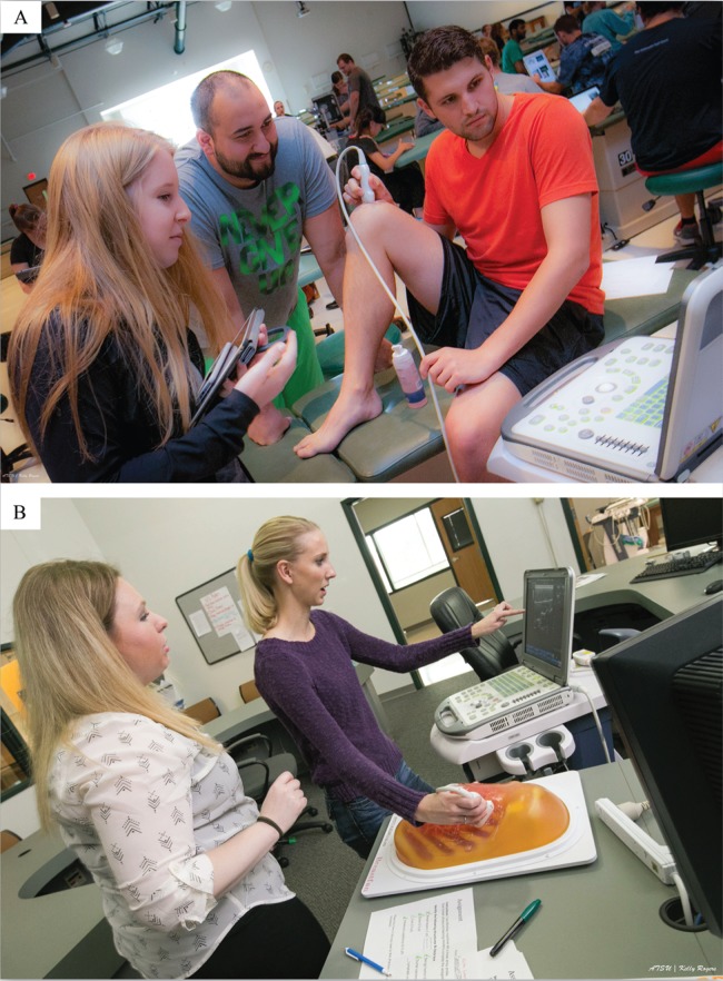
Many ultrasound laboratories included clinical cases, which utilized a clinical topic that was demonstrated through ultrasound imaging. For example, when studying ultrasound imaging of the neck vessels, students were first presented a case on carotid artery disease with a related carotid endarterectomy surgery video, which illustrated related anatomical structures. Then students were presented with the ultrasound demonstration on how to perform the carotid artery ultrasound examination and identify possible atherosclerotic plaques. After the demonstration, students scanned each other and mastered scanning skills (See Figure 1). The introduction of clinical cases provided correlations between the ultrasound laboratories and clinical courses, such as Internal Medicine and The Complete Doctor. These correlations allowed students to visualize and learn the imaging material better and in a more clinically relevant context.
Figure 1.
- A - Medical students learning knee anatomy during the lower limb musculoskeletal ultrasound laboratory.
- B - Medical students learning breast normal anatomy and pathology using Breast Ultrasound Training Model (Kyoto Kagaku).
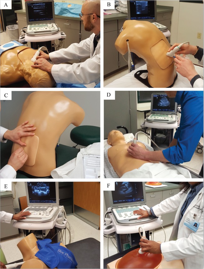
In planning the new Clinical Ultrasound course, significant assets were committed by the college to add ultrasound phantoms to the ultrasound laboratories, including simulators with pathological conditions that allow a realistic approach to teaching ultrasound imaging during the clinical blocks (See Figure 2, Table 2). Some phantoms simulate pathological conditions, while others allow for needle-guidance training.
Figure 2.
- A - Gen II Ultrasound Central Line Training Model (CAE Healthcare).
- B - Midscapular Thoracentesis Ultrasound Training Model (CAE Healthcare).
- C - Lumbar Puncture and Spinal Epidural Training Model (CAE Healthcare).
- D - Focused Assessment with Sonography for Trauma (FAST) Exam Real Time Ultrasound Training Model (CAE Healthcare).
- E - Transvaginal Sonohysterography and Sonosalpingography Ultrasound Training Model (CAE Healthcare).
- F - Fetus Ultrasound Examination Phantom SPACEFAN-ST (Kyoto Kagaku).
Table 2.
Ultrasound Laboratories for the Clinical Ultrasound Course Using Ultrasound Phantoms
| Ultrasound Laboratory | Ultrasound Training Model | Structures of Interest/Objectives |
|---|---|---|
| Obstetrics ultrasound | Fetus Ultrasound Examination Phantom SPACEFAN-ST (Kyoto Kagaku) |
|
| Gynecology ultrasound | Transvaginal SonoHysterography and Sonosalpingography Ultrasound Training Model (CAE Healthcare) |
|
| Breast ultrasound | Breast Ultrasound Training Model (Kyoto Kagaku) |
|
| Lumbar puncture | Lumbar Puncture and Spinal Epidural Training Model (CAE Healthcare) |
|
| Central line | Gen II Ultrasound Central Line Training Model (CAE Healthcare) |
|
| Thoracentesis | Midscapular Thoracentesis Ultrasound Training Model (CAE Healthcare) |
|
| FAST exam and abdominal aorta | FAST Exam Real Time Ultrasound Training Model (CAE Healthcare) Abdominal Aortic Aneurysm Ultrasound Training Model (CAE Healthcare) |
|
Abbreviation: FAST, focused assessment with sonography for trauma.
While integrating ultrasound into the first and second years of osteopathic medical education, we also identified the curricular areas of anatomy, osteopathic manipulative medicine (OMM), physiology, and internal medicine as ones that would benefit most from the inclusion of ultrasound. A workshop combining electrocardiography and echocardiography has been integrated into the cardiology block, and has been well received by students. Importantly, it significantly improved their understanding of the electrophysiology of the heart.11 Students were able to better correlate electrical activity with cardiac mechanical events and understand cardiac physiology.11
While integrating ultrasound into the ATSU-KCOM curriculum, an ultrasound component was added to the OMM course.21 It included six ultrasound assignments that required students to obtain ultrasound images of musculoskeletal structures, which were correlated with the body regions presented in the OMM course. Students scanned cranial and cervical structures and lumbar, sacral, and thoracic regions. Students were required to obtain images of the spinous processes of L4 and L5 and the base of the sacrum, laminae, and erector spinae muscles. Students also had to find the atlas (C1), its posterior arch, and the vertebral artery and vein.
Dental Ultrasound Laboratories
The integration of ultrasound has also opened new opportunities for dental medical education22 because we introduced ultrasound technology to first-year dental students at A.T. Still University’s Missouri School of Dentistry & Oral Health. The ultrasound laboratories focused on head, neck, and abdominal anatomy. Laboratories introduced dental students to a new, noninvasive imaging modality that does not use ionizing radiation. Survey results indicated that students enjoyed the ultrasound laboratory exercise and felt ultrasound was an effective learning tool because it provided a better understanding of maxillofacial anatomy.22
Student-Initiated Research Resulting from the Ultrasound Curriculum
Successful implementation of the ultrasound curriculum triggered significant research interest among medical students on a variety of topics, such as determining whether an ultrasound experience reinforced learning in Gross Anatomy 5 or the efficiency of assessing ultrasonography skills using a practical exam.23 Students also researched 3-dimensional/4-dimensional ultrasound technology and its impact on medical education and rural health (unpublished study). Students also began to study how point-of-care ultrasonography has been used in urban versus rural areas.
Conclusions
Medical imaging presents significant challenges to medical students and, despite its importance, only 5% of total teaching time is dedicated to radiology in medical education.24 The training that students receive through the KCOM ultrasound curriculum may help them better understand other diagnostic imaging modalities that are widely used in medical practice and become more versed in medical imaging in general. Ultrasound training has the potential to enhance the learning experience for students and improve the quality of their future patient care.
Acknowledgments
The authors thank Patricia Sexton, DHEd, Associate Dean for Curriculum at A.T. Still University for manuscript review and Deborah Goggin, MA, Scientific Writer, at A.T. Still University for help with manuscript preparation.
Biography
Tatyana Kondrashova, MD, PhD, (left), Department of Family Medicine, Preventive Medicine, and Community Health, and Peter Kondrashov, PhD, (right), Anatomy Department, are at A.T. Still University—Kirksville College of Osteopathic Medicine, Kirksville, Missouri.
Contact: tkondrashova@atsu.edu


Footnotes
Disclosure
None reported.
References
- 1.Syperda VA, Trivedi PN, Melo LC, Freeman ML, Ledermann EJ, Smith TM, Alben JO. Ultrasonography in preclinical education: a pilot study. J Am Osteopath Assoc. 2008;108:601–605. [PubMed] [Google Scholar]
- 2.Zoll K, Kondrashov P, Pazdernik V, Beatty D, Arseneaux M, Atieh T, Kondrashova T. Medical student perception of the impact of early ultrasonography education on experiences during clinical rotations. Med Sci Educ. 2017 Ahead of print. [Google Scholar]
- 3.Dreher SM, DePhilip R, Bahner D. Ultrasound exposure during gross anatomy. J Emerg Med. 2014;46:231–240. doi: 10.1016/j.jemermed.2013.08.028. [DOI] [PubMed] [Google Scholar]
- 4.Brown B, Adhikari S, Marx J, Lander L, Todd GL. Introduction of ultrasound into gross anatomy curriculum: perceptions of medical students. J Emerg Med. 2012;43:1098–1102. doi: 10.1016/j.jemermed.2012.01.041. [DOI] [PubMed] [Google Scholar]
- 5.Kondrashov P, Johnson JC, Boehm K, Rice D, Kondrashova T. Impact of the clinical ultrasound elective course on retention of anatomical knowledge by second-year medical students in preparation for board exams. Clin Anat. 2015;28:156–163. doi: 10.1002/ca.22494. [DOI] [PubMed] [Google Scholar]
- 6.Swamy M, Searle RF. Anatomy teaching with portable ultrasound to medical students. BMC Med Educ. 2012;12:99. doi: 10.1186/1472-6920-12-99. [DOI] [PMC free article] [PubMed] [Google Scholar]
- 7.Teichgräber UKM, Meyer JMA, Nautrup CP, von Rautenfeld DB. Ultrasound anatomy: a practical teaching system in human gross anatomy. Med Educ. 1996;30:296–298. doi: 10.1111/j.1365-2923.1996.tb00832.x. [DOI] [PubMed] [Google Scholar]
- 8.Tshibwabwa ET, Groves HM. Integration of ultrasound in the education programme in anatomy. Med Educ. 2005;39:1148–1148. doi: 10.1111/j.1365-2929.2005.02288.x. [DOI] [PubMed] [Google Scholar]
- 9.So S, Patel RM, Orebaugh SL. Ultrasound imaging in medical student education: impact on learning anatomy and physical diagnosis. Anat Sci Educ. 2017;10:176–189. doi: 10.1002/ase.1630. [DOI] [PubMed] [Google Scholar]
- 10.Brunner M, Moeslinger T, Spieckermann PG. Echocardiography for teaching cardiac physiology in practical student courses. Am J Physiol. 1995;268:S2–9. doi: 10.1152/advances.1995.268.6.S2. [DOI] [PubMed] [Google Scholar]
- 11.Kondrashova T, Sexton W, Baer R, Kondrashov P. Integration of didactic knowledge with procedural skill: learning cardiac electrophysiology and electrocardiography using ultrasound. Med Sci Educ. 2015;25:141–147. [Google Scholar]
- 12.Dinh VA, Frederick J, Bartos R, Shankel TM, Werner L. Effects of ultrasound implementation on physical examination learning and teaching during the first year of medical education. J Ultrasound Med. 2015;34:43–50. doi: 10.7863/ultra.34.1.43. [DOI] [PubMed] [Google Scholar]
- 13.Fox JC, Schlang JR, Maldonado G, Lotfipour S, Clayman RV. Proactive medicine: the “UCI 30,” an ultrasound-based clinical initiative from the University of California, Irvine. Acad Med. 2014;89:1–3. doi: 10.1097/ACM.0000000000000292. [DOI] [PubMed] [Google Scholar]
- 14.Griksaitis MJ, Scott MP, Finn GM. Twelve tips for teaching with ultrasound in the undergraduate curriculum. Med Teach. 2014;36:19–24. doi: 10.3109/0142159X.2013.847909. [DOI] [PubMed] [Google Scholar]
- 15.Hoppmann R, Cook T, Hunt P, Fowler S, Paulman L, Wells J, Richeson N, Thomas L, Wilson B, Neuffer F, McCallum J, Smith S. Ultrasound in medical education: a vertical curriculum at the University of South Carolina School of Medicine. J S C Med Assoc. 2006;102:330–334. [PubMed] [Google Scholar]
- 16.Bernard S, Richardson C, Hamann CR, Lee S, Dinh VA. Head and neck ultrasound education: a multimodal educational approach in the predoctoral setting. J Ultrasound Med. 2015;34:1437–1443. doi: 10.7863/ultra.34.8.1437. [DOI] [PubMed] [Google Scholar]
- 17.Hoppmann RA, Rao VV, Poston MB, Howe DB, Hunt PS, Fowler SD, Paulman LE, Wells JR, Richeson NA, Catalana PV, Thomas LK, Britt Wilson L, Cook T, Riffle S, Neuffer FH, McCallum JB, Keisler BD, Brown RS, Gregg AR, Sims KM, Powell CK, Garber MD, Morrison JE, Owens WB, Carnevale KA, Jennings WR, Fletcher S. An integrated ultrasound curriculum (iUSC) for medical students: 4-year experience. Crit Ultrasound J. 2011;3:1–12. doi: 10.1007/s13089-011-0052-9. [DOI] [PMC free article] [PubMed] [Google Scholar]
- 18.Rao S, van Holsbeeck L, Musial JL, Parker A, Bouffard JA, Bridge P, Jackson M, Dulchavsky SA. A pilot study of comprehensive ultrasound education at the Wayne State University School of Medicine: a pioneer year review. J Ultrasound Med. 2008;27:745–749. doi: 10.7863/jum.2008.27.5.745. [DOI] [PubMed] [Google Scholar]
- 19.Royer DF. The role of ultrasound in graduate anatomy education: current state of integration in the United States and faculty perceptions. Anat Sci Educ. 2016;9:453–467. doi: 10.1002/ase.1598. [DOI] [PubMed] [Google Scholar]
- 20.Royer DF, Kessler R, Stowell JR. Evaluation of an innovative hands-on anatomy-centered ultrasound curriculum to supplement graduate gross anatomy education. Anat Sci Educ. 2016 doi: 10.1002/ase.1670. Ahead of print. [DOI] [PubMed] [Google Scholar]
- 21.Kondrashova T, Lockwood MD. Innovative approach to teaching osteopathic manipulative medicine: the integration of ultrasonography. J Am Osteopath Assoc. 2015;115:212–220. doi: 10.7556/jaoa.2015.043. [DOI] [PubMed] [Google Scholar]
- 22.Kondrashova T, De Wan D, Briones MU, Kondrashov P. Integration of ultrasound imaging into pre-clinical dental education. Eur J Dent Educ. 2016 doi: 10.1111/eje.12205. Ahead of print. [DOI] [PubMed] [Google Scholar]
- 23.Petty JL, Kondrashov P, Johnson JC, Kondrashova T. Performance assessment in medical school curricula: an innovative method of evaluating sonographic skills using ultrasound practical examinations. Med Sci Educ. 2016;26:749–757. [Google Scholar]
- 24.Heptonstall N, Ali T, Mankad K. Integrating radiology and anatomy teaching in medical education in the UK: the evidence, current trends, and future scope. Acad Radiol. 2016;23:521–526. doi: 10.1016/j.acra.2015.12.010. [DOI] [PubMed] [Google Scholar]


