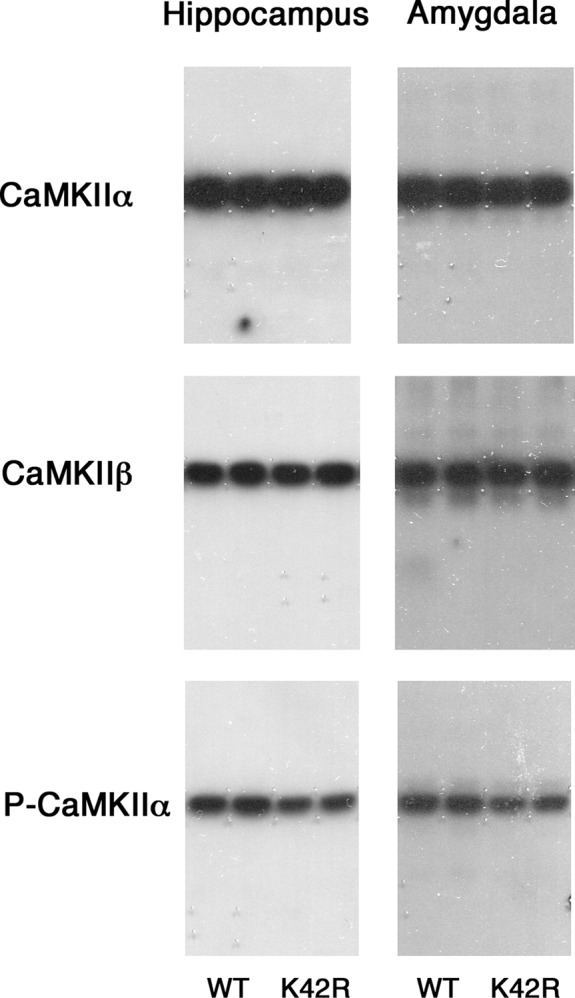
Figure 2.

Representative immunoblots showing the CaMKIIα, CaMKIIβ and phospho-T286-CaMKIIα levels in the hippocampus and amygdala of the kinase-dead CaMKIIα (K42R)-KI mouse as compared to the wild-type mouse. The amounts of protein used were 2, 4, and 8 μg for the detection of CaMKIIα, CaMKIIβ, and phospho-T286-CaMKIIα (P-CaMKIIα), respectively, from hippocampal or amygdala homogenates. Autoradiography of duplicated samples from a pair of wild-type (WT) and kinase-dead CaMKIIα (K42R)-KI mice in the same experimental group used for quantitative immunoblot analyses are shown. See also Table 1.
