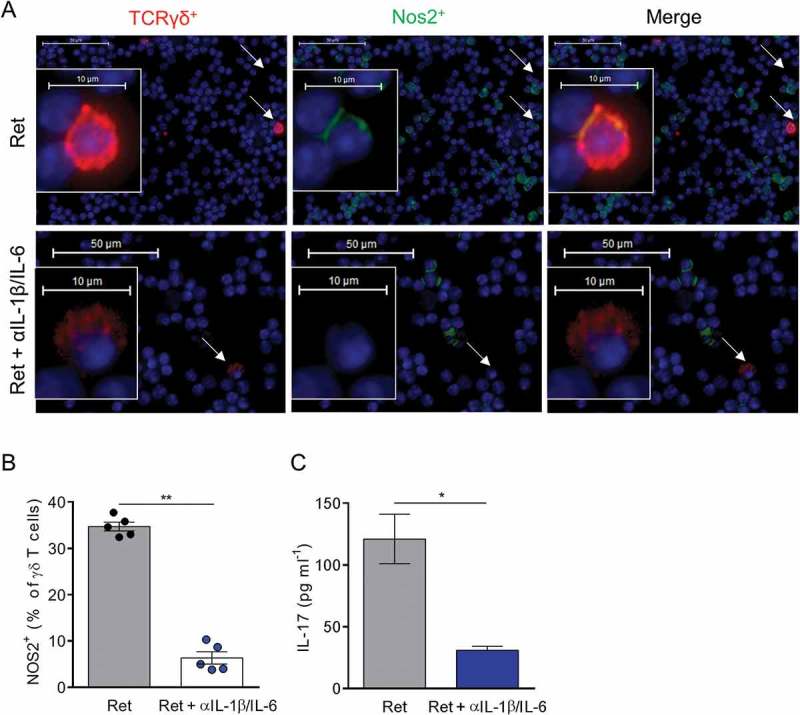
Figure 5.

In vivo neutralization of IL-1β and IL-6 suppresses NOS2 expression in γδ T cells.
(A-C) 3 month old Ret mice were treated or not during 2 weeks with antibodies neutralizing IL-1β and IL-6. (A, B) NOS2 expression was analyzed by microscopy in TdLNs from 5 mice in each group. Representative images showing γδ T cells positive for NOS2 derived either from untreated or treated Ret mice and stained with antibodies to TCR γδ (red), NOS2 (green) and counterstained with DAPI (blue). Bars 10 µM. 40 X objective. Arrows indicate γδ T cells. (B) NOS2 positive γδ T cells were quantified from 500 to 1500 γδ T cells. (C) Protein levels of IL-17 in primary tumor extracts, from untreated (n = 5) and treated (n = 5) Ret mice, were determined by ELISA. Each point represents individual mouse. Bars are mean ± SEM. *P < 0.05, **P < 0.01. (Mann-Whitney test).
