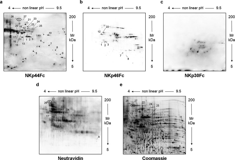
Figure 1.

Analysis of culture supernatant from HEK293T cells by two-dimensional electrophoresis (2-DE). Concentrated HEK293T-SN was analyzed by two-dimensional electrophoresis (2-DE). After blotting, membranes were probed with NKp44Fc (A), NKp46Fc (B), or NKp30Fc (C) followed by HRP-conjugated anti-human IgG mAb. HEK293T-SN-biot was subjected to the same procedure and the membrane was incubated with HRP-conjugated Neutravidin (D). In parallel, HEK293T-SN (600 μg) was separated by 2-DE and proteins were stained with “blue silver” colloidal Coomassie (E). Numbers indicate the spots selected for mass spectrometry analysis; in panels (A), (D), and (E) spot 26 is highlighted with a black circle.
