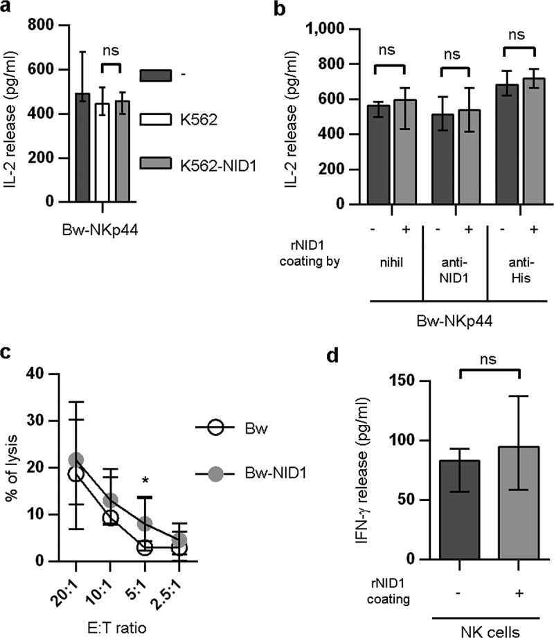
Figure 8.

Functional effects of cell surface-associated NID1 or plastic-bound rNID1 in Bw-NKp44 and in NK cells. (A) Bw-NKp44 cells were cultured alone or in the presence of wild type or NID1-transfected K562 cells for 20 h at 37°C. IL-2 release in the SN was evaluated by ELISA. Data are medians of duplicates ± interquartile range and are the pooled results of three independent experiments. ns = 0.5714 by two-tailed Mann-Whitney test. (B) Bw-NKp44 cells were incubated on plates coated with rNID1, anti-NID1 + rNID1, or anti-His + rNID1 for 20 h at 37°C. IL-2 release in the SN was evaluated by ELISA. Data are medians of duplicates ± interquartile range and are the pooled results of two independent experiments. ns = 0.8286 (−) or ns = 0.6571 (anti-NID1, anti-His) by two-tailed Mann-Whitney test. (C) Polyclonal NK cell lines were evaluated for their cytolytic activity in a 4-h 51Cr release assay against Bw and Bw-NID1 cells at the indicated E:T ratios. Data are medians of duplicates ± interquartile range and are the pooled results of fifteen experiments performed with NK cells derived from five donors. *p = 0.0277 by one-tailed Wilcoxon test. (D) Polyclonal NK cell lines were incubated on plates coated with rNID1 for 20 h at 37°C. IFN-γ release in the SN was evaluated by ELISA. Data are medians of eight independent experiments ± interquartile range performed with NK cells derived from three donors. ns = 0.2305 by one-tailed Wilcoxon test.
