Abstract
Evofosfamide (TH-302) is a clinical-stage hypoxia-activated prodrug of a DNA-crosslinking nitrogen mustard that has potential utility for human papillomavirus (HPV) negative head and neck squamous cell carcinoma (HNSCC), in which tumor hypoxia limits treatment outcome. We report the preclinical efficacy, target engagement, preliminary predictive biomarkers and initial clinical activity of evofosfamide for HPV-negative HNSCC. Evofosfamide was assessed in 22 genomically characterized cell lines and 7 cell line–derived xenograft (CDX), patient-derived xenograft (PDX), orthotopic, and syngeneic tumor models. Biomarker analysis used RNA sequencing, whole-exome sequencing, and whole-genome CRISPR knockout screens. Five advanced/metastatic HNSCC patients received evofosfamide monotherapy (480 mg/m2 qw × 3 each month) in a phase 2 study. Evofosfamide was potent and highly selective for hypoxic HNSCC cells. Proliferative rate was a predominant evofosfamide sensitivity determinant and a proliferation metagene correlated with activity in CDX models. Evofosfamide showed efficacy as monotherapy and with radiotherapy in PDX models, augmented CTLA-4 blockade in syngeneic tumors, and reduced hypoxia in nodes disseminated from an orthotopic model. Of 5 advanced HNSCC patients treated with evofosfamide, 2 showed partial responses while 3 had stable disease. In conclusion, evofosfamide shows promising efficacy in aggressive HPV-negative HNSCC, with predictive biomarkers in development to support further clinical evaluation in this indication.
Keywords: Oncology, Therapeutics
Keywords: Cancer, Head & neck cancer, hypoxia
The clinical-stage hypoxia-targeting agent evofosfamide (TH-302) shows promising efficacy in poor-prognosis, biomarker-selected HPV-negative head and neck carcinoma in combination with radiotherapy or immunotherapy.
Introduction
Hypoxia has been explored as a therapeutic target due to its prevalence and severity in tumors and its contributions to aggressive disease (1). The evidence supporting hypoxia as an oncology target is strongest in head and neck squamous cell carcinoma (HNSCC) (2–4), a malignancy that accounts for 5% of global cancer mortality (5). Risk of HNSCC is strongly linked to tobacco and alcohol exposure (6) and infection with high-risk human papillomavirus (HPV) serotypes (7). HPV-associated disease represents a distinct entity with a lower mutational burden (8, 9), greater lymphocytic and myeloid infiltration (10), and favorable prognosis (11, 12). Locally advanced HNSCC is treated with surgery and definitive radiotherapy, often with concurrent platinum-based chemotherapy (13, 14). Overexpression of the epidermal growth factor receptor (EGFR) in a subset of tumors (15) led to the development of cetuximab with radiotherapy for locally advanced HNSCC (16) and with chemotherapy at first-line for metastatic/recurrent disease (17). Immunotherapy has also shown efficacy for HNSCC (18, 19), with nivolumab and pembrolizumab both approved at second-line for recurrent/metastatic HNSCC, though response rates to anti–PD-1 monotherapy remain disappointing at 15%.
These advances notwithstanding, HNSCC survival has not markedly improved in recent decades and distant metastasis, locoregional recurrence, second primaries, and therapy resistance remain major challenges. Hypoxia is a marker of poor prognosis (3) and predictor of radiotherapy failure in HNSCC (2), specifically in HPV-negative disease (4). Accordingly, a voluminous literature has investigated hypoxic modification to improve HNSCC outcomes (20). Notable approaches include modified oxygen breathing (21), oxygen-mimetic radiosensitizers (22), and direct ablation of hypoxic cells with selective cytotoxins (23), though such strategies have not seen widespread adoption. Compelling evidence also points to a role for hypoxia in immune evasion (24). Hypoxia-inducible factor 1 (HIF-1) is a transcriptional activator of PD-L1 in myeloid and tumor cells (25) and HIF transcriptional signatures inversely correlate with T cell infiltration in HNSCC (26). Hypoxia promotes the recruitment of regulatory T cells (27, 28) and tumor-associated macrophages (TAMs) (29), production of PGE2, IL-6, and IL-10 (30), and the immunosuppressive activity of myeloid cells (31, 32). Additionally, hypoxia results in extracellular accumulation of adenosine and impairment of T cell–mediated immunity (33). Extracellular acidification associated with hypoxic microenvironments also creates a formidable barrier to T cell function and persistence (34–36).
Such considerations highlight the rationale for targeting hypoxia in HNSCC, both in the context of radiation oncology for local disease and with T cell–directed immunotherapies for advanced disease. Evofosfamide is a clinical-stage hypoxia-activated prodrug designed to target the DNA-crosslinking nitrogen mustard bromo-iso-phosphoramide (Br-IPM) to regions of hypoxia (37), leading to DNA damage, γH2AX phosphorylation, cell cycle arrest, and cleavage of caspase-3 and -6 (38–40). Despite narrowly missing its primary phase 3 overall survival (OS) endpoint with gemcitabine for advanced pancreatic adenocarcinoma (41), evofosfamide shows abundant evidence of preclinical (40, 42–51) and clinical activity (52, 53). However, evofosfamide has not been comprehensively investigated for HNSCC. Here, we report the preclinical efficacy, target engagement, development of predictive biomarkers, and initial clinical activity of evofosfamide for this indication.
Results
Evofosfamide is potent and highly selective for hypoxic HNSCC cells.
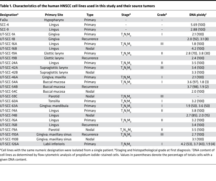
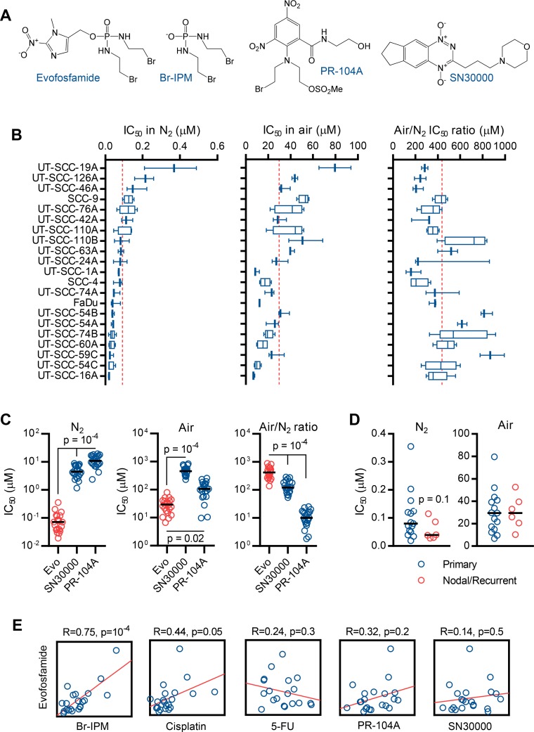
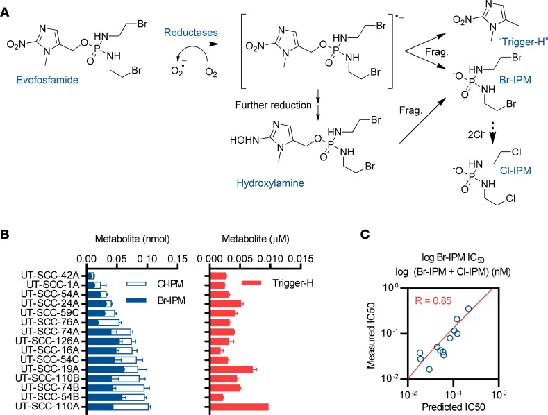
To explore the potential of evofosfamide for HNSCC, we assembled 27 HPV-negative cell lines derived from HNSCC of varying primary site, histopathological grade, and TNM stage (Table 1) and characterized 22 of these by whole-exome sequencing (Supplemental File 1; supplemental material available online with this article; https://doi.org/10.1172/jci.insight.122204DS1). In a subset of cases, paired cell lines isolated from primary, nodal, and/or recurrent sites in the same patient were studied. We compared the in vitro antiproliferative potency and hypoxic selectivity of evofosfamide to other hypoxia-activated prodrugs PR-104A (54) and SN30000 (55) (Supplemental Figure 1). We also compared Br-IPM, the active metabolite of evofosfamide, to the standard chemotherapy agents cisplatin and 5-fluorouracil (Supplemental Figure 2). This study examined 21 HNSCC cell lines challenged with compounds under anoxia (N2) or 20% oxygen (henceforth referred to as Air, see Figure 1A for structures and Supplemental File 2 for the full dataset) to define IC50 values (drug concentrations for 50% inhibition of cell growth). Evofosfamide showed nanomolar potency for HNSCC cells under anoxia and was strongly suppressed by oxygen, with a median Air/N2 IC50 ratio of 360-fold (Figure 1B). Evofosfamide was significantly more potent and selective than PR-104A or SN30000 (Figure 1C) and was equivalently active against lines derived from primary, nodal, or recurrent lesions (Figure 1D). Cell line sensitivity to evofosfamide was strongly correlated with sensitivity to Br-IPM under anoxia and weakly correlated with cisplatin but not with other agents (Figure 1E), in keeping with a DNA-crosslinking mechanism of action for evofosfamide and suggesting that variation in the intrinsic sensitivity of HNSCC cell lines to Br-IPM accounts for a significant component of the 19-fold spread in anoxic IC50 values for evofosfamide. To investigate this further, we measured the reductive activation of evofosfamide (see Figure 2A for the metabolic pathway) by liquid chromatography–tandem mass spectrometry (LC–MS/MS) in 15 cell lines (Figure 2B). Evofosfamide activation rates were variable between cell lines and combining reductive activation (measured as the concentration of metabolites produced) with Br-IPM sensitivity (anoxic IC50) as independent variables in a multiple linear regression model — log10(evofosfamide IC50) = a + (b × log10[Br-IPM IC50]) + (c × log10[Br-IPM + Cl-IPM concentration]) — improved the fit between predicted and measured evofosfamide IC50 from R = 0.73 to R = 0.85 (Figure 2C and Supplemental Table 1). These data confirmed that evofosfamide responsiveness is significantly determined by sensitivity to DNA crosslinking, with the rate of evofosfamide activation serving as an additional determinant.
Table 1. Characteristics of the human HNSCC cell lines used in this study and their source tumors.
Figure 1. Potency and hypoxic selectivity of evofosfamide and reference agents for head and neck squamous cell carcinoma (HNSCC) cell lines.
(A) Chemical structures of evofosfamide, its DNA crosslinking metabolite bromo-iso-phosphoramide mustard (Br-IPM), and the comparator hypoxia-activated prodrugs PR-104A (a dinitrobenzamide) and SN30000 (a benzotriazine di-N-oxide). (B) Antiproliferative activity of evofosfamide — measured as the concentration required for 50% inhibition of cell growth (IC50) — in 21 HNSCC cell lines assessed by sulforhodamine B assay after 4-hour drug exposure under anoxia (N2) or ambient oxygen (air) followed by 5-day regrowth, with hypoxia selectivity represented as the Air-N2 IC50 quotient. The Air/N2 ratio is plotted as the mean ± SEM from 3 or more intraexperiment quotients calculated from anoxic and normoxic assays performed on the same days. Boxes represent the median and interquartile range, whereas whiskers mark the minimum and maximum IC50 determinations from 3 or more independent experiments. Dashed lines denote the mean IC50 values for the cell line panel. (C) Comparison of the in vitro antiproliferative potency and hypoxic selectivity of evofosfamide (evo), PR-104A, and SN30000 in 21 HNSCC cell lines. Data points denote mean IC50 values for individual cell lines computed from 3 or more experiments, with IC50 values and Air/N2 IC50 quotients defined as per panel B. Horizontal lines mark the median values. The statistical significance of differences in the potency and selectivity of drugs was assessed by 1-way ANOVA with Dunnett’s correction. (D) Comparison of the antiproliferative potency (as IC50 under N2 or air) of evofosfamide in HNSCC cell lines derived from primary (n = 15) or nodal/recurrent (n = 6) lesions. Statistical significance of differences in IC50 values between these groups was assessed by Mann-Whitney test. (E) Comparison of the pattern of HNSCC cell line sensitivity (as IC50 values) to evofosfamide and to Br-IPM, cisplatin, 5-FU, PR-104A, and SN30000 under anoxia. IC50 values were defined as per panel B and data points correspond to individual cell lines. Axes are linear and functions are Pearson’s correlations.
Figure 2. Enzymatic activation of evofosfamide and its contribution to antiproliferative activity in a subset of 15 cultured head and neck squamous cell carcinoma (HNSCC) cell lines.
(A) Route of metabolic activation of evofosfamide in hypoxic tumor cells by enzymatic 1-electron reduction. As described in literature (39), evofosfamide undergoes enzyme-catalyzed 1-electron reduction to a transient radical anion that fragments to release Br-IPM and the free 2-nitroimidazole trigger (“Trigger-H”). In the presence of oxygen, the radical anion is back-oxidized to evofosfamide, preventing accumulation of Br-IPM and thus conferring hypoxic selectivity. Fragmentation at the 1-electron (radical anion) stage competes with further reduction to the corresponding hydroxylamine, which also fragments to release Br-IPM but does not generate Trigger-H. Halide exchange results in the conversion of Br-IPM to more stable Cl-IPM. Br-IPM, Cl-IPM, and Trigger-H are thus all diagnostic of the reductive activation of evofosfamide. Frag, fragmentation. (B) Comparison of the facility of reductive activation of evofosfamide in HNSCC cell lines by liquid chromatography–tandem mass spectrometry (LC-MS/MS) measurement of intracellular Br-IPM, Cl-IPM, and Trigger-H concentrations at the endpoint of 1-hour exposures to 30 μM evofosfamide under anoxia. Data are the mean ± range from 2 independent determinations. (C) Multiple linear regression of measured evofosfamide antiproliferative activity (as the concentration required for 50% inhibition of cell growth, IC50) in 15 HNSCC cell lines under anoxia compared to IC50 values predicted in the same lines using log Br-IPM IC50 under anoxia as a measure of sensitivity to the active metabolite and the log of the sum of the intracellular concentrations of Br-IPM and Cl-IPM formed from evofosfamide (i.e., the values plotted in panel B) as a measure of its reductive metabolism.
A proliferation metagene correlates with evofosfamide sensitivity.
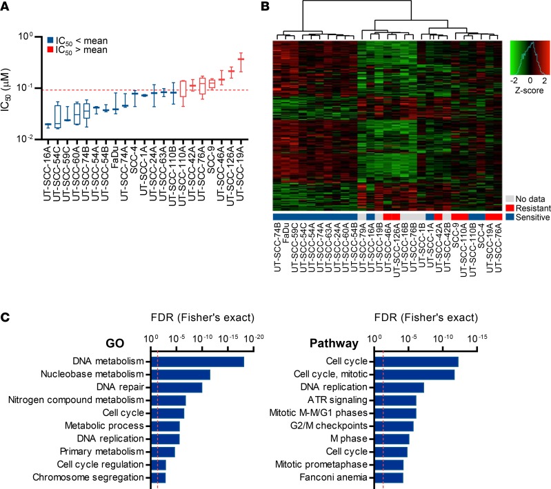
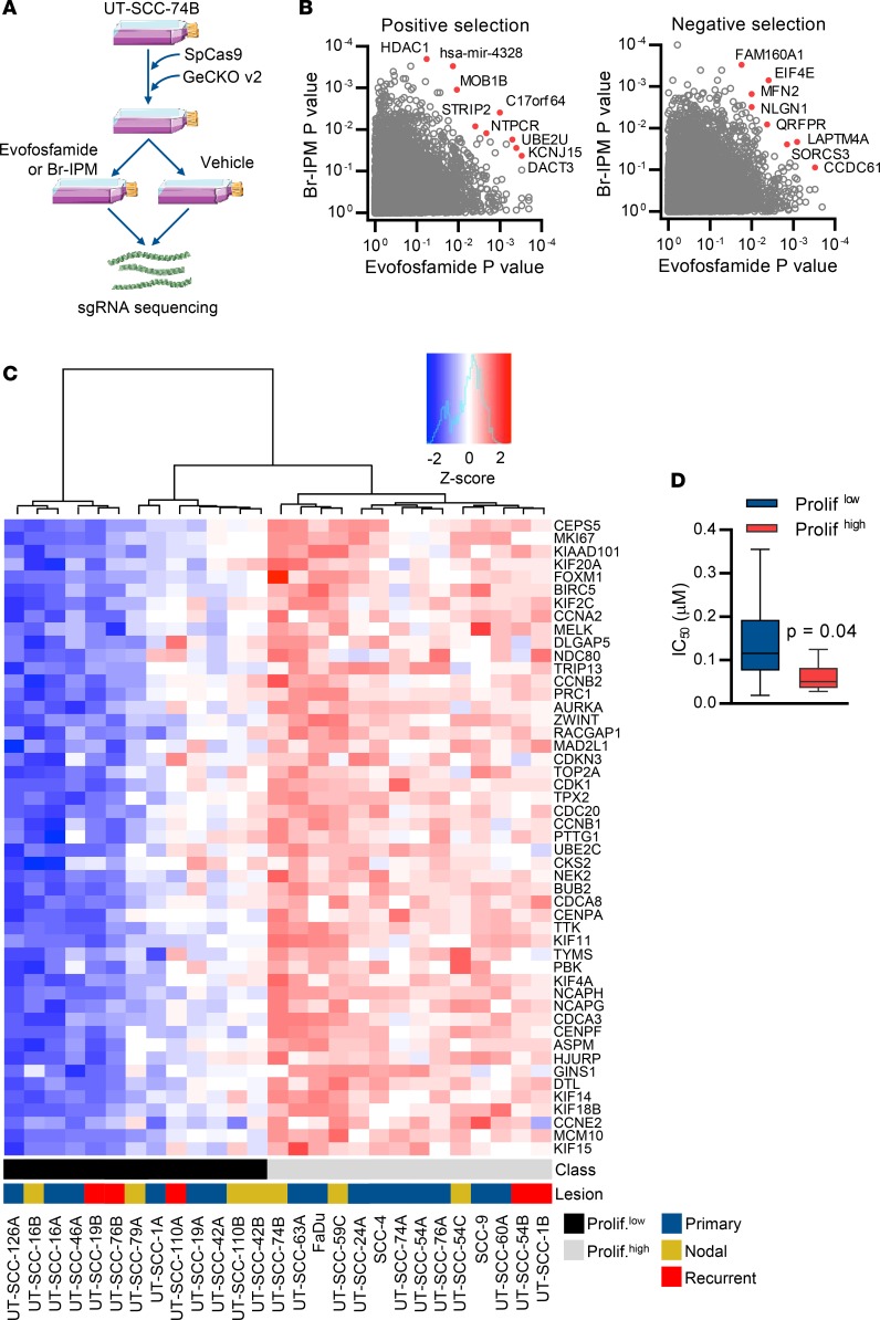
To explore molecular correlates of evofosfamide sensitivity, we performed RNA sequencing (RNAseq) on the HNSCC cell lines (Supplemental Figure 3) and differential expression analysis of evofosfamide-sensitive and -resistant lines dichotomized by the arithmetic mean anoxic IC50 value (Figure 3A), defining genes differentially expressed in evofosfamide-sensitive and -resistant cell lines (Figure 3B and Supplemental File 3). Querying the latter for enrichments of gene ontology and pathway classifiers identified overrepresentation of terms relating to the cell cycle (Benjamini-Hochberg–adjusted Fisher’s exact P value, false discovery rate [FDR] < 10–12), DNA repair (FDR < 10–10), and DNA metabolism (FDR < 10–18; Figure 3C). This result was reproduced when cell lines were dichotomized by the geometric mean IC50 value or separated into tertiles (not shown). Related terms were similarly enriched among genes that correlated with evofosfamide sensitivity when anoxic IC50 values were treated as a continuous variable (Supplemental Figure 4). As an orthogonal approach, we performed whole-genome CRISPR knockout screens in UT-SCC-74B cells transduced with the GeCKOv2 single guide RNA (sgRNA) library (56). Cas9-expressing UT-SCC-74B cells were transduced to generate a knockout library that was highly complex (Supplemental Figure 5, A–C) and functionally validated by screening with 6-thioguanine, where drug exposure resulted in outgrowth of cells carrying mutations in the known 6-thioguanine sensitivity genes HPRT1 and NUDT5 (Supplemental Figure 5, D and E). Challenging this library separately with evofosfamide and Br-IPM (Figure 4A) selected for differential survival of clones carrying sgRNA targeted to putative modifiers of drug sensitivity (Figure 4B). Functional analysis of the latter revealed overrepresentation of genes involved in cell proliferation (gene ontology classifier GO0042127; FDR < 0.05). To develop an initial predictive biomarker for evofosfamide from these findings, we hierarchically clustered the HNSCC cell lines using a published (57) 61-gene proliferation signature — 49 of which were expressed in the cell lines (Supplemental File 4) — to define proliferationlo and proliferationhi classes (Figure 4C). The proliferationlo and proliferationhi classes showed differential population doubling times (Supplemental Figure 6) and in vitro sensitivity to evofosfamide (Figure 4D), with rapidly proliferating lines more susceptible to treatment.
Figure 3. Sensitivity of HNSCC cell lines to evofosfamide is associated with expression of proliferation-related genes.
(A) Head and neck squamous cell carcinoma (HNSCC) cell lines dichotomized by the arithmetic mean of anoxic evofosfamide IC50 values into sensitive and resistant groups for differential expression analysis using RNA sequencing. Data are defined as in Figure 1B. (B) Unsupervised hierarchical clustering of HNSCC cell lines according to their expression of the top 200 most differentially expressed genes (by P value) between evofosfamide-sensitive and evofosfamide-resistant cell lines. The gene list was defined using limma and the clustering performed using the ward.D method with Euclidean distance. (C) Statistical overrepresentation of gene ontology (GO) terms (assessed using PANTHER [ref. 80]; left) or molecular pathways (assessed using GeneSetDB [ref. 81], right) among the 200 genes most differentially expressed between evofosfamide-sensitive and evofosfamide-resistant cell lines (as per panel B). False discovery rates (FDRs) arising from Benjamini–Hochberg adjustment of P values from Fisher’s exact tests are shown for the 10 most significant GO and pathway findings. Dashed lines denote the 5% FDR.
Figure 4. A proliferation expression signature correlates with evofosfamide sensitivity in head and neck squamous cell carcinoma (HNSCC) cell lines.
(A) The workflow for functional genomic screens for modifiers of evofosfamide or Br-IPM sensitivity. SpCas9, Streptococcus pyogenes Cas9. (B) Enrichment (positive selection) or depletion (negative selection) of single guide RNA (sgRNA) in evofosfamide- and Br-IPM-treated UT-SCC-74B cells (2 replicates per condition). The screen was deconvoluted using the RIGER method (83) with weighted-sum aggregation to output gene-level P values that were adjusted using the Benjamini-Hochberg method. (C) Unsupervised hierarchical clustering (ward.D method with Euclidean distance) of HNSCC cell lines according to their expression (measured by RNAseq) of a published tumor proliferation metagene (57). Of 61 genes in the proliferation cluster, 49 were expressed in HNSCC cells. The resulting proliferation metagene class assignments and whether cell lines were derived from primary, nodal, or recurrent lesions are indicated. (D) Differential in vitro sensitivity to evofosfamide per antiproliferative IC50 assay in HNSCC cell lines separated by proliferation metagene status, where the grouping of cell lines as proliferationhi or proliferationlo was accomplished by assignment of cell lines to binary clusters defined by unsupervised hierarchical clustering according to expression values of the metagene as shown in C. Box plots show the mean and interquartile range, whereas whiskers show the maximum and minimum IC50 values for metagene-high (n = 13) and metagene-low (n = 8) cell lines (≥3 independent experiments). Statistical significance was assessed by Mann–Whitney test.
Evofosfamide is active against biomarker-selected HNSCC cell line–derived xenograft models.
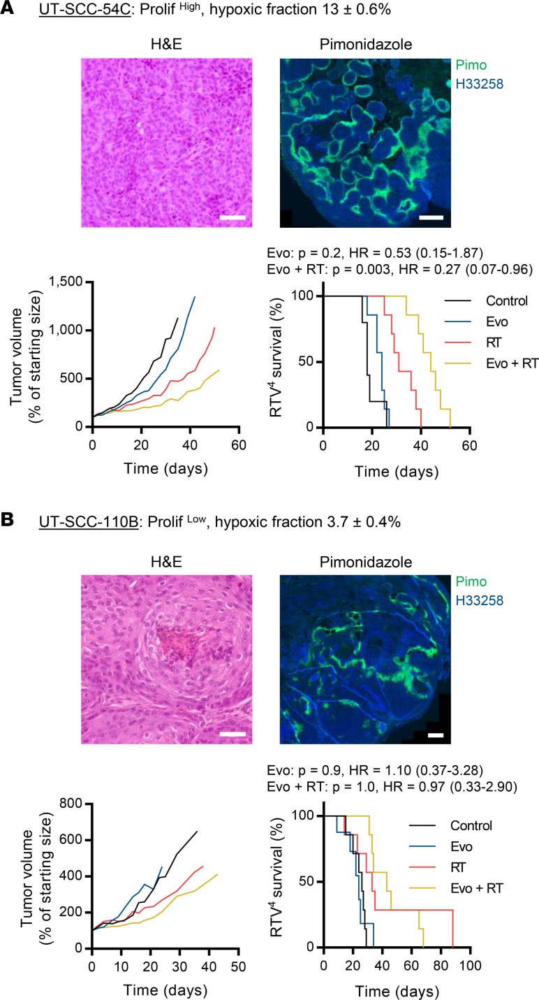
We compared the efficacy of evofosfamide monotherapy or in combination with radiotherapy in CDX models predicted to be differentially sensitive by biomarker analysis. UT-SCC-54C xenografts (proliferationhi) were highly hypoxic, with a pimonidazole-positive fraction of 13% ± 0.6% (mean ± SEM, n = 4), while tumors grown from proliferationlo UT-SCC-110B cells were less hypoxic (3.7% ± 0.4%, n = 4). Despite having limited monotherapy activity in UT-SCC-54C, evofosfamide slowed median tumor growth when administered concurrently with radiotherapy and significantly prolonged the time to 4-fold increase in tumor volume (RTV4 44 vs. 31 days; log-rank P = 0.003, hazard ratio [HR] = 0.3 vs. radiotherapy; Figure 5A). The addition of evofosfamide to radiotherapy offered no additional benefit in the UT-SCC-110B model predicted to be treatment refractory (Figure 5B).
Figure 5. Efficacy of evofosfamide alone or with radiotherapy in biomarker-selected head and neck squamous cell carcinoma (HNSCC) cell line–derived xenograft models.
Evofosfamide (evo) was assessed as a single agent (50 mg/kg i.p. qd × 5 weekly for 3 cycles) or in combination with radiotherapy (RT, 1 × 10 Gy on day 5 of the first cycle) by growth delay in the proliferation metagene-high, highly hypoxic cell line–derived xenograft (CDX) UT-SCC-54C (A) and the metagene-low, less hypoxic CDX UT-SCC-110B (B). Growth plots show the volume of the median tumor at each assessment. Survival analyses used the time to 4-fold increase in starting tumor volume (RTV4) as the experimental endpoint, with treatment effects assessed by log-rank test (evo vs. control vehicle, evo + RT vs. RT alone). Cohort sizes were 6–7 animals per group for UT-SCC-54C and 7–8 animals per group for UT-SCC-110B. Representative H&E and pimonidazole (pimo) immunostains are shown for each model. Hypoxic fractions relative to the Hoechst 33258–positive (H33258-positive) tumor area are expressed as the mean ± SEM of whole sections from 4 tumors per model. Scale bars: 50 μm (H&E) and 500 μm (pimonidazole). HR, hazard ratio.
Evofosfamide is active against HNSCC patient–derived xenograft models.
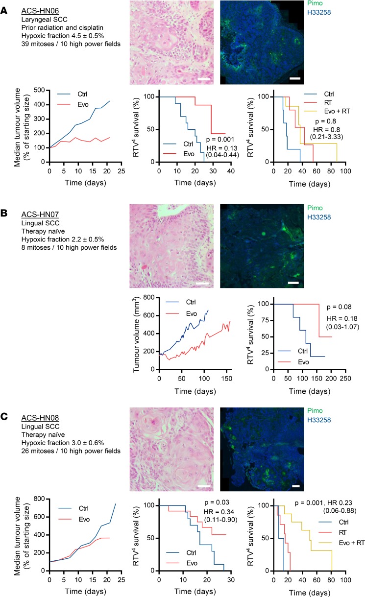
Next, we evaluated the activity of evofosfamide in 3 patient-derived xenograft (PDX) models derived from laryngeal and lingual squamous cell carcinomas (Figure 6; see supplement section 4 for clinical history and histopathology). In all cases, the histology of the PDX tumors closely resembled the original clinical specimen (not shown). The PDX models all had inactivating missense mutations in TP53, while ACS-HN07 was a PIK3CAE545K mutant and ACS-HN08 an HRASG13V and NOTCH1C1536Y mutant (Supplemental Table 2). The pimonidazole-positive hypoxic fractions of the PDX models were 4.5% ± 0.5% (n = 7), 2.5% ± 0.5% (n = 4), and 3.0% ± 0.6% (n = 5) for ACS-HN06, ACS-HN07, and ACS-HN08, respectively. Evofosfamide monotherapy significantly prolonged survival in 2 PDX models (ACS-HN06: median time to RTV4 29 vs. 18 days, log-rank P = 0.001, HR = 0.13; ACS-HN08: median time to RTV4 undefined vs. 17 days, log-rank P = 0.03, HR = 0.34), with the least proliferative and hypoxic model, ACS-HN07, not achieving statistical significance for survival (median time to RTV4 181 vs. 112 days, log-rank P = 0.08, HR = 0.18). In combination with radiotherapy, evofosfamide significantly prolonged survival in ACS-HN08 versus radiation alone (median time to RTV4 50 vs. 10 days, log-rank P = 0.001, HR = 0.23), but provided no additional benefit to radiotherapy in ACS-HN06.
Figure 6. Efficacy of evofosfamide alone or with radiotherapy in head and neck squamous cell carcinoma (HNSCC) patient–derived xenograft models.
Evofosfamide (evo) was assessed as a single agent (50 mg/kg i.p. qd × 5 weekly for 3 cycles) or in combination with radiotherapy (RT, 1 × 10 Gy on day 5 of the first cycle) by growth delay in the laryngeal patient-derived xenograft (PDX) ACS-HN06 (A) and the lingual PDXs ACS-HN07 (B) and ACS-HN08 (C). The patient from whom ACS-HN06 was derived had received prior radiotherapy and high-dose cisplatin, whereas ACS-HN07 and ACS-HN08 were therapy-naive. Growth plots show the volume of the median tumor at each assessment. Survival analyses used the time to 4-fold increase in starting tumor volume (RTV4) as the experimental endpoint, with treatment effects assessed by log-rank test (evo vs. control vehicle [ctrl], evo + RT vs. RT alone). Cohort sizes were 7–10 per group. Representative H&E and pimonidazole (pimo) immunostains are shown for each model. Hypoxic fractions relative to the Hoechst 33258–positive (H33258-positive) tumor area are expressed as the mean ± SEM of whole sections from 4–7 tumors per model. The number of mitotic cells per 10 high-power fields was scored by a consultant pathologist (DK). Scale bars: 50 μm (H&E) and 500 μm (pimo). SCC, squamous cell carcinoma; HR, hazard ratio.
Evofosfamide reduces nodal hypoxia and augments CTLA-4 blockade.
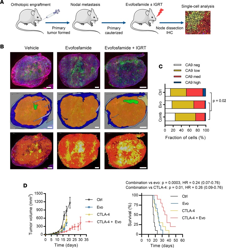
As clinical testing of evofosfamide would initially be for advanced HNSCC, we evaluated target engagement (as a reduction in hypoxic fraction) in spontaneously disseminated nodal metastases in an orthotopic UT-SCC-74B model (Figure 7A). Evofosfamide as monotherapy or with concurrent image-guided cervical node irradiation reduced the fraction of cytokeratin-positive tumor cells showing medium or high immunostaining for carbonic anhydrase 9 (CA9; Figure 7, B and C), an endogenous marker of hypoxia, consistent with target engagement in regionally metastatic disease.
Figure 7. Evofosfamide reduces nodal hypoxia and augments CTLA-4 blockade in head and neck squamous cell carcinoma (HNSCC) tumor models.
(A) Experimental design for assessing target engagement (as a reduction in the CA9-positive hypoxic tumor cell fraction) in nodal lesions disseminated from an orthotopic UT-SCC-74B tumor model. (B) Definiens TissueStudio analysis of CA9 staining in nodal lesions from treated and control animals. The top row shows CA9 staining (red), pan-cytokeratin staining (green), and DAPI staining (blue) in representative sections from each treatment group. The middle row shows tissue segmentation into tumor (orange), normal tissue (blue), necrosis (green), and artifact (gray). The bottom row shows the cellular classification, in which individual cells (identified by DAPI staining) are assigned 1 of 4 categories according to CA9 expression: negative (white), low (yellow), medium (orange), or high (red). Scale bars: 200 μm. (C) Comparison of the proportion of viable tumor cells showing negative, low, medium, or high expression of CA9 in nodal lesions dissected from animals treated with control vehicle (n = 2), evofosfamide monotherapy (evo, n = 3), or evofosfamide plus image-guided radiotherapy (IGRT; comb, combination n = 3) was analyzed using machine learning in the Definiens TissueStudio environment. Statistical significance of the reduction in CA9 staining in treated tumor sections was assessed by χ2 test. (D) Efficacy of evofosfamide (50 mg/kg i.p. qd × 5 weekly for 2 cycles interspaced by 1-week treatment holiday) alone or with concurrent anti–CTLA-4 antibody (9H10, 100 μg/dose i.p. q3d × 3 weekly) in the syngeneic HNSCC model, SCC-7. The tumor growth plot (left panel) shows the mean ± SEM tumor volume for 10 animals per treatment group. Survival analysis (right panel) used log-rank tests with time to tumor volume ≥1,000 mm3 to define events. HR, hazard ratio.
Given the increasing use of immunotherapy for advanced HNSCC (18, 19) and the potential contributions of hypoxia to immunotherapy resistance (24), we evaluated evofosfamide in combination with CTLA-4 blockade in the syngeneic SCC-7 model, which was refractory to PD-1 blockade (not shown). Evofosfamide and anti–CTLA-4 antibody both moderately prolonged survival as single agents (Figure 7D; log-rank for evofosfamide P = 0.002, HR = 0.3; for anti–CTLA-4, P = 0.003, HR = 0.3), whereas concurrent combination therapy further improved survival (log-rank vs. anti–CTLA-4 alone, P = 0.01, HR = 0.3). These data highlighted the potential of combining evofosfamide with T cell–directed therapies for advanced HNSCC.
Evofosfamide shows initial evidence of activity in heavily pretreated HNSCC.
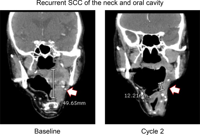
We undertook initial clinical testing of evofosfamide for HNSCC as part of a phase 2 solid tumor expansion cohort to a previously published (52) phase 1a monotherapy trial (Supplemental Figure 7, NCT00495144). The phase 2 component of the study enrolled 72 subjects, including 5 with histologically confirmed locally advanced or metastatic HNSCC who had failed standard-of-care, including surgery and radiotherapy with concurrent cisplatin or cetuximab. These patients received single-agent evofosfamide by i.v. infusion at the recommended phase 2 dose of 480 mg/m2 qw × 3 (1-week rest) for up to 6 cycles or until progression. Of the 5 HNSCC patients treated, 2 showed confirmed partial responses per Response Evaluation Criteria In Solid Tumors (RECIST) 1.0 lasting 113 and more than 176 days, while 3 had stable disease for a disease-control rate of 100%. Median progression-free survival (PFS) was 169 days (range 113 to 314 days), while median OS was not reached (range >113 to >316 days) after 316-day maximum follow-up. Safety was consistent with previous reports (52), with adverse events including nausea, skin rash fatigue, and emesis. A presented case study of a 71-year-old white female with poorly differentiated neck and oral cavity squamous cell carcinoma locally recurrent after prior surgery, radiotherapy, and cetuximab showed an excellent partial response of bulky disease at cycle 2 (Figure 8), which was maintained at cycle 4 prior to target lesion progression at cycle 6.
Figure 8. Partial response (PR) to evofosfamide monotherapy in an example head and neck squamous cell carcinoma (HNSCC) case.
This 71-year-old woman presented with a recurrent, poorly differentiated squamous cell carcinoma (SCC) of the neck and oral cavity metastatic to lymph nodes that was previously treated with cetuximab, radiotherapy, and surgery. At baseline, the coronal CT (left panel) shows recurrent tumor centered in the left oropharynx, extending from the region of the palate to the lateral and posterior pharyngeal wall, with inferior extension into the hypopharynx. The patient received evofosfamide (480 mg/m2 qw × 3 per 28-day cycle), with PR noted at cycles 2 (right panel) and 4. Disease progression based on target lesions was observed at cycle 6.
Discussion
Systemic drugs that interact favorably with radiotherapy and immunotherapy are needed for HNSCC and other malignancies. We show that evofosfamide provides promise in this context by targeting hypoxic tumor cell populations that are refractory to radiation (2) and T cell–directed therapy (24), though varying the administered doses of these agents would be necessary to establish whether effects are synergistic or additive. The initial clinical activity and efficacy seen in tumor models (which showed hypoxic fractions comparable to clinical disease; ref. 58) establish compelling evidence for clinical development of evofosfamide in HPV-negative HNSCC. Attractive indications in the current treatment landscape include evofosfamide in combination with PD-1 blockade for platinum-resistant disease, with salvage radiation for locally recurrent disease or with first-line chemoradiation. Our observation that evofosfamide reduces hypoxia in nodal lesions is consistent with the possibility of using this agent for locally advanced HNSCC presentations (such as extracapsular disease), though it will be important to confirm our observation using a more direct measure of tumor hypoxia than CA9. While combining evofosfamide with radiotherapy or chemoradiation enjoys a strong scientific rationale, the dose-limiting mucositis of evofosfamide at the monotherapy maximum tolerated dose (MTD) portends a toxicological interaction with oral/neck irradiation, as one-third of HNSCC patients treated with fractionated radiotherapy experience grade 3/4 mucositis (59). Whether dose reduction (the evofosfamide dose used with gemcitabine in the phase 3 MAESTRO trial (41) was 40% lower than monotherapy MTD) or non–concurrent scheduling can ameliorate such toxicity warrants investigation. Evofosfamide treatment has been reported to inhibit suppressive myeloid activity and enhance T cell infiltration in syngeneic tumor models (60, 61), suggesting that immunotherapy combinations may be attractive. Considering the well-described role for CTLA-4 blockade in enhancing T cell priming (62) and recent reports of synergy between CTLA-4 blockade and T cell mobilization following immunogenic nitrogen mustard–based chemotherapy in cutaneous melanoma (63), combining evofosfamide with anti–CTLA-4 antibodies may be particularly tractable. A current phase 1b trial of evofosfamide with ipilimumab, recruiting patients with defined solid tumors including HPV-negative HNSCC (NCT03098160), will provide an initial view of the safety and efficacy profile of an evofosfamide–immunotherapy combination. Given the role of hypoxia in TAM and myeloid-derived suppressor cell (MDSC) survival and recruitment (29, 31, 32), combining evofosfamide with emerging myeloid-targeted modalities, such as CSF1R inhibitors (64), may also yield therapeutic synergy.
Evofosfamide has been the subject of 26 clinical trials, most notably failing in 2 phase 3 studies — in combination with doxorubicin for unresectable or metastatic soft-tissue sarcoma (SARC021 trial) and with gemcitabine for first-line treatment of unresectable or metastatic pancreatic adenocarcinoma (PDAC; MAESTRO trial). While SARC021 was definitively negative (65) — potentially owing in part to unanticipated antagonism between evofosfamide and doxorubicin caused by intermolecular electron transfer (66) — MAESTRO only narrowly missed primary endpoint (P = 0.059 for OS) (41). Interestingly, recent data show that an ethanol-based formulation newly implemented for MAESTRO resulted in unexpected pharmacokinetics of evofosfamide, with systemic exposure at the 340 mg/m2 MAESTRO dose equivalent to those achieved at 240 mg/m2 in the preceding randomized phase 2 PDAC trial (53, 67). Oncologic outcomes were also consistent between the 2 cohorts (median OS 8.7 months in both cases) and these findings may explain why the efficacy seen at 340 mg/m2 in the phase 2 trial were not reproduced in MAESTRO. Additionally, a recent retrospective molecular analysis of MAESTRO (68), in which evaluable tissue was divided into training (n = 12) and test sets (n = 172) to define a 20-protein predictive biomarker, suggested that MAESTRO would have succeeded with patient stratification when the impact of the biomarker was simulated 1,000 times across the full study cohort (n = 693). These observations suggest that evofosfamide may well be an active agent despite its clinical setbacks, given appropriate dosing and patient stratification strategies.
Failure of the prototypical hypoxic cytotoxin, tirapazamine, to extend OS with chemoradiation in HNSCC unselected for hypoxia or HPV status (23), despite evidence of specific activity in hypoxic (69), HPV-negative tumors (70), also underscores the importance of precision medicine strategies for evofosfamide (71). Using correlative and functional genomic approaches, we demonstrated evofosfamide sensitivity in HNSCC models to be associated with a proliferation-related transcriptional program, though the predictive utility of this metagene is yet to be formally defined in an independent validation setting. The latter is particularly important given our observation that the ACS-HN08 PDX model showed benefit from the addition of evofosfamide to radiotherapy, whereas ACS-HN06 did not, which could not be explained by differences in hypoxic fraction or mitotic count. The need for independent validation notwithstanding, the fact that gene expression signatures of proliferation and hypoxia have been associated with adverse prognosis (72–74) is significant insofar as it defines a rational target population for evofosfamide. While severely hypoxic cells themselves undergo cell cycle arrest, the single-agent efficacy of evofosfamide we observed implies that this agent has substantial activity outside this severely hypoxic fraction. However, intratumor heterogeneity in proliferation- and hypoxia-related gene expression revealed by single-cell RNAseq in HNSCC (75), in addition to the known macroregional heterogeneity in hypoxia in many cancers (76), demands caution in the development of companion diagnostics that require tissue sampling. In this regard, functional imaging of tumor hypoxia such as positron emission tomography using fluorinated 2-nitroimidazole radiopharmaceuticals (77) remains attractive, with progressive improvements in this technology yielding better reproducibility, signal intensity, and resolution (78). Intriguingly, while specific prodrug-activating reductases were a dominant sensitivity modifier for tirapazamine analogues (79), this was not observed in the present study, suggesting that differences in prodrug activation (outside of hypoxia) may be smaller for evofosfamide. Notably, patients in the phase 1/2 evofosfamide monotherapy trial were not consented for genomic analysis; thus, it will be of interest to prospectively evaluate whether the biomarkers explored here are predictive of clinical benefit in HNSCC. The findings reported in the present study provide a strong rationale for the ongoing clinical development of this agent.
Methods
Compounds.
Evofosfamide and Br-IPM were gifted by Threshold Pharmaceuticals. SN30000, PR-104A, and deuterated standards were synthesized at the University of Auckland, New Zealand. Cisplatin, 5-fluorouracil, and 6-thioguanine were procured from Sigma-Aldrich. Hamster anti–mouse CTLA-4 antibody (clone 9H10) was acquired from Bio X Cell. Pimonidazole was purchased from NPI Inc.
Cell lines and culture.
SCC-4, SCC-9, and FaDu were sourced from the American Type Culture Collection). SCC-7 was gifted by Duska Separovic (Wayne State University, Detroit, Michigan, USA). UT-SCC cell lines were derived by Reidar Grénman at the University of Turku, Finland and short-tandem repeat (STR) profiles established in-house (Supplemental Table 3). Cells were cultured at 37°C in humidified 5% CO2 incubators using media described in Supplemental Table 4. Cultures were maintained for less than 3 months cumulative passage from frozen stocks authenticated by STR profiling (DNA Diagnostics) and confirmed to be Mycoplasma negative by PlasmoTest (InvivoGen). Cell lines were confirmed HPV negative by aligning RNAseq reads to the HPV E6 and E7 genes. Ploidy of cell lines was measured as described in Supplemental Methods 1.1.
RNAseq.
RNA was extracted from cell lines in logarithmic growth under ambient oxygen and TruSeq Stranded mRNA libraries were then prepared from 500 ng input RNA using v4 chemistry. PCR enrichment was limited to 10 cycles. Final libraries were quantified using a Qubit HS DNA Assay Kit (Thermo Fisher Scientific) and quality was assessed with a 2100 Bioanalyzer (Agilent Technologies). Libraries were normalized, pooled equimolarly, and sequenced on 3 lanes of a HiSeq 2500 flow cell with 125-bp paired-end sequencing (Illumina). Reads were aligned to hg19 with STAR and mRNA abundance estimated using RSEM. FPKM data were log2 transformed and comparison of count distributions demonstrated that further between-library normalization was unnecessary (Supplemental Figure 8). Differential expression analysis, correlation with IC50 data, and hierarchical clustering were performed in R and used limma, Spearman, and the ward.D method with Euclidean distance. Statistical enrichment of gene ontology (GO) and pathway classifications was assessed using the PANTHER (80) and GeneSetDB (81) databases, respectively. RNAseq data are available from the NCBI Sequence Read Archive (accession number PRJNA477597).
Whole-exome sequencing.
Genomic DNA was extracted from early-passage cells using a QIAamp DNA Blood Mini Kit (Qiagen) and assessed electrophoretically and by Qubit high-sensitivity double-stranded DNA assay (Thermo Fisher Scientific). Exome libraries were prepared from 1 μg DNA using the SureSelectXT2 system (Agilent Technologies) with v4 chemistry following the manufacturer’s protocol and sequenced on a NextSeq500 (Illumina) using high-output, 2 × 150 bp flow cells. Reads were aligned to hg19 using BWA-MEM with default parameters and variants called using VarScan-2 mpileup2snp/indel specifying maximum depth 500, mapping quality ≥15, base quality ≥10, coverage ≥20, reads2 ≥4, and variant frequency ≥0.1. The resulting vcf files were annotated using ANNOVAR, including annotation with the ljb26 ANNOVAR gene position annotations and COSMIC v81. Variants of interest were then filtered using the following criteria: (a) read depth (reference + alternate) at the variant site ≥50, (b) ≥10 reads alternate, (c) ≥20% of reads alternate, (d) the site of mutation not within the Encode Dac Mappability blacklist, (e) variant is exonic (mRNA or ncRNA) or splicing, and (f) single-nucleotide variations not synonymous. Variants were considered of interest when less frequent than 1:1,000 in healthy humans (gnomAD exome) and either (a) predicted deleterious by PROVEAN or SIFT and the variant position was mutated in ≥1 tumor in COSMIC v81 or (b) noted as pathogenic in ClinVar by at least one investigator. Further annotations were added using the solid tumor dataset for curation of potential driver mutations in Cancer Genome Interpreter (cancergenomeinterpreter.org). Exome sequencing data are available from the NCBI Sequence Read Archive (accession number PRJNA477597).
Antiproliferative (IC50) assays.
Cells in logarithmic growth were seeded in 96-well plates, using densities optimized for linear dynamic range (Supplemental Table 5), and allowed to attach over 2 hours. Three-fold dilutions of compounds (10 increments in duplicate per compound) were then added to the plates and incubated for 4 hours prior to wash-out effected by 3 media changes. For anoxic treatments, cells were plated, attached, and treated in an H2/Pd-scrubbed HEPA Hypoxystation (Don Whitley Scientific) using media and plasticware equilibrated for ≥3 days. The plates were then incubated in 20% oxygen for 5 days before assessing culture density by sulforhodamine B colorimetry. Four-parameter logistic regressions were fitted to the concentration-response curves and solved to compute IC50 values, which were defined as drug concentrations that reduced cell number to 50% of untreated wells on the same plate.
LC-MS/MS assays of evofosfamide metabolism.
Cells were prepared for drug metabolism studies and intracellular and extracellular samples were extracted with methanol, pooled, and evofosfamide, Br-IPM, Cl-IPM, and Tr-H were analyzed by LC-MS/MS as reported (82).
Whole-genome CRISPR knockout screens.
Whole-genome CRISPR knockout screens for modifiers of sensitivity to evofosfamide and Br-IPM in UT-SCC-74B were performed as described in Supplemental Methods 1.2.
CDX models.
Age-matched, female NIH-III mice (NIH-Lystbg-JFoxn1nuBtkxid; Charles River) were inoculated with 5 × 10⁶ UT-SCC-110B or UT-SCC-54C cells subcutaneously into the spinal midline 10 mm above the tail base. Tumor volume was monitored by electronic calipers using the function π/6 × width × length2. Once tumors reached 200 mm3, they were randomized to receive control vehicle (saline), 50 mg/kg evofosfamide, saline with 10 Gy radiation, or 50 mg/kg evofosfamide with 10 Gy radiation. Evofosfamide and saline were administered i.p. qd × 5 for 3 weekly cycles. Radiation was administered on day 5 of cycle 1 locally to the tumor while animals were restrained and shielded in a custom-designed jig and using a 60Co Eldorado 78 radiotherapy unit at a dose rate of 1.7 Gy/min. Hypoxic fractions of CDX models were assessed as described in Supplemental Methods 1.4.
PDX models.
Fresh tumor specimens were obtained from resections performed at Auckland City Hospital of histologically confirmed HNSCC presumed HPV-negative by p16 immunostaining and extraoropharyngeal primary site. Tissue was engrafted into anesthetized (100 mg/kg ketamine, Ceva Animal Health; 10 mg/kg xylazine, Phoenix Pharm), 6- to 8-week old, female NSG (NOD.Cg-PrkdcscidIl2rγtm1Wjl/SzJ; Jackson Laboratory) or NOD scid (NOD.CB17-Prkdcscid/NCrCrl; Charles River) mice as 1- to 2-mm3 fragments through an incision on bilateral subcutaneous flanks. Upon reaching 1,500 mm3, tumors were fragmented and collected for cryopreservation or engraftment into additional mice. Second-generation PDXs were grown to 1,500 mm3, and then the mice were treated i.p. with 60 mg/kg pimonidazole and tumors collected for histology, DNA extraction, or subcutaneous engraftment on the midline (as above) for radiotherapy or bilaterally on the flanks for evofosfamide monotherapy. NIH-III mice with third-generation PDXs were randomized to receive drug treatment as above once tumors reached 200 mm3. Targeted sequencing of 409 genes using the Ion AmpliSeq Comprehensive Cancer Panel (Thermo Fisher Scientific) was performed for the PDX models and originating clinical specimens as described in Supplemental Methods 1.3. Histopathological characterization of the PDX models is described in Supplemental Information Section 4. Hypoxic fractions of PDX models were assessed as described in Supplemental Methods 1.4.
Orthotopic model.
UT-SCC-74B cells (0.5 × 10⁶ in 30 μl) were injected into the tongue tip of female, 8- to 12-week-old NSG mice and palpable tumors resected thereafter under anesthesia (5% isoflurane induction; 2% maintenance). For analgesia, 5 mg/kg meloxicam and 0.1 mg/kg buprenorphine (both s.c.) were administered before surgery, then meloxicam was given qd × 2. Once the animals reached the surgical plane, tumors were removed using a microvessel cauterization tool. Enrofloxacin (2.27 mg/ml) was administered in drinking water for 7 days after surgery and animals were weighed regularly. Twelve days after resection, animals were randomized to receive evofosfamide alone or in combination with cervical lymph node irradiation delivered using an X-RAD 320 image-guided radiotherapy unit (Precision X-Ray). Evofosfamide was administered i.p. at 50 mg/kg qd × 5 in saline in a weekly schedule for 13 doses. Beginning on the same day, radiation was delivered as 3 × 2 Gy daily fractions to anesthetized (isoflurane as above) mice in 2 lateral beams directed at the cervical lymph nodes using an 8-mm collimator and a dose rate of 2.53 Gy/min calculated for an isocentric depth of 6 mm. Six days after completing treatment, mice were administered 1 mg i.v. H33258 (Sigma-Aldrich) and nodal tumors harvested for CA9 and pan-cytokeratin immunohistochemistry, imaging, and quantification as described in Supplemental Methods 1.5.
Syngeneic model.
SCC-7 cells (105, ATCC) were engrafted in the right flank subcutis of female, 6- to 8-week-old C3H/HeJ (Tlr4Lps-d) mice (Jackson Laboratory). Beginning 7 days after inoculation, evofosfamide was administered i.p. at 50 mg/kg qd × 5 for 2 weekly cycles spaced by a 1-week treatment holiday. Concurrent αCTLA-4 (9H10, Bio X Cell) was administered i.p. at 100 μg/dose on days 1, 4, and 7 of each cycle. Tumor growth was monitored by electronic calipers as above and events for survival analysis (log-rank test) recorded when tumors reached 1,000 mm3.
Clinical evaluation.
Five patients with unresectable or metastatic, histologically confirmed HNSCC who had failed standard of care were recruited, with informed consent, to a phase 2, multicenter, open-label, nonrandomized solid tumor dose expansion cohort to a phase 1a evofosfamide monotherapy trial (NCT00495144). The dose-escalation phase of the trial, which recruited 57 patients and defined dose-limiting toxicities and an evofosfamide MTD of 575 mg/m2 as 30- to 60-minute i.v. infusion administered on days 1, 8, and 15 of 28-day cycles, has already been published (52). Patients in the unpublished phase 2 expansion cohort, which recruited 72 subjects with malignant melanoma, small-cell lung cancer, non–small-cell lung cancer, hepatocellular carcinoma, or malignancies with squamous or transitional cell histology, received evofosfamide at 575 mg/m2 or 480 mg/m2 using the same treatment schedule. Inclusion criteria were age ≥18 years, measurable disease by RECIST 1.0, Eastern Cooperative Oncology Group (ECOG) performance status of 0 or 1, resolution of reversible toxicity from prior therapies, serum creatinine at or below the upper limit of normal, and adequate liver and hematologic function. Prior high-dose chemotherapy or radiotherapy to >25% of the bone marrow were exclusion criteria. All 5 HNSCC who were recruited had progressed locally or distantly after prior surgery and fractionated radiotherapy with concurrent cisplatin or cetuximab. These subjects received up to 6 cycles of evofosfamide at 480 mg/m2, with further cycles available at the discretion of the investigator. The primary endpoint was objective response rate (ORR, per RECIST 1.0 assessed at each cycle), while PFS and duration of response were secondary endpoints. Toxicity was graded according to the National Cancer Institute Common Toxicity Criteria for Adverse Events (NCI CTCAE) version 3.0.
Statistics.
Statistical tests were performed in R, Prism (v7.03; GraphPad) or SigmaPlot (v13.0; Systat Software) and were 2-tailed. Two-tailed Student’s t tests, Mann-Whitney tests, and 1-way ANOVA with Dunnett’s correction were used for assessment of differences between 2 or more groups, respectively, except for differences in CA9 staining, which were assessed by χ2 test with integer-rounded percentages. Linear regression analysis used Pearson’s correlation testing. Differential expression analyses used the limma method, with gene expression clusters generated using the ward.D method with Euclidean distance. Enrichment of gene ontology and molecular pathway terms in gene lists were assessed using Fisher’s exact tests. Adjustment for multiple hypothesis testing used the Benjamini–Hochberg method. Survival analyses used log-rank tests with events defined as a 4-fold increase in tumor volume from baseline (CDX and PDX models) or time to 1,000-mm3 tumor volume (SCC-7). P values less than 0.05 were considered significant. Animal experiments were randomized and nonblinded. In all box-and-whisker plots, boxes denote the median, interquartile range, and minimum and maximum observations from independent replication of experiments. Replicate numbers and details of statistical tests used are described in the figure legends.
Study approvals.
The clinical study was performed after institutional review boards’ approval and in accord with assurances filed with and approved by the U.S. Department of Health and Human Services. The collection of patient specimens for PDX engraftment was approved by the New Zealand Health and Disability Ethics Committee and Auckland City Hospital. Written informed consent was received from all participants prior to their inclusion in the clinical study of for collection of tumor tissue. All animal experiments observed institutional guidelines at the University of Auckland, Princess Margaret Centre and MD Anderson Cancer Center, and in accord with animal welfare laws in New Zealand, Canada, and the United States.
Author contributions
SMFJ, MKK, TDM, KOH, SKB, BGW, CPH, WRW, MAC, and FWH designed and supervised experiments. PB, AL, AS, WWW, DL, PMK, CRHL, CRH, TWL, LC, MZ, SB, and FWH performed experiments and collected experimental data. SMFJ, PT, DK, NPL, PMK, NSK, CRH, TWL, TDM, KOH, CGP, MAC, and FWH analyzed experimental and/or bioinformatic data. NNS, EGC, SIJ, JJN, DK, AMJM, NPM, and JMC consented patients, treated patients, and provided tissue samples. SMFJ, SKB, BGW, CPH, CGP, WRW, and FWH conceived and led the study. RAG developed the UT-SCC cell lines. All authors contributed to writing and editing the manuscript.
Supplementary Material
Acknowledgments
The authors are grateful to the patients and their families who participated in this research through clinical trial involvement or tissue donation. We thank New Zealand Genomics Ltd. for RNAseq services, Emma Wrightson for the generation of PDX models, Michael Hay and Benjamin Dickson for drug synthesis, Sarah McManaway and Julia Harms for STR profiling and the quantification of hypoxic fractions in PDX models, Umaiyaall Shanmugaraajah for the measurement of cell line doubling times, and Chandrakanth Bhoothpur for the targeted sequencing of PDX models. Assistance from the Auckland Regional Tissue Bank is gratefully acknowledged. This work was supported by the Cancer Research Trust New Zealand (John Gavin Postdoctoral Fellowship GOT-1438-JGPDF to F.W. Hunter); the Cancer Society of New Zealand (project grant 15.16 to F.W. Hunter, W.R. Wilson, and C.G. Print); the Health Research Council of New Zealand (Programme grant 14/538 to W.R. Wilson, S.M.F. Jamieson, K.O. Hicks, and C.G. Print); the Maurice Wilkins Centre for Molecular Biodiscovery (specialized equipment access grants to F.W. Hunter and S.M.F. Jamieson); Threshold Pharmaceuticals, Inc. (commercial sponsorship of clinical trial NCT00495144); the University of Auckland (early-career research excellence award to F.W. Hunter and Biopharma Thematic Research Initiative to S.M.F. Jamieson); the Royal Society Te Apārangi (Marsden grant UOA1401 to W.R. Wilson, S.K. Bohlander, F.W. Hunter, and C.G. Print); and the MD Anderson Stiefel Oropharyngeal Research Fund (to M.A. Curran). P.M. Kakadia and S.K. Bohlander were supported by Leukaemia & Blood Cancer New Zealand and the family of Marijanna Kumerich.
Version 1. 08/23/2018
Electronic publication
Footnotes
Conflict of interest: SMJ and WRW are named as inventors on a patent (US9101632B2) relating to the clinical-stage hypoxia-targeting prodrug tarloxotinib bromide. MAC has stock or other ownership interests in Oncoresponse, ImmunOS, Esperion, Molecular Templates and CytomX. MAC received honoraria from Pfizer and Amgen. MAC has consulting or advisory roles with ImmunoMet, ImmunOS, Innovio, Agenus, Nurix, Aptevo, Xencor, and Pieris. MAC received research funding from AstraZeneca and ImmunoMet. EGC received research funding from Celgene, Incyte, Igynta, Stemline, Boehringer-Ingelheim, Lily, and Merck. EGC has Advisory Board roles for Celgene, Vicus Therapeutics, Five Prime, Halozyme, Genentech, NovoCure, Merrimack, and Pfizer. SIJ received research funding from AstraZeneca. CGP is on the Board of Directors of the New Zealand Institute of Environmental Science and Research, which has interests in human health diagnostics. CPH was an employee and stockholder of Threshold Pharmaceuticals (now Molecular Templates), the developer of evofosfamide.
Reference information: JCI Insight. 2018;3(16):e122204. https://doi.org/10.1172/jci.insight.122204.
Contributor Information
Peter Tsai, Email: p.tsai@auckland.ac.nz.
Pratha Budhani, Email: pratha.budhani@gmail.com.
Arthur Liu, Email: ALiu2@mdanderson.org.
Dennis Kee, Email: DennisK@adhb.govt.nz.
Avik Shome, Email: a.shome@auckland.ac.nz.
Dan Li, Email: d.li@auckland.ac.nz.
Nooriyah Poonawala-Lohani, Email: n.poonawala@auckland.ac.nz.
Tet Woo Lee, Email: tw.lee@auckland.ac.nz.
Laura Caporiccio, Email: Laura.Caporiccio@uhnresearch.ca.
Mark Zaidi, Email: markzaidi@yahoo.ca.
Sehrish Butt, Email: sehrish.butt@rmp.uhn.ca.
Charles P. Hart, Email: charles.hart@berkeley.edu.
William R. Wilson, Email: wr.wilson@auckland.ac.nz.
Michael A. Curran, Email: MCurran@mdanderson.org.
Francis W. Hunter, Email: f.hunter@auckland.ac.nz.
References
- 1.Wilson WR, Hay MP. Targeting hypoxia in cancer therapy. Nat Rev Cancer. 2011;11(6):393–410. doi: 10.1038/nrc3064. [DOI] [PubMed] [Google Scholar]
- 2.Nordsmark M, et al. Prognostic value of tumor oxygenation in 397 head and neck tumors after primary radiation therapy. An international multi-center study. Radiother Oncol. 2005;77(1):18–24. doi: 10.1016/j.radonc.2005.06.038. [DOI] [PubMed] [Google Scholar]
- 3.Brizel DM, Sibley GS, Prosnitz LR, Scher RL, Dewhirst MW. Tumor hypoxia adversely affects the prognosis of carcinoma of the head and neck. Int J Radiat Oncol Biol Phys. 1997;38(2):285–289. doi: 10.1016/S0360-3016(97)00101-6. [DOI] [PubMed] [Google Scholar]
- 4.Lassen P, et al. HPV-associated p16-expression and response to hypoxic modification of radiotherapy in head and neck cancer. Radiother Oncol. 2010;94(1):30–35. doi: 10.1016/j.radonc.2009.10.008. [DOI] [PubMed] [Google Scholar]
- 5.Ferlay J, et al. Cancer incidence and mortality worldwide: sources, methods and major patterns in GLOBOCAN 2012. Int J Cancer. 2015;136(5):E359–E386. doi: 10.1002/ijc.29210. [DOI] [PubMed] [Google Scholar]
- 6.Blot WJ, et al. Smoking and drinking in relation to oral and pharyngeal cancer. Cancer Res. 1988;48(11):3282–3287. [PubMed] [Google Scholar]
- 7.Gillison ML, et al. Evidence for a causal association between human papillomavirus and a subset of head and neck cancers. J Natl Cancer Inst. 2000;92(9):709–720. doi: 10.1093/jnci/92.9.709. [DOI] [PubMed] [Google Scholar]
- 8.Agrawal N, et al. Exome sequencing of head and neck squamous cell carcinoma reveals inactivating mutations in NOTCH1. Science. 2011;333(6046):1154–1157. doi: 10.1126/science.1206923. [DOI] [PMC free article] [PubMed] [Google Scholar]
- 9.Stransky N, et al. The mutational landscape of head and neck squamous cell carcinoma. Science. 2011;333(6046):1157–1160. doi: 10.1126/science.1208130. [DOI] [PMC free article] [PubMed] [Google Scholar]
- 10.Partlová S, et al. Distinct patterns of intratumoral immune cell infiltrates in patients with HPV-associated compared to non-virally induced head and neck squamous cell carcinoma. Oncoimmunology. 2015;4(1):e965570. doi: 10.4161/21624011.2014.965570. [DOI] [PMC free article] [PubMed] [Google Scholar]
- 11.Ang KK, et al. Human papillomavirus and survival of patients with oropharyngeal cancer. N Engl J Med. 2010;363(1):24–35. doi: 10.1056/NEJMoa0912217. [DOI] [PMC free article] [PubMed] [Google Scholar]
- 12.Fakhry C, et al. Improved survival of patients with human papillomavirus-positive head and neck squamous cell carcinoma in a prospective clinical trial. J Natl Cancer Inst. 2008;100(4):261–269. doi: 10.1093/jnci/djn011. [DOI] [PubMed] [Google Scholar]
- 13.Cooper JS, et al. Postoperative concurrent radiotherapy and chemotherapy for high-risk squamous-cell carcinoma of the head and neck. N Engl J Med. 2004;350(19):1937–1944. doi: 10.1056/NEJMoa032646. [DOI] [PubMed] [Google Scholar]
- 14.Adelstein DJ, et al. An intergroup phase III comparison of standard radiation therapy and two schedules of concurrent chemoradiotherapy in patients with unresectable squamous cell head and neck cancer. J Clin Oncol. 2003;21(1):92–98. doi: 10.1200/JCO.2003.01.008. [DOI] [PubMed] [Google Scholar]
- 15.Ozanne B, Richards CS, Hendler F, Burns D, Gusterson B. Over-expression of the EGF receptor is a hallmark of squamous cell carcinomas. J Pathol. 1986;149(1):9–14. doi: 10.1002/path.1711490104. [DOI] [PubMed] [Google Scholar]
- 16.Bonner JA, et al. Radiotherapy plus cetuximab for squamous-cell carcinoma of the head and neck. N Engl J Med. 2006;354(6):567–578. doi: 10.1056/NEJMoa053422. [DOI] [PubMed] [Google Scholar]
- 17.Vermorken JB, et al. Platinum-based chemotherapy plus cetuximab in head and neck cancer. N Engl J Med. 2008;359(11):1116–1127. doi: 10.1056/NEJMoa0802656. [DOI] [PubMed] [Google Scholar]
- 18.Ferris RL, et al. Nivolumab for recurrent squamous-cell carcinoma of the head and neck. N Engl J Med. 2016;375(19):1856–1867. doi: 10.1056/NEJMoa1602252. [DOI] [PMC free article] [PubMed] [Google Scholar]
- 19.Seiwert TY, et al. Safety and clinical activity of pembrolizumab for treatment of recurrent or metastatic squamous cell carcinoma of the head and neck (KEYNOTE-012): an open-label, multicentre, phase 1b trial. Lancet Oncol. 2016;17(7):956–965. doi: 10.1016/S1470-2045(16)30066-3. [DOI] [PubMed] [Google Scholar]
- 20.Overgaard J. Hypoxic modification of radiotherapy in squamous cell carcinoma of the head and neck--a systematic review and meta-analysis. Radiother Oncol. 2011;100(1):22–32. doi: 10.1016/j.radonc.2011.03.004. [DOI] [PubMed] [Google Scholar]
- 21.Kaanders JH, Bussink J, van der Kogel AJ. ARCON: a novel biology-based approach in radiotherapy. Lancet Oncol. 2002;3(12):728–737. doi: 10.1016/S1470-2045(02)00929-4. [DOI] [PubMed] [Google Scholar]
- 22.Overgaard J, et al. A randomized double-blind phase III study of nimorazole as a hypoxic radiosensitizer of primary radiotherapy in supraglottic larynx and pharynx carcinoma. Results of the Danish Head and Neck Cancer Study (DAHANCA) Protocol 5-85. Radiother Oncol. 1998;46(2):135–146. doi: 10.1016/S0167-8140(97)00220-X. [DOI] [PubMed] [Google Scholar]
- 23.Rischin D, et al. Tirapazamine, cisplatin, and radiation versus cisplatin and radiation for advanced squamous cell carcinoma of the head and neck (TROG 02.02, HeadSTART): a phase III trial of the Trans-Tasman Radiation Oncology Group. J Clin Oncol. 2010;28(18):2989–2995. doi: 10.1200/JCO.2009.27.4449. [DOI] [PubMed] [Google Scholar]
- 24.Chouaib S, Noman MZ, Kosmatopoulos K, Curran MA. Hypoxic stress: obstacles and opportunities for innovative immunotherapy of cancer. Oncogene. 2017;36(4):439–445. doi: 10.1038/onc.2016.225. [DOI] [PMC free article] [PubMed] [Google Scholar]
- 25.Noman MZ, et al. PD-L1 is a novel direct target of HIF-1α, and its blockade under hypoxia enhanced MDSC-mediated T cell activation. J Exp Med. 2014;211(5):781–790. doi: 10.1084/jem.20131916. [DOI] [PMC free article] [PubMed] [Google Scholar]
- 26.Ottensmeier CH, et al. Upregulated glucose metabolism correlates inversely with CD8+ T-cell infiltration and survival in squamous cell carcinoma. Cancer Res. 2016;76(14):4136–4148. doi: 10.1158/0008-5472.CAN-15-3121. [DOI] [PubMed] [Google Scholar]
- 27.Facciabene A, et al. Tumour hypoxia promotes tolerance and angiogenesis via CCL28 and T(reg) cells. Nature. 2011;475(7355):226–230. doi: 10.1038/nature10169. [DOI] [PubMed] [Google Scholar]
- 28.Deng B, et al. Intratumor hypoxia promotes immune tolerance by inducing regulatory T cells via TGF-β1 in gastric cancer. PLoS ONE. 2013;8(5):e63777. doi: 10.1371/journal.pone.0063777. [DOI] [PMC free article] [PubMed] [Google Scholar]
- 29.Lewis C, Murdoch C. Macrophage responses to hypoxia: implications for tumor progression and anti-cancer therapies. Am J Pathol. 2005;167(3):627–635. doi: 10.1016/S0002-9440(10)62038-X. [DOI] [PMC free article] [PubMed] [Google Scholar]
- 30.Lewis CE, Pollard JW. Distinct role of macrophages in different tumor microenvironments. Cancer Res. 2006;66(2):605–612. doi: 10.1158/0008-5472.CAN-05-4005. [DOI] [PubMed] [Google Scholar]
- 31.Corzo CA, et al. HIF-1α regulates function and differentiation of myeloid-derived suppressor cells in the tumor microenvironment. J Exp Med. 2010;207(11):2439–2453. doi: 10.1084/jem.20100587. [DOI] [PMC free article] [PubMed] [Google Scholar]
- 32.Doedens AL, et al. Macrophage expression of hypoxia-inducible factor-1 alpha suppresses T-cell function and promotes tumor progression. Cancer Res. 2010;70(19):7465–7475. doi: 10.1158/0008-5472.CAN-10-1439. [DOI] [PMC free article] [PubMed] [Google Scholar]
- 33.Ohta A, et al. A2A adenosine receptor protects tumors from antitumor T cells. Proc Natl Acad Sci USA. 2006;103(35):13132–13137. doi: 10.1073/pnas.0605251103. [DOI] [PMC free article] [PubMed] [Google Scholar]
- 34.Lardner A. The effects of extracellular pH on immune function. J Leukoc Biol. 2001;69(4):522–530. [PubMed] [Google Scholar]
- 35.Nakagawa Y, Negishi Y, Shimizu M, Takahashi M, Ichikawa M, Takahashi H. Effects of extracellular pH and hypoxia on the function and development of antigen-specific cytotoxic T lymphocytes. Immunol Lett. 2015;167(2):72–86. doi: 10.1016/j.imlet.2015.07.003. [DOI] [PubMed] [Google Scholar]
- 36.Payen VL, Porporato PE, Baselet B, Sonveaux P. Metabolic changes associated with tumor metastasis, part 1: tumor pH, glycolysis and the pentose phosphate pathway. Cell Mol Life Sci. 2016;73(7):1333–1348. doi: 10.1007/s00018-015-2098-5. [DOI] [PMC free article] [PubMed] [Google Scholar]
- 37.Duan JX, et al. Potent and highly selective hypoxia-activated achiral phosphoramidate mustards as anticancer drugs. J Med Chem. 2008;51(8):2412–2420. doi: 10.1021/jm701028q. [DOI] [PubMed] [Google Scholar]
- 38.Saggar JK, Tannock IF. Activity of the hypoxia-activated pro-drug TH-302 in hypoxic and perivascular regions of solid tumors and its potential to enhance therapeutic effects of chemotherapy. Int J Cancer. 2014;134(11):2726–2734. doi: 10.1002/ijc.28595. [DOI] [PubMed] [Google Scholar]
- 39.Meng F, et al. Molecular and cellular pharmacology of the hypoxia-activated prodrug TH-302. Mol Cancer Ther. 2012;11(3):740–751. doi: 10.1158/1535-7163.MCT-11-0634. [DOI] [PubMed] [Google Scholar]
- 40.Sun JD, et al. Selective tumor hypoxia targeting by hypoxia-activated prodrug TH-302 inhibits tumor growth in preclinical models of cancer. Clin Cancer Res. 2012;18(3):758–770. doi: 10.1158/1078-0432.CCR-11-1980. [DOI] [PubMed] [Google Scholar]
- 41.Van Cutsem E, et al. MAESTRO: A randomized, double-blind phase III study of evofosfamide (Evo) in combination with gemcitabine (Gem) in previously untreated patients (pts) with metastatic or locally advanced unresectable pancreatic ductal adenocarcinoma (PDAC) J Clin Oncol. 2016;34(15_suppl):4007 [Google Scholar]
- 42.Liu Q, et al. TH-302, a hypoxia-activated prodrug with broad in vivo preclinical combination therapy efficacy: optimization of dosing regimens and schedules. Cancer Chemother Pharmacol. 2012;69(6):1487–1498. doi: 10.1007/s00280-012-1852-8. [DOI] [PMC free article] [PubMed] [Google Scholar]
- 43.Duran R, et al. Preclinical benefit of hypoxia-activated intra-arterial therapy with evofosfamide in liver cancer. Clin Cancer Res. 2017;23(2):536–548. doi: 10.1158/1078-0432.CCR-16-0725. [DOI] [PMC free article] [PubMed] [Google Scholar]
- 44.Peeters SG, et al. TH-302 in combination with radiotherapy enhances the therapeutic outcome and is associated with pretreatment [18F]HX4 hypoxia PET imaging. Clin Cancer Res. 2015;21(13):2984–2992. doi: 10.1158/1078-0432.CCR-15-0018. [DOI] [PubMed] [Google Scholar]
- 45.Hu J, et al. Synergistic induction of apoptosis in multiple myeloma cells by bortezomib and hypoxia-activated prodrug TH-302, in vivo and in vitro. Mol Cancer Ther. 2013;12(9):1763–1773. doi: 10.1158/1535-7163.MCT-13-0123. [DOI] [PubMed] [Google Scholar]
- 46.Portwood S, et al. Activity of the hypoxia-activated prodrug, TH-302, in preclinical human acute myeloid leukemia models. Clin Cancer Res. 2013;19(23):6506–6519. doi: 10.1158/1078-0432.CCR-13-0674. [DOI] [PubMed] [Google Scholar]
- 47.Hunter FW, Hsu HL, Su J, Pullen SM, Wilson WR, Wang J. Dual targeting of hypoxia and homologous recombination repair dysfunction in triple-negative breast cancer. Mol Cancer Ther. 2014;13(11):2501–2514. doi: 10.1158/1535-7163.MCT-14-0476. [DOI] [PubMed] [Google Scholar]
- 48.Lohse I, et al. Targeting hypoxic microenvironment of pancreatic xenografts with the hypoxia-activated prodrug TH-302. Oncotarget. 2016;7(23):33571–33580. doi: 10.18632/oncotarget.9654. [DOI] [PMC free article] [PubMed] [Google Scholar]
- 49.Benito J, et al. Hypoxia-activated prodrug TH-302 targets hypoxic bone marrow niches in preclinical leukemia models. Clin Cancer Res. 2016;22(7):1687–1698. doi: 10.1158/1078-0432.CCR-14-3378. [DOI] [PMC free article] [PubMed] [Google Scholar]
- 50.Zhang L, Marrano P, Wu B, Kumar S, Thorner P, Baruchel S. Combined antitumor therapy with metronomic topotecan and hypoxia-activated prodrug, evofosfamide, in neuroblastoma and rhabdomyosarcoma preclinical models. Clin Cancer Res. 2016;22(11):2697–2708. doi: 10.1158/1078-0432.CCR-15-1853. [DOI] [PubMed] [Google Scholar]
- 51.Nytko KJ, et al. The hypoxia-activated prodrug evofosfamide in combination with multiple regimens of radiotherapy. Oncotarget. 2017;8(14):23702–23712. doi: 10.18632/oncotarget.15784. [DOI] [PMC free article] [PubMed] [Google Scholar]
- 52.Weiss GJ, et al. Phase 1 study of the safety, tolerability, and pharmacokinetics of TH-302, a hypoxia-activated prodrug, in patients with advanced solid malignancies. Clin Cancer Res. 2011;17(9):2997–3004. doi: 10.1158/1078-0432.CCR-10-3425. [DOI] [PubMed] [Google Scholar]
- 53.Borad MJ, et al. Randomized phase II trial of gemcitabine plus TH-302 versus gemcitabine in patients with advanced pancreatic cancer. J Clin Oncol. 2015;33(13):1475–1481. doi: 10.1200/JCO.2014.55.7504. [DOI] [PMC free article] [PubMed] [Google Scholar]
- 54.Patterson AV, et al. Mechanism of action and preclinical antitumor activity of the novel hypoxia-activated DNA cross-linking agent PR-104. Clin Cancer Res. 2007;13(13):3922–3932. doi: 10.1158/1078-0432.CCR-07-0478. [DOI] [PubMed] [Google Scholar]
- 55.Hicks KO, et al. Pharmacokinetic/pharmacodynamic modeling identifies SN30000 and SN29751 as tirapazamine analogues with improved tissue penetration and hypoxic cell killing in tumors. Clin Cancer Res. 2010;16(20):4946–4957. doi: 10.1158/1078-0432.CCR-10-1439. [DOI] [PMC free article] [PubMed] [Google Scholar]
- 56.Sanjana NE, Shalem O, Zhang F. Improved vectors and genome-wide libraries for CRISPR screening. Nat Methods. 2014;11(8):783–784. doi: 10.1038/nmeth.3047. [DOI] [PMC free article] [PubMed] [Google Scholar]
- 57.Nagalla S, et al. Interactions between immunity, proliferation and molecular subtype in breast cancer prognosis. Genome Biol. 2013;14(4):R34. doi: 10.1186/gb-2013-14-4-r34. [DOI] [PMC free article] [PubMed] [Google Scholar]
- 58.Wijffels KI, et al. Vascular architecture and hypoxic profiles in human head and neck squamous cell carcinomas. Br J Cancer. 2000;83(5):674–683. doi: 10.1054/bjoc.2000.1325. [DOI] [PMC free article] [PubMed] [Google Scholar]
- 59.Trotti A, et al. Mucositis incidence, severity and associated outcomes in patients with head and neck cancer receiving radiotherapy with or without chemotherapy: a systematic literature review. Radiother Oncol. 2003;66(3):253–262. doi: 10.1016/S0167-8140(02)00404-8. [DOI] [PubMed] [Google Scholar]
- 60.Ai M, et al. Tumor hypoxia drives immune suppression and immunotherapy resistance. J Immunother Cancer. 2015;3(Suppl_2):P392 [Google Scholar]
- 61.et al. Combination hypoxia-specific chemotherapy and immunotherapy of prostate cancer. Cancer Res. 2014;74(19):A629 [Google Scholar]
- 62.Sotomayor EM, Borrello I, Tubb E, Allison JP, Levitsky HI. In vivo blockade of CTLA-4 enhances the priming of responsive T cells but fails to prevent the induction of tumor antigen-specific tolerance. Proc Natl Acad Sci USA. 1999;96(20):11476–11481. doi: 10.1073/pnas.96.20.11476. [DOI] [PMC free article] [PubMed] [Google Scholar]
- 63.Ariyan CE, et al. Robust antitumor responses result from local chemotherapy and CTLA-4 blockade. Cancer Immunol Res. 2018;6(2):189–200. doi: 10.1158/2326-6066.CIR-17-0356. [DOI] [PMC free article] [PubMed] [Google Scholar]
- 64.Cannarile MA, Weisser M, Jacob W, Jegg AM, Ries CH, Rüttinger D. Colony-stimulating factor 1 receptor (CSF1R) inhibitors in cancer therapy. J Immunother Cancer. 2017;5(1):53. doi: 10.1186/s40425-017-0257-y. [DOI] [PMC free article] [PubMed] [Google Scholar]
- 65.Tap WD, et al. Doxorubicin plus evofosfamide versus doxorubicin alone in locally advanced, unresectable or metastatic soft-tissue sarcoma (TH CR-406/SARC021): an international, multicentre, open-label, randomised phase 3 trial. Lancet Oncol. 2017;18(8):1089–1103. doi: 10.1016/S1470-2045(17)30381-9. [DOI] [PMC free article] [PubMed] [Google Scholar]
- 66.Anderson RF, Li D, Hunter FW. Antagonism in effectiveness of evofosfamide and doxorubicin through intermolecular electron transfer. Free Radic Biol Med. 2017;113:564–570. doi: 10.1016/j.freeradbiomed.2017.10.385. [DOI] [PubMed] [Google Scholar]
- 67.Higgins JP, Sarapa N, Kim J, Poma E. Unexpected pharmacokinetics of evofosfamide observed in phase III MAESTRO study. J. Clin. Oncol. 2018;36(15_suppl):2568 [Google Scholar]
- 68.Domenyuk V, et al. Poly-ligand profiling differentiates pancreatic cancer patients according to treatment benefit from gemcitabine+placebo versus gemcitabine+evofosfamide and identifies candidate targets. Ann Oncol. 2018;29(suppl_5):mdy151.131 [Google Scholar]
- 69.Rischin D, et al. Prognostic significance of [18F]-misonidazole positron emission tomography-detected tumor hypoxia in patients with advanced head and neck cancer randomly assigned to chemoradiation with or without tirapazamine: a substudy of Trans-Tasman Radiation Oncology Group Study 98.02. J Clin Oncol. 2006;24(13):2098–2104. doi: 10.1200/JCO.2005.05.2878. [DOI] [PubMed] [Google Scholar]
- 70.Rischin D, et al. Prognostic significance of p16INK4A and human papillomavirus in patients with oropharyngeal cancer treated on TROG 02.02 phase III trial. J Clin Oncol. 2010;28(27):4142–4148. doi: 10.1200/JCO.2010.29.2904. [DOI] [PMC free article] [PubMed] [Google Scholar]
- 71.Hunter FW, Wouters BG, Wilson WR. Hypoxia-activated prodrugs: paths forward in the era of personalised medicine. Br J Cancer. 2016;114(10):1071–1077. doi: 10.1038/bjc.2016.79. [DOI] [PMC free article] [PubMed] [Google Scholar]
- 72.Zhang L, et al. Genomic analysis of nasopharyngeal carcinoma reveals TME-based subtypes. Mol Cancer Res. 2017;15(12):1722–1732. doi: 10.1158/1541-7786.MCR-17-0134. [DOI] [PubMed] [Google Scholar]
- 73.Giri U, et al. Molecular signatures associated with clinical outcome in patients with high-risk head-and-neck squamous cell carcinoma treated by surgery and radiation. Int J Radiat Oncol Biol Phys. 2006;64(3):670–677. doi: 10.1016/j.ijrobp.2005.08.032. [DOI] [PubMed] [Google Scholar]
- 74.Toustrup K, et al. Development of a hypoxia gene expression classifier with predictive impact for hypoxic modification of radiotherapy in head and neck cancer. Cancer Res. 2011;71(17):5923–5931. doi: 10.1158/0008-5472.CAN-11-1182. [DOI] [PubMed] [Google Scholar]
- 75.Puram SV, et al. Single-cell transcriptomic analysis of primary and metastatic tumor ecosystems in head and neck cancer. Cell. 2017;171(7):1611–1624.e24. doi: 10.1016/j.cell.2017.10.044. [DOI] [PMC free article] [PubMed] [Google Scholar]
- 76.Dhani NC, et al. Analysis of the intra- and intertumoral heterogeneity of hypoxia in pancreatic cancer patients receiving the nitroimidazole tracer pimonidazole. Br J Cancer. 2015;113(6):864–871. doi: 10.1038/bjc.2015.284. [DOI] [PMC free article] [PubMed] [Google Scholar]
- 77.Horsman MR, Mortensen LS, Petersen JB, Busk M, Overgaard J. Imaging hypoxia to improve radiotherapy outcome. Nat Rev Clin Oncol. 2012;9(12):674–687. doi: 10.1038/nrclinonc.2012.171. [DOI] [PubMed] [Google Scholar]
- 78.Silvoniemi A, Suilamo S, Laitinen T, Forsback S. Repeatability of tumour hypoxia imaging using [18 F]-EF5 PET/CT in head and neck cancer. Eur J Nucl Med Mol Imaging. 2017;45(2):161–169. doi: 10.1007/s00259-017-3857-3. [DOI] [PMC free article] [PubMed] [Google Scholar]
- 79.Hunter FW, et al. Identification of P450 oxidoreductase as a major determinant of sensitivity to hypoxia-activated prodrugs. Cancer Res. 2015;75(19):4211–4223. doi: 10.1158/0008-5472.CAN-15-1107. [DOI] [PubMed] [Google Scholar]
- 80.Mi H, et al. PANTHER version 11: expanded annotation data from Gene Ontology and Reactome pathways, and data analysis tool enhancements. Nucleic Acids Res. 2017;45(D1):D183–D189. doi: 10.1093/nar/gkw1138. [DOI] [PMC free article] [PubMed] [Google Scholar]
- 81.Araki H, Knapp C, Tsai P, Print C. GeneSetDB: A comprehensive meta-database, statistical and visualisation framework for gene set analysis. FEBS Open Bio. 2012;2:76–82. doi: 10.1016/j.fob.2012.04.003. [DOI] [PMC free article] [PubMed] [Google Scholar]
- 82.Hong CR, et al. Cellular pharmacology of evofosfamide (TH-302): A critical re-evaluation of its bystander effects. Biochem Pharmacol. doi: 10.1016/j.bcp.2018.08.027. [DOI] [PubMed] [Google Scholar]
- 83.Luo B, et al. Highly parallel identification of essential genes in cancer cells. Proc Natl Acad Sci USA. 2008;105(51):20380–20385. doi: 10.1073/pnas.0810485105. [DOI] [PMC free article] [PubMed] [Google Scholar]
Associated Data
This section collects any data citations, data availability statements, or supplementary materials included in this article.


