Abstract
Background:
The high incidence of venous leg ulcers and the difficult to give a complete healing involves in an increase of costs for National Health System. Main therapies to obtain a fast healing are compressive bandages, treatment of abnormal venous flow and in-situ-strategies of wound care. Negative pressure therapy does not conventionally used, because these systems not allow the use of compression bandages. Recently the development of ultraportable devices has improved the compliance and the results.
Methods:
Ten patients with venous chronic ulcer on the lower extremities were recruited for this study: all patients had venous leg ulcers from at least one year. We treated the patients with autologous partial thickness skin graft and subsequently we applied NANOVA device included in compressive bandage. We used NANOVA for fourteen days and after we made traditional medications. We submitted a questionnaire to evaluate the impact of dressing and NANOVA device in the quality of life of patients.
Results:
The device contributed to the formation of granulation tissue and increased the success rate of autologous skin graft without limiting mobility of patient. In addition to this, we have been able to perform compression bandages thanks to small size of this device. Eight ulcers healed within 90 days of medication.
Conclusions:
We believe that ultraportable negative pressure systems are useful devices for treatment of venous leg ulcers because them allows to realize a compressive bandage without mobility limitations. (www.actabiomedica.it)
Keywords: negative pressure therapy, venous leg ulcers, wound care, nanova
Introduction
Venous leg ulcers represent an important and complex challenge for physician and for wound-care practitioners. They also have a high incidence, so to involve an exorbitant costs for National Health System (1, 2). Treatment of venous leg ulcers is based on three main elements:
- Compressive therapy
- Correction of pathological venous flow
- Wound care
Negative pressure therapy has used rarely as main treatment because the devices are cumbersome and reduce the patients’ mobility (3, 4). Furthermore the encumbrance of these devices is incompatible with compression bandage, a key point for the treatment of venous leg ulcers. In the past few year new ultraportable Negative Pressure Wound Therapy (NPWT) system have been introduced: PICO, UNO, NANOVA. These devices had small weight and haven’t a reservoir for wound fluids: canisters are represented by adsorbent dressings directly connected to suction pump (5, 6).
In particular NANOVA is a mechanical device consisting of a pump that is actuated manually by pressing superior top half ho device.
In this research the author tries to verify:
- The impact of ultraportable systems of negative pressure therapy NANOVA in the treatment of venous leg ulcers
- the impact of this device in the activities of daily life
- the possibility of to perform compression bandages and negative pressure therpy, improving the management of patients.
Materials and methods
We used the negative pressure therapy device NANOVA in 10 patients with venous leg ulcers present at least one year localized to the malleolar regions after skin graft.
We tried to increase the maximum local conditions of the wounds before the use of graft and of devices: a series of dressing changes were made in order to reduce the excess of fibrin and bacterial load. These change dressings were conducted as below every day:
- disinfection with iodopovidone
- debridement of fibrin with a scalpel and with the aid of hydrogel on paraffin gauze
- compression bandages.
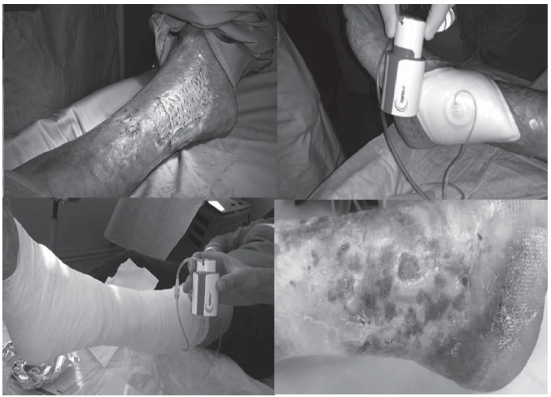
When the wounds appeared free from fibrin and when granulation tissue was visible, the patients were admitted to the day hospital and all wounds were covered by partial thickness sking graft. Local anesthesia was performed and the wound was cleaned mechanically to remove all traces of any remaining fibrin. Skin graft was sutured to the wound (7, 8). At the end of surgical procedure, we applied paraffin gauze and absorbent gauze connected to NANOVA devices on the wound covered by skin graft. A compressive dressing with bandage was made including the tube of NANOVA. All devices were linked to the bandage with an adhesive plaster, so to be easily disconnected and checked by the patient and by physician in the following days.
Dressing change were conducted every two days in order to verify:
- the state of vitality of the graft
- the presence of maceration or infection
We used NANOVA device for fourteen days of therapy (7 change dressing)
After fourteen days of negative pressure therapy, we performed medications with siliconate polyurethane foam and compressive bandage on the unhealed wounds.
A questionnaire to evaluate the impact of these devices in quality of life was submitted during the therapy (4) (Table 1).)
Table 1.
Questionnaire on the quality of life with the use of negative pressure therapy
|
Results
NANOVA had allowed to drain effectively the secretions of venous ulcers, contributing to the engraftment of skin graft. Thanks to the combination of compression therapy and negative pressure therapy we have registered no graft necrosis in eight patient (Figure 1). In two patients we registered an incomplete engraftment, with half wound surface not healed. These two patients were medicated with polyurethane foam and compressive bandage respectively for 27 and 43 days.
Figure 1.
All patients had no limitation in walking and the compressive dressing improved clinical management since the device was portable and included in the bandage.
Analysis of questionnaire
The analysis of questionnaires showed a good impact of Nanova device during the daily activities. The device was included in the dressing and the patients had no difficulty to perform flights of stairs or walking in the days of therapy. All patients were able to perform moderate activities and were not limited in the dress of going to work.
Discussion
In this clinical study we observed the double benefit of treatment with NPWT carried out with ultraportable system. We tested Nanova system in secreting lesions with good results (9). The benefits of NPWT are known for some time: these devices allows the reduction of edema and exudate removing inflammatory mediators in the immediate post-operative. This improves local conditions for wound healing (10, 11). The use of negative pressure provides an intermittent or continuous subatmospheric pressure through by a pump connected to a reservoir that collects wound fluids. The maintenance of the negative pressure is achieved with the use of a specific dressing to form a closed, airtight environmental. First use of negative pressure drainage with occlusive dressing was in 1993 by Fleischmann (12). In later years this technique has been widely studied in vivo and in vitro. Negative pressure wound therapy (NPWT) provide a rapid wound healing by different mechanism such as contraction of the edges (13, 14), stimulating the angiogenesis (15), increase the formation of granulation tissue (16, 17). The use of NPWT has greatly improved the management and outcomes of critical wounds wounds such as diabetic ulcers, pressure ulcers, venous ulcers and traumatic wounds (13). Over the years, the NPWT devices have become increasingly sophisticated, small and portable. First devices were large and heavy, in need of electrical current network and compromised the mobility of the patient. Then smaller machines were later introduced (18). Currently there are devices with rechargeable batteries that guarantee a good autonomy (19). This feature has a considerable impact on the possibilities of treatment, allowing patients to return home after the dressing change (in outpatient room), increasing the compliance and reducing the coast of hospitalization (20).
Nanova is a mechanical device consisting of a pump that is actuated manually by pressing the top. It has no electrical components and the patient must be instructed on the correct operation: in case of loss of vacuum, the upper part tends to rise on the sides showing a yellow line (21, 22).
Due to the mechanical operation no alarms are present, therefore in case of loss of vacuum during the night, this is not reported. The dressing can absorb about 45 cc of fluid. The granulation tissue recruitment is higher as NANOVA as the reached value is of -125 mm Hg.
NANOVA system has the advantage of being ultraportable. The vacuum pump can be included in the bandage and the patient can immediately start to move without difficulty. The lack of a container significantly reduces the volumetric bulk of the device. The novelty of this ultraportable is to be included in the dressing. The ability to perform a compression bandage in venous ulcers is very important from a therapeutic point. We want to remember that the treatment of venous ulcers includes the correction of anomalous venous flow, compression therapy, physical activity (when possible) and local therapy of the ulcer.
The benefits of negative pressure therapy, were confirmed in this study. The device NANOVA has proven to be effective and well tolerated. Its small size allowed for a greater tolerance by patients with higher freedom of movement compared to other devices of the same type. With this study, we believe that the NANOVA device can greatly increase the therapeutic possibilities without limiting the mobility of patients
Conclusions
The use of ultraportable devices for negative pressure therapy allows an effective treatment of venous leg ulcers improving the local condition with suction of secretion and without sacrificing compression therapy, the walking of patients and with an acceptable impact in daily activities.
References
- 1.Brafa A, Campana M, Grimaldi L, et al. Management of gynecomastia: an outcome analysis in a multicentric study. Minerva chirurgica. 2011;66(5):375–84. [PubMed] [Google Scholar]
- 2.Cuomo R, Nisi G, Grimaldi L, Brandi C, Sisti A, D’Aniello C. Immunosuppression and abdominal wall defects: use of autologous dermis. In vivo. 2015;29(6):753–5. [PubMed] [Google Scholar]
- 3.Cuomo R, Russo F, Sisti A, et al. Abdominoplasty in mildly obese patients (BMI 30-35 kg/m2): metabolic, biochemical and complication analysis at one year. In vivo. 2015;29(6):757–61. [PubMed] [Google Scholar]
- 4.Cuomo R, Zerini I, Botteri G, Barberi L, Nisi G, D’Aniello C. Postsurgical pain related to breast implant: reduction with lipofilling procedure. In vivo. 2014;28(5):993–6. [PubMed] [Google Scholar]
- 5.Cuomo R, Sisti A, Grimaldi R, et al. Modified Arrow Flap Technique for Nipple Reconstruction. 2016;22(6):710–1. doi: 10.1111/tbj.12659. [DOI] [PubMed] [Google Scholar]
- 6.D’Aniello C, Cuomo R, Grimaldi L, et al. Superior Pedicle Mammoplasty without Parenchymal incisions after Massive Weight Loss. 2016:1–11. doi: 10.1080/08941939.2016.1240837. [DOI] [PubMed] [Google Scholar]
- 7.D’Antonio A, Cuomo R, Angrisani B, Memoli D, Angrisani P. Desmoplastic cellular neurothekeoma mimicking a desmoplastic melanocytic tumor. Journal of the American Academy of Dermatology. 2011;65(2):e57–8. doi: 10.1016/j.jaad.2010.12.029. [DOI] [PubMed] [Google Scholar]
- 8.Grimaldi L, Cuomo R, Brandi C, Botteri G, Nisi G, D’Aniello C. Octyl-2-cyanoacrylate adhesive for skin closure: eight years experience. In vivo. 2015;29(1):145–8. [PubMed] [Google Scholar]
- 9.Cuomo R, Addesso M, Altieri R, D’Antonio A. A bone fide atypical fibroxanthoma of penis. 2014;6(4):381–3. doi: 10.4103/0974-7796.141009. [DOI] [PMC free article] [PubMed] [Google Scholar]
- 10.Webb LX. New techniques in wound management: vacuum-assisted wound closure. The Journal of the American Academy of Orthopaedic Surgeons. 2002;10(5):303–11. doi: 10.5435/00124635-200209000-00002. [DOI] [PubMed] [Google Scholar]
- 11.Young SR, Hampton S, Martin R. Non-invasive assessment of negative pressure wound therapy using high frequency diagnostic ultrasound: oedema reduction and new tissue accumulation. International wound journal. 2013;10(4):383–8. doi: 10.1111/j.1742-481X.2012.00994.x. [DOI] [PMC free article] [PubMed] [Google Scholar]
- 12.Fleischmann W, Strecker W, Bombelli M, Kinzl L. Vacuum sealing as treatment of soft tissue damage in open fractures. Der Unfallchirurg. 1993;96(9):488–92. [PubMed] [Google Scholar]
- 13.Borgquist O, Gustafsson L, Ingemansson R, Malmsjo M. Micro- and macromechanical effects on the wound bed of negative pressure wound therapy using gauze and foam. Annals of plastic surgery. 2010;64(6):789–93. doi: 10.1097/SAP.0b013e3181ba578a. [DOI] [PubMed] [Google Scholar]
- 14.Malmsjo M, Ingemansson R, Martin R, Huddleston E. Negative-pressure wound therapy using gauze or open-cell polyurethane foam: similar early effects on pressure transduction and tissue contraction in an experimental porcine wound model. Wound repair and regeneration : official publication of the Wound Healing Society [and] the European Tissue Repair Society. 2009;17(2):200–5. doi: 10.1111/j.1524-475X.2009.00461.x. [DOI] [PubMed] [Google Scholar]
- 15.Chen SZ, Li J, Li XY, Xu LS. Effects of vacuum-assisted closure on wound microcirculation: an experimental study. Asian journal of surgery/Asian Surgical Association. 2005;28(3):211–7. doi: 10.1016/S1015-9584(09)60346-8. [DOI] [PubMed] [Google Scholar]
- 16.Hudson DA, Adams KG, Van Huyssteen A, Martin R, Huddleston EM. Simplified negative pressure wound therapy: clinical evaluation of an ultraportable, no-canister system. International wound journal. 2015;12(2):195201. doi: 10.1111/iwj.12080. [DOI] [PMC free article] [PubMed] [Google Scholar]
- 17.Malmsjo M, Huddleston E, Martin R. Biological effects of a disposable, canisterless negative pressure wound therapy system. Eplasty. 2014;14:e15. [PMC free article] [PubMed] [Google Scholar]
- 18.D’Antonio A, Addesso M, Memoli D, et al. Lymph nodebased disease and HHV-8/KSHV infection in HIV seronegative patinets: report of three new cases of a heterogeous group of diseases. 2011;93(3):795–801. doi: 10.1007/s12185-011-0849-0. [DOI] [PubMed] [Google Scholar]
- 19.Nisi G, Cuomo R, Brandi C, Grimaldi L, Sisti A, D’Aniello C. Carbon dioxide therapy and hyaluronic acid for cosmetic correction of the nasolabial folds. Journal of cosmetic dermatology. 2016;15(2):169–75. doi: 10.1111/jocd.12213. [DOI] [PubMed] [Google Scholar]
- 20.Nisi G, Barberi L, Ceccaccio L, et al. Effect of repeated subcutaneous injections of carbon dioxide (CO2) on inflammation linked to hypoxia in adipose tissue graft. European review for medical and pharmacological sciences. 2015;19(23):4501–6. [PubMed] [Google Scholar]
- 21.Nisi G, Grimaldi L, Brandi C, et al. Cutaneous metastasis of the superior lip from adenocarcinoma of the gastro-oesophageal junction. A case report. Chirurgia italiana. 2007;59(6):883–6. [PubMed] [Google Scholar]
- 22.Rosato L, Lazzeri D, Campana M, et al. Mesotherapy should not replace the surgical approach in the treatment of benign symmetric lipomatosis. Aesthetic plastic surgery. 2011;35(2):278–80. doi: 10.1007/s00266-010-9571-1. [DOI] [PubMed] [Google Scholar]


