Abstract
Runting-stunting syndrome (RSS) is one of the diseases associated with many detected viruses. In Brazil, there were reports of several enteric disease outbreaks in chickens in which avian nephritis virus (ANV) was detected; however, the role of ANV in the outbreaks and whether the virus was a causative agent of these cases of enteric diseases were not determined. The aim of this study was to isolate ANV in specific pathogen-free (SPF) chicken embryonated eggs (CEE) from the enteric contents of chickens showing signs of RSS. For this purpose, 22 samples of chicken enteric contents that were positive only for ANV were inoculated into 7 and 14-day-old SPF-CEE via the yolk sac route and incubated for 5 d, with a total of 3 passages. Virus isolation was confirmed by the presence of embryo injuries, detection of viral RNA by RT-PCR, and visualization of viral particles using electron microscopy. Therefore, the 7-day-old inoculated embryos showed dwarfism, gelatinous consistency, hemorrhage, and edema in the embryos, whereas the 14-day-old did not show any alteration. Viral RNA was detected in the embryos of both ages of inoculation, and the same viral particles were visualized. The embryos from the mock group showed no alteration and were negative for all the tests. The viral cDNA was sequenced, and the molecular and phylogenetic analyses showed that the Brazilian isolates are more related with the ANV-1 serotype group; the sequences of these isolates showed a high percentage of nucleotide (86.4 to 94.9%) and amino acid (92.3 to 98.7%) similarity with other sequences from China, Japan, Australia, and the United States that belong to this serotype previously classified group. In this study, we isolated 8 samples of ANV in SPF-CEE from enteric content samples from chickens with RSS. In doing so, we showed the pathological injuries to the embryo caused by the virus and the molecular characterization of a part of the ORF 1b gene of the virus.
Keywords: chicken, avian nephritis virus, viral isolation, chicken embryo, molecular characterization
INTRODUCTION
Enteric diseases are a big problem for animal health as well as in the poultry industry. Infected birds are affected from the first day of birth showing early clinical signs of enteric diseases, principally diarrhea; however, chickens of different ages are also affected, whether or not they are showing clinical signs (Pantin-Jackwood et al., 2007; Smyth et al., 2009; Todd et al., 2010; Mettifogo et al., 2014; Veen et al., 2017; Xue et al., 2017; De la Torre et al., 2018; Kang et al., 2018). In poultry, runting-stunting syndrome (RSS) is of great relevance to avian intestinal health, and many viruses are involved (Pantin-Jackwood et al., 2008; Day et al., 2015; Smyth, 2017). The syndrome is also known as infectious stunting syndrome, pale-bird syndrome, malabsorption syndrome, malassimilation, and helicopter disease (Goodwin et al., 1993; Kang et al., 2012, 2018; Bulbule et al., 2013; Day and Zsak, 2013; Nuñez and Ferreira, 2013; Smyth, 2017), and was first described by Olsen (1977). The syndrome is characterized in animals by dwarfism, poor development, diarrhea, and mortality (Kang et al., 2012, 2018; Day and Zsak, 2013; Zsak et al., 2013; Domaska-Blicharz et al., 2017). Chicken parvovirus (ChPV), avian rotavirus (ARTv-A), avian reovirus (AReo), chicken astrovirus (CAstV), and avian nephritis virus (ANV) have been detected in the enteric contents of chickens showing enteric diseases (Goodwin et al., 1993; Otto et al., 2006; Pantin-Jackwood et al., 2007, 2011; Nuñez and Ferreira, 2013; Mettifogo et al., 2014). ANV was reported and isolated for the first time by Yamaguchi et al. (1977) and is genetically related with the family Astroviridae, and classified as an astrovirus (Imada et al., 2000). ANV was associated with growth suppression, kidney lesions, and mortality in young chickens (Yamaguchi et al., 1977; Imada et al., 1979; Pantin-Jackwood et al., 2008). ANV was known to be reported in many countries around the world, including Japan, the United States, Australia, India, and South America, although mainly reported only in Brazil (Mettifogo et al., 2014). ANV has been associated with many symptoms beyond kidney injuries, with enteric problems appearing in naturally and experimentally infected animals. These animals experience apathy, weight loss, ruffled feathers, and mortality (Pantin-jackwood et al., 2008; Todd et al., 2010; Kang et al., 2012). ANV is a single-stranded RNA virus with a genome composed of 3 open reading frames (ORFs). ORF 1a is located close to the 5΄ terminus of the genome and encodes the viral protease. ORF 1a is followed by ORF 1b that encodes an RNA polymerase (Imada et al., 2000). ORF 2 encodes the capsid protein precursors and is located after ORF 1b and before the 3΄ untranslated region (UTR) (Imada et al., 2000; Todd et al., 2011). ORF 1b is a very conserved gene in ANV genome and is widely used for molecular amplification for diagnosis of ANV (Day et al., 2007; Chu et al., 2008). This polyprotein together with ORF 1a is cleaved into the non-structural proteins required for transcription and viral replication (Marvin, 2017). The sequencing of ORF 1b gene is not used for astrovirus genotyping where the capsid gene (ORF2) is used (Todd et al., 2011; Smyth et al., 2017; Luna et al., 2016), but could show similarities or differences between sequences from different serotypes of ANV. Recently, the presence of ANV in chickens, layer hens, and breeders showing signs of enteric diseases was reported in Brazil (Nuñez and Ferreira, 2013; Mettifogo et al., 2014; Luna et al., 2016; De la Torre et al., 2018), and many outbreaks of enteric diseases have also been observed (unpublished data). The role of ANV in enteric disease in Brazilian chicken flocks is not well understood, and because there is a lack of viral isolates, it is difficult to study this virus.
The aim of the present work was to isolate ANV in CEEs and to characterize molecularly a part of ORF 1b gene of ANV from enteric content samples of chickens showing RSS.
MATERIAL AND METHODS
Samples
Enteric contents (280) were collected between 2008 and 2010 from broilers or broiler breeders from farms located in São Paulo, Santa Catarina, and Rio Grande do Sul. These animals showed signs of enteric diseases, such as apathy, feather ruffles, and diarrhea. Samples were sent to the avian pathology laboratory and submitted for molecular analyses and screening for enteric viruses, such as CAstV, ANV, infectious bronchitis virus (IBV), ARTv-A, AReo, ChPV, and fowl adenovirus group 1 (FAdV-1), as described previously by Mettifogo et al. (2014). From these samples, 22 were positive for ANV only (Table 1) and were used in this study for the tentative isolation of this virus.
Table 1.
Samples used in the present study, including their origin, geographical localization, chicken ages, clinical signs, and isolation in SPF-CEE.
| Designation of samples | Origin of sample | Age (d) | Localization Brazilian State | Chickens presenting clinical signs | Type of sample | Isolation of ANV in SPF-CEE of 2 ages | GenBank Accession Number | ||
|---|---|---|---|---|---|---|---|---|---|
| Diarrhea | Runting-Stunting | 7 d | 14 d | ||||||
| 162 | NI | 40 | NI | No | Yes | Feces | – | – | NR |
| 182 | NI | 27 | NI | NI | Yes | Feces | + | – | MF509885.1 |
| 243 | NI | 52 | SP | No | No | Feces | + | – | MF509884.1 |
| 336-3 | NI | NI | NI | Yes | Yes | Intestine | + | – | MF509883.1 |
| 336-8 | NI | NI | NI | Yes | Yes | Feces | – | – | NR |
| 354-6 | BC | 29 | SC | Yes | Yes | Intestine | – | – | NR |
| 358-1 | BC | 8 | SC | No | No | Intestine | – | – | NR |
| 358-2 | BC | 8 | SC | No | No | Intestine | – | – | NR |
| 358-8 | BC | 10 | SC | No | No | Intestine | – | – | NR |
| 358-9 | BC | 10 | SC | Yes | Yes | Intestine | + | – | MF509881.1 |
| 362-1 | BC | 3 | RS | No | Yes | Intestine | – | – | NR |
| 365-2 | LH | 53 | SP | Yes | No | Feces | – | – | NR |
| 365-5 int | LH | 87 | SP | No | No | Feces | – | – | NR |
| 369-G | NI | NI | NI | No | Yes | Feces | – | – | NR |
| 372-2 | BC | 28 | RS | No | Yes | Intestine | – | – | NR |
| 400-2 | BC | 7 | SP | Yes | Yes | Intestine | + | + | MF509882.1 |
| 400-3 | BC | 14 | SP | Yes | Yes | Intestine | – | – | NR |
| 400-5 | BC | 21 | SP | Yes | Yes | Intestine | – | – | NR |
| 400-6 | BC | 21 | SP | Yes | Yes | Intestine | + | + | MF509886.1 |
| 400-7 | BC | 28 | SP | Yes | Yes | Intestine | – | – | NR |
| 400-8 | BC | 28 | SP | Yes | Yes | Intestine | – | – | NR |
| 401-4 | BC | 21 | SP | Yes | Yes | Intestine | – | – | NR |
SP: São Paulo; SC: Santa Catarina; RS: Rio Grande do Sul; BC = Broiler Chickens; NI = not informed; LH = Layer Hens; – = negative; + = positive.
Viral Isolation
Preparation of Inoculum for Isolation of ANV in SPF-CEE
A 1:1 suspension of samples positive only for ANV and 0.1 M pH 7.4 phosphate-buffered solution (PBS) was made, and 750 μL of sample and 750 μL of PBS were added to a 1.5-mL sterile microfuge, homogenized, and centrifuged at 12,000 g for 30 min to 4°C. The supernatant was collected and filtered with a 0.22-μM filter, and 0.2 mL of filtered inoculum of each 22 samples was inoculated simultaneously into 3 specific pathogen-free chicken embryonated egg (SPF-CEE) via the yolk sac route of 7- and 14-day-old White Leghorn chicken. The same number of SPF-CEE of the same ages was inoculated with PBS using the same conditions as the viral samples to generate a mock group. The sealed inoculated eggs were maintained for 5 d in an automated hatchery that provides both stable temperature (37.5 to 38°C) and humidity (40 to 55%). Embryo livability was checked every 24 h after inoculation, and the dead embryos were discarded in the first 24 h. Afterward, the eggs that died were maintained at 4°C, and at the fifth day post-infection, a necropsy of the live and dead embryos was performed. Three passages of each sample were performed. After each necropsy, a 1:1 suspension of the macerated embryo and PBS was made. The suspension was homogenized and centrifuged for 30 min at 12 000 × g and 4°C. The supernatant was then filtered and used as an inoculum, which was inoculated in the same quantity using the same route of inoculation as described above in the subsequent passages. The SPF-CEE were kindly donated by CEVA Animal Health, Brazil.
Viral Confirmation
Post-mortem Examination
At the fifth day after inoculation, the SPF-CEE were subjected to necropsies after natural death or euthanasia. The embryos and the egg membrane supports were subjected to post-mortem examination for pathological alterations present in the embryos, such as dwarfism, gelatinous consistency, edema, or hemorrhaging. The coelomic cavity was also opened, and the organs were analyzed to find any pathological alterations.
Molecular Detection of ANV Nucleic Acid Extraction
The extraction of the nucleic acids was performed using 250 μL of the supernatant from the SPF-CEE suspension described above at each passage using TRIzol (Invitrogen, Life Technologies, Carlsbad, CA) following the manufacturer's instructions.
RT-PCR for Amplification of the UTR Gene of ANV
After each passage, all samples were submitted for detection of the ANV viral RNA by the RT-PCR assay. RT-PCR was performed according to the procedure described by Todd et al. (2010), with many modifications. The reverse transcription was done using 1 μg of extracted RNA in a mixed reaction of 20 μL containing 1X buffer, 0.1 M DTT, and 0.5 μM of each primer (Table 2), 10 mM of each dNTP, 200 U M-MLV enzyme (Invitrogen by Life Technologies), and ultrapure DNase- and RNase-free distilled water (Invitrogen by Life Technologies). The reaction was performed at 37°C for 50 min followed by 70°C for 15 min. The cDNA obtained was used in a PCR reaction, using 23 μL of 1x buffer, 0.5 μM of each primer (Table 2), 37.5 mM MgCl2, 1 mM of each DNTP, ultrapure DNase- and RNase-free distilled water (Invitrogen by Life Technologies), and 2 μL of cDNA. PCR was run at 94°C for 4 min followed by 40 cycles of 95°C for 1 min, 58°C for 1 min, and 72°C for 30 s and a final step of 72°C for 10 min.
Table 2.
Virus, gene, amplicon, and primer sequences used in the present study.
| Virus | Gene | Amplicon | Primers | Sequence | Reference |
|---|---|---|---|---|---|
| Avian Nephritis Virus | ORF 1 | 473 bp | ANV Pol 1F | 5΄-GYTGGGCGCYTCYTTTGAYAC-3΄ | Day et al., 2007 |
| ANV Pol 1R | 5΄-CRTTTGCCCKRTARTCTTTRT-3΄ | ||||
| UTR | 182 bp | ANV F | 5΄-ACG GCG AGT ACC ATC GAG-3΄ | Todd et al., 2010 | |
| ANV R | 5΄-AAT GAA AAG CCC ACT TTC GG-3΄ | ||||
| Chicken Astrovirus | ORF 1b | 362 bp | Cas Pol 1F | 5΄-GAYCARCGAATGCGRAGRTTG-3΄ | Day et al., 2007 |
| Cas Pol 1R | 5΄-TCAGTGGAAGTGGGKARTCTAC-3΄ | ||||
| Chicken Parvovirus | NS | 561 bp | PV 1F | 5΄-TTCTAATAACGATATCACT-3΄ | Zsak et al., 2009 |
| PV 1R | 5΄-TTTGCGCTTGCGGTGAAGTCTGGCTCG-3΄ | ||||
| Infectious Bronchitis Virus | UTR | 179 bp | UTR 11 | 5΄-ATGTCTATCGCCAGGGAAATGTC-3΄ | Cavanagh et al., 2002 |
| UTR 41 | 5΄-GGGCGTCCAAGTGCTGTACCC-3΄ | ||||
| UTR 31 | 5΄-GCTCTAACTCTATACTAGCCTA-3΄ | ||||
| Avian Rotavirus | NSP4 | 630 bp | NSP4-F30 | 5΄-GTGCGGAAAGATGGAGAAC-3΄ | Day et al., 2007 |
| NSP4-R660 | 5΄-GTTGGGGTACCAGGGATTAA-3΄ | ||||
| Fowl Adenovirus-1 | HEXON | 897 bp | Hexon A | 5΄-CAARTTCAGRCAGACGGT-3΄ | Meulemans et al., 2001 |
| Hexon B | 5΄-TAGTGATGMCGSGACATCAT-3΄ | ||||
| Avian Reovirus | S4 | 1120 bp | S4-F13 | 5΄-GTGCGTGTTGGAGTTTCCCG-3΄ | Pantin-Jackwood et al., 2008 |
| S4-1133 | 5΄-TACGCCATCCTAGCTGGA-3΄ | ||||
| Newcastle Diseases virus | F | 324 bp | NEW S | 5΄-GGAGGATGTTGGCAGCATT-3΄ | Pang et al., 2002 |
| NEW AS |
bp = base pair.
Molecular Analysis
RT-PCR for Amplification of the Fragment of the OFR 1b Gene of ANV
The samples where ANV was detected in the assay described above were submitted for molecular analysis of the ORF 1b fragment ANV genes using an RT-PCR assay, and the cDNA was sequenced. The RT-PCR reaction was performed according to Day et al. (2007), with some modifications. The RT and PCR reactions were conducted in the same conditions as were described above except for the present reactions that used specific primers for the partial amplification of the ORF 1b gene (Table 2) and the annealing temperature was changed to 56°C.
Sequencing of the Fragment of the OFR 1b Gene of ANV
The amplified fragments corresponding to a part of ORF 1b gene of ANV were purified using the Illustra GFX PCR DNA and Gel Band Purification Kit (GE Healthcare Bio-Sciences, Piscataway) according to the manufacturer’s instructions. The obtained DNA was sequenced using BigDye Terminator v3.1 Cycle Sequencing Kit (Applied Biosystem, Thermo Fisher Scientific) according to the manufacturer's instructions (Invitrogen Life Technologies) in an ABI 3730 DNA Analyzer (Applied Biosystems, Thermo Fisher Scientific). The obtained electropherograms were edited using the software package CLC Main Work Blench 7.7.3. The sequences were aligned and analyzed with other similar sequences present in GenBank that corresponded to sequences from other parts of world using the software package CLUSTAL X 2.0.11. The molecular and phylogenetic analyses were done using the software package MEGA 7 (Kumar et al., 2016).
Differential Diagnosis
All embryo samples were screened for other enteric viruses: ChPV as described by Zsak et al. (2009); CAstV and ART-A as described by Day et al. (2007); AReo as described by Pantin-Jacwood et al. (2007); IBV as described by Cavanagh et al. (2002); and FAdV-1 according to Meulemans et al. (2001). These were also screened for Newcastle disease virus as described by Pang et al. (2002) in Table 2.
Negative Staining for Electron Microscopy
The supernatant obtained from the embryo suspension described above was clarified and stained as described previously by Nunez et al. (2015). The samples were examined by an electron microscope (Philips, Model EM 208, Amsterdam, Netherlands) to check for the presence of viral particles.
RESULTS
Isolation of ANV in SPF CCE
In this study, 22 samples were positive only for ANV and were inoculated in 7- and 14-day-old SPF-CEE using the yolk sac route. From these inoculated samples, we propagated 6 samples in 7-day-old SPF-CEE and 2 samples in 14-day-old SPF-CEE. ANV isolation was validated by the presence of pathological alteration in the embryo, ANV positivity based on RT-PCR, and visualization of viral particles using electron microscopy.
Post-mortem Examination of Embryos Infected with ANV
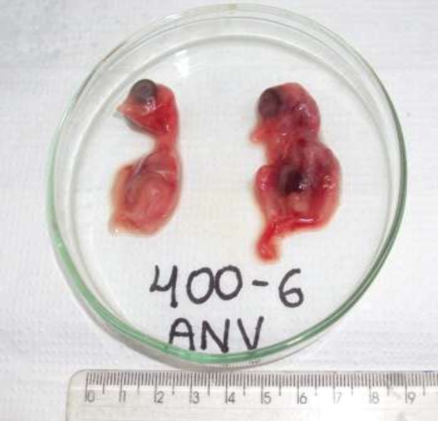
The SPF-CEE inoculated with 7-day-olds showed problems such as hemorrhage, dwarfism, edema, and gelatinous consistency in whole embryos body (Figure 1) features present on the embryo since second passage, at the first passage any injuries were observed, following the second passage were quantified 10% (1/9) mortality. The neck, wings, legs, and claws showed intense edema and hemorrhage. The embryo skin appears more transparent, and the internal organs were easily seen. The alive infected embryos showed lethargic movements and were poorly feathered. The death embryo showed the same pathological alteration found in the slaughtered embryos. In the 14-day-old inoculated SPF-CEE, no injuries were observed either on the body or in the internal organs, in any of the 3 passages. At this age of inoculation, no mortality was observed. The negative control 7- and 14-day-old embryos showed no signs of pathological alteration.
Figure 1.

Embryos from which ANV was isolated showing dwarfism, gelatinous consistency, edema, and hemorrhage.
Viral Confirmation
Molecular Detection of ANV
The molecular analyses used in the amplification of the UTR gene of ANV in the embryos inoculated with the ANV-positive samples showed an amplicon of approximately 182 bp. The tests detected 8 samples positive for ANV in the second and third passages. Every sample was positive in the first passage. From these samples, 6 were isolated in 7-day-old SPF-CEEs, and 2 samples were isolated in 14-day-old SPF-CEEs (Table 1). Samples 400-2 and 400-6 were positive in the embryos inoculated at both ages. The negative control was negative for the analyzed virus in all 3 passages.
Molecular Analyses
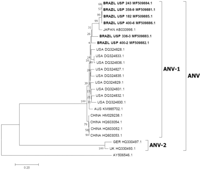
In the samples from which the UTR was amplified, part of the ORF 1b gene was also amplified and sequenced. The molecular analyses of the fragment were approximately 473 bp and were carried out using other sequences present in GenBank. The analysis of the sequences obtained here is reported to have 87.7 to 99.5% nucleotide (NT) similarity and 94.9 to 100% amino acid (AA) similarity between them. The sequences obtained in the present work were compared with the sequences from the ANV-1 serotype previously classified group and shared high similarity of NT and AA with sequences from Australia, Japan, China, and United States (Table 3). The sequences from the ANV-2 serotype previously classified group had 40.2 to 41.8% NT similarity and 10.9 to 11.5% AA similarity with the sequence from Germany and 40.8 to 41.6% NT similarity and 10.9 to 12.1% AA similarity with the sequence from the United Kingdom (Table 3). The molecular analyses showed that the samples isolated from SPF-CEEs have more similarity with sequences of the ANV-1 serotype group, suggesting that the isolates share high similarity to this group. The phylogenetic tree showed 2 well-defined groups: one group (clade bootstrap value = 100%) that clustered the sequences previously serotyped as ANV-1 group and the second group (clade bootstrap value = 100%) that grouped the sequences previously serotyped as ANV-2. The sequences from the isolates obtained in this work were aligned with ANV-1 serotype group, clustered with the sequence from Japan. The sequences from China, the United States, and Australia were also grouped in the ANV-1 group, and the sequences from Germany and the United Kingdom were grouped in the ANV-2 serotype group (Figure 2). The present results show that the isolated ANV strains in this study share high similarity of NT and AA with the sequences of strain previously serotyped as ANV-1.
Table 3.
Comparison of the nucleotide and amino acid identities of the sequences of Brazilian ANV isolates with other sequences.
| N. | Sequence identification | Amino acids identity | |||||||||||||||||||||||
|---|---|---|---|---|---|---|---|---|---|---|---|---|---|---|---|---|---|---|---|---|---|---|---|---|---|
| ANV-1 | ANV-2 | ||||||||||||||||||||||||
| 1 | 2 | 3 | 4 | 5 | 6 | 7 | 8 | 9 | 10 | 11 | 12 | 13 | 14 | 15 | 16 | 17 | 18 | 19 | 20 | 21 | 22 | 23 | |||
| ANV-1 | 1 | BR USP 182 MF509885.1 | – | 100 | 94.9 | 100 | 94.9 | 100 | 95.5 | 96.8 | 92.9 | 95.5 | 94.2 | 94.9 | 94.9 | 93.6 | 92.9 | 92.3 | 94.9 | 94.2 | 94.2 | 93.6 | 94.2 | 11.5 | 10.9 |
| 2 | BR USP 243 MF509884.1 | 99.5 | – | 94.9 | 100 | 94.9 | 100 | 95.5 | 96.8 | 92.9 | 95.5 | 94.2 | 94.9 | 94.9 | 93.6 | 92.1 | 92.3 | 94.9 | 94.2 | 94.2 | 93.6 | 94.2 | 11.5 | 10.9 | |
| 3 | BR USP 336.3 MF509883.1 | 87.7 | 88.1 | – | 94.9 | 97.4 | 94.9 | 96.8 | 92.9 | 94.2 | 96.8 | 95.5 | 96.1 | 98.7 | 97.4 | 96.8 | 94.9 | 96.8 | 94.2 | 94.9 | 95.5 | 94.9 | 10.9 | 12.1 | |
| 4 | BR USP 358.9 MF509881.1 | 99.5 | 100 | 88.1 | – | 94.9 | 100 | 95.5 | 96.8 | 92.9 | 95.5 | 94.2 | 94.9 | 94.9 | 93.6 | 92.9 | 92.3 | 94.9 | 94.2 | 94.2 | 93.6 | 94.2 | 11.5 | 10.9 | |
| 5 | BR USP 400.2 MF509882.1 | 87.7 | 88.1 | 91.3 | 88.1 | – | 94.9 | 96.8 | 92.9 | 95.5 | 96.8 | 95.5 | 96.1 | 97.4 | 96.1 | 95.5 | 94.9 | 96.8 | 95.5 | 96.1 | 96.8 | 96.1 | 10.9 | 12.1 | |
| 6 | BR USP 400.6 MF509886.1 | 99.3 | 99.3 | 87.9 | 99.3 | 87.9 | – | 95.5 | 96.8 | 92.9 | 95.5 | 94.2 | 94.9 | 94.9 | 93.6 | 92.9 | 92.3 | 94.9 | 94.2 | 94.2 | 93.6 | 94.2 | 11.5 | 10.9 | |
| 7 | AUS KM985702-1 | 87.7 | 87.7 | 90.2 | 87.7 | 92.1 | 87.9 | – | 94.9 | 94.9 | 94 | 96.1 | 95.5 | 98 | 96.8 | 96.1 | 95.5 | 98 | 95.5 | 96.1 | 96.8 | 96.1 | 10.9 | 12.1 | |
| 8 | JAP AB033998.1 | 94.5 | 94.9 | 86.8 | 94.9 | 87.9 | 94.7 | 87.7 | – | 92.3 | 94.9 | 94.9 | 94.2 | 94.2 | 94.2 | 92.3 | 91.7 | 94.2 | 93.6 | 93.6 | 92.9 | 93.6 | 11.5 | 12.1 | |
| 9 | USA DQ324831.1 | 86.4 | 86.4 | 90.2 | 86.4 | 91.7 | 86.2 | 92.1 | 87.3 | – | 97.4 | 96.1 | 94.2 | 95.5 | 94.2 | 93.6 | 94.9 | 96.1 | 94.2 | 94.2 | 93.6 | 94.2 | 10.9 | 12.8 | |
| 10 | USA DQ324832.1 | 87.3 | 87.7 | 89.2 | 87.7 | 91.9 | 87.5 | 92.6 | 88.1 | 94.5 | – | 98.7 | 96.8 | 98 | 96.8 | 96.1 | 96.8 | 98.7 | 96.8 | 96.8 | 96.1 | 96.8 | 10.9 | 12.8 | |
| 11 | USA DQ324829.1 | 87.1 | 87.1 | 90.2 | 87.1 | 92.1 | 87.3 | 92.6 | 87.1 | 93.6 | 94 | – | 95.5 | 96.8 | 95.5 | 95.5 | 95.5 | 97.4 | 95.5 | 95.5 | 94.9 | 95.5 | 11.5 | 13.4 | |
| 12 | USA DQ324830.1 | 86 | 86.4 | 89.8 | 86.4 | 91.7 | 86.2 | 90.9 | 87.1 | 92.3 | 93.6 | 93.6 | – | 97.4 | 96.1 | 95.5 | 93.6 | 95.5 | 94.9 | 94.9 | 95.5 | 94.9 | 10.9 | 12.8 | |
| 13 | USA DQ324833.1 | 87.7 | 87.7 | 90.9 | 87.7 | 91.7 | 87.5 | 91.7 | 87.3 | 93.8 | 92.8 | 93.4 | 92.8 | – | 98.7 | 98 | 96.1 | 98 | 95.5 | 96.1 | 96.8 | 96.1 | 10.9 | 12.8 | |
| 14 | USA DQ324836.1 | 87.5 | 87.5 | 90.2 | 87.5 | 91.1 | 87.3 | 91.3 | 87.1 | 93 | 92.3 | 93 | 92.3 | 98.9 | – | 96.8 | 94.9 | 96.8 | 94.2 | 94.9 | 95.5 | 94.9 | 10.3 | 12.8 | |
| 15 | USA DQ324828.1 | 87.5 | 87.5 | 91.9 | 87.5 | 92.1 | 87.7 | 91.7 | 87.9 | 92.6 | 92.3 | 92.8 | 92.6 | 94.5 | 93.4 | – | 94.2 | 96.1 | 93.6 | 94.2 | 94.9 | 94.2 | 10.9 | 12.8 | |
| 16 | USA DQ324827.1 | 87.3 | 87.3 | 90 | 87.3 | 91.7 | 87.1 | 91.5 | 87.5 | 93.6 | 93.8 | 94.2 | 91.5 | 93.8 | 93 | 93.2 | – | 96.8 | 94.2 | 94.9 | 94.2 | 94.9 | 10.9 | 12.8 | |
| 17 | USA DQ324835.1 | 89.4 | 89.4 | 89.9 | 89.4 | 92.3 | 89.2 | 93.4 | 88.5 | 92.8 | 94 | 93.4 | 91.1 | 93 | 92.3 | 91.9 | 94.2 | – | 96.8 | 97.4 | 96.8 | 97.4 | 10.9 | 12.8 | |
| 18 | CH HQ603052.1 | 87.7 | 87.7 | 90.2 | 87.7 | 91.1 | 87.5 | 90.9 | 87.3 | 91.7 | 91.7 | 92.8 | 90.2 | 91.7 | 91.1 | 91.1 | 93 | 92.3 | – | 99.3 | 98.7 | 99.3 | 11.5 | 13.4 | |
| 19 | CH HQ603053.1 | 87.9 | 87.9 | 90.4 | 87.9 | 91.3 | 87.7 | 91.1 | 87.5 | 91.9 | 91.9 | 93 | 90.4 | 91.9 | 91.3 | 91.3 | 93.2 | 92.6 | 99.7 | – | 99.3 | 100 | 11.5 | 13.4 | |
| 20 | CH HQ603054.1 | 87.5 | 87.5 | 90.4 | 87.5 | 91.3 | 87.3 | 91.1 | 87.1 | 91.5 | 91.5 | 92.8 | 90.6 | 92.3 | 91.7 | 91.7 | 92.8 | 92.1 | 99.3 | 99.5 | – | 99.3 | 11.5 | 13.4 | |
| 21 | CH HQ603038.1 | 87.1 | 87.5 | 90 | 87.5 | 90.9 | 86.8 | 90.4 | 87.1 | 91.9 | 92.1 | 92.8 | 91.1 | 91.9 | 91.5 | 91.3 | 92.3 | 92.1 | 98.9 | 99.1 | 99.1 | – | 11.5 | 13.4 | |
| ANV-2 | 22 | GER HQ330497.1 | 41.8 | 41.4 | 40.2 | 41.4 | 40.2 | 41.2 | 40.4 | 40.6 | 41 | 40.2 | 42 | 40.2 | 41.6 | 41.2 | 41.2 | 41.8 | 40.2 | 42 | 42.2 | 42.2 | 42.2 | – | 69.5 |
| 23 | GER HQ330493.1 | 41.6 | 41.6 | 40.8 | 41.6 | 41.6 | 41.4 | 41 | 40.6 | 41.6 | 40.8 | 42.8 | 40.6 | 42.2 | 41.8 | 42.2 | 42.8 | 41.4 | 41.8 | 42 | 42 | 42 | 92 | – | |
| Nucleotides identity | |||||||||||||||||||||||||
BR: Brazil; AUS: Australia; JAP: Japan; USA: United States of America; CH: China; GER: Germany
Figure 2.
The phylogenetic tree was inferred using MEGA7 software on the alignments of the partial ORF 1b sequences of ANV using a neighbor-joining phylogeny method joined with the maximum composite likelihood model with 1000 bootstraps of replication. The tree showed the phylogenetic relationships of the Brazilian ANV isolates in SPF CEEs with other sequences present in GenBank. The numbers along the branches show the bootstrap value for every 1000 replicates. The scale bar represents the number of substitutions per site. The GenBank accession numbers of the sequences used here are shown in the tree. The Goose Parvovirus sequence (AY506546.1) was used as an outgroup.
Differential Diagnosis
All SPF-CEEs inoculated in this study in the 3 passages were negative for ChPV, CAstV, FAdV-1, ARTv-A, AReo, IBV, and Newcastle disease virus by PCR or RT-PCR.
Electron Microscopy
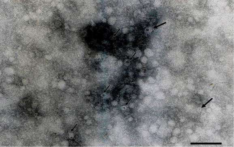
The electron microscopic images of the negatively stained samples from which ANV was isolated show viral particles in the supernatant (Figure 3).
Figure 3.
Transmission electron micrograph showing non-enveloped, icosahedral particles of ANV in chicken embryo supernatant suspension, showing a star relief (arrows), measuring on average 30 nm in diameter. Micrographs of strain 400-6 isolated in 7-day-old SPF CEE. Bar = 280 nm.
DISCUSSION
This paper describes the isolation of ANV in SPF-CEE from chicken enteric content samples with signs of RSS in Brazilian chicken flocks. These samples showed pathological features caused by the virus in the embryo. The studied virus isolated in 7- and 14-day-old SPF-CEE confirmed to be infected based on the presence of pathological alterations in the embryos, detection of viral RNA, and visualization of viral particles. Viral isolation in SPF-CEE is an old technique that has been used for several years to propagate and isolate viral agents to permit the study of their pathogenicity and virus behavior or to make vaccines oriented to protect the chickens against diseases (Guy, 2008; Alemnesh et al., 2012; Nuñez et al., 2015). Several viruses have been isolated in SPF-CEE either using primary cultures cells from many organs or using cell lines, where some viruses grow better than in eggs or primary cultures (McNulty et al., 1979; Baxendale and Mebatsion, 2004). Astrovirus of chicken origin, CAstV, and ANV grew well in the Leghorn Male Hepatoma (LMH) cell line, showing cytopathic effect with plaque formation after 6 d of inoculation (Baxendale and Mebatsion, 2004). ANV was cultivated and propagated in 11-day-old SPF-CEE onto the chorioallantoic membrane (Imada et al., 1979), and CAstV was isolated in 7- and 14-day-old SPF-CEE using the yolk sac route (Nuñez et al., 2015). The isolation of ANV in SPF-CEE using the yolk sac route is another alternative for replication as described in this study. These results are consistent with previous studies in which ANV was isolated using 7-day-old eggs (Imada et al., 1979), but isolation using 14-day-old eggs is not described.
ANV is a virus that is widely studied. It has been present in poultry since 1976; it was described for the first time by Yamaguchi et al. (1977) and characterized as a picornavirus based on its physicochemical and morphological properties. It was characterized as an astrovirus using genetic characterization and analysis of its complete genome, which classified it as a new avian astrovirus (Imada et al., 2000). Actually, ANV has been reported around the world and has been detected in many countries and on all continents. In recent years, ANV was detected in Brazil in chickens and turkeys showing signs of enteric diseases, principally signs of RSS (Hewson et al., 2010; Mettifogo et al., 2014; Moura-Alvarez et al., 2014; Luna et al., 2016; De la Torre et al., 2018). The implication or relation of this virus in RSS pathogeneses is not well clear. Here, ANV was isolated from enteric content samples from chickens showing RSS in the SPF-CEE, results that would help in understanding the behavior of the virus.
Isolation of viruses from the enteric contents is always a challenge due to the presence of several viruses in the same sample, which makes it difficult to find a sample where a unique virus is present. This presents a bigger challenge in the case of viral isolation from RSS because several viruses are involved with this syndrome and these viruses are shed through the feces (Day et al., 2007; Pantin-Jackwood et al., 2007; Oluwayelu et al., 2012; Day and Zsak, 2013; Bennett and Gunson, 2017; Veen et al., 2017; Kang et al., 2018). In this study, the isolation of ANV was possible from the starting enteric content samples where the 7-day-old embryos show pathological alterations, such as hemorrhage, edema, and dwarfism when inoculated. Other studies when CAstV was isolated from the enteric contents also reported the presence of embryos and with alterations, such as hemorrhage and dwarfism (Bulbule et al., 2013; Nuñez et al., 2015), but mortality was not reported in these studies. ANV, as the name indicates, is a virus that is commonly associated with nephritis and injuries in the kidneys. The ANV isolated here in 14-day-old SPF-CEE did not show any macroscopic alteration of the kidneys, neither in the other organs of the embryo. Due to a possible presence of a low viral concentration, only 3 passages were carried out in this study, and the virus present in the kidneys was not quantified as well as no histopathological analyses were carried out to determine microscopical alteration in the kidneys.
ANV inoculated in 1-day-old SPF chickens produced dwarfism, enteric alterations, diarrhea, and a mild yellowish-tan discoloration in kidneys. From day 3 to 21, the virus was also detected in the bursa, jejunum, and spleen, but no alterations were found (Imada et al., 1979, 1981; Shirai et al., 1991), showing that the target organ of this virus is kidneys, but it could be present in other organs, such as gut. Actually, ANV is related to one of the principal causative agents of enteric problems, more specifically those related with RSS (Kang et al., 2012; Day and Zsak, 2013; Mettifogo et al., 2014; Day et al., 2015; Smyth, 2017; De la Torre et al., 2018). The samples used here were from chickens showing signs of enteric diseases that were positive only for ANV after molecular screening, suggesting that the principal agent is involved in the genesis of some enteric problems may be ANV. During the last decade, several outbreaks of RSS were reported in commercial chicken flocks in several states of Brazil (unpublished data), but the presence of ANV in Brazilian poultry was reported previously in chickens (Mettifogo et al., 2014) showing that the virus is present in the 45% of samples analyzed alone or in combination with other viruses. ANV was also reported in 56% of turkeys (Moura-Alvarez et al., 2013). These results showed the importance of ANV in the enteric problems, and how important is the study and isolation of the ANV that is circulating in Brazilian chicken flocks as it would help in the understanding of the virus behavior and would bring more information about its pathogenicity.
Many viruses were identified and related to RSS, such as ChPV, avian rotavirus, avian reovirus, CAstV, FAdV-1, as well as ANV principally related with renal infections. However, their role in enteric diseases is not well understood; however, chickens inoculated orally with ANV produce dwarfism and diarrhea (Kang et al., 2012; Day and Zsak, 2013; Smyth, 2017). The study of enteric diseases or, more specifically, RSS is a big challenge because some viral agents are involved in their pathogenesis. The application of the next generation nucleic acid sequencing and metagenomics studies of the viral population in the intestinal contents of chickens with RSS shows the presence of other viruses beyond the commonly reported virus, such as picobirnaviruses and caliciviruses (Day and Zsak, 2013; Day et al., 2015; Smyth, 2017). This shows the viral diversity and possible viral combinations that could be in the chicken gut, which complicates the study of enteric diseases and syndromes. However, there are studies showing that when a virus is inoculated alone, it produces RSS (Kang et al., 2012, 2018; Zsak et al., 2013). ANV inoculated alone could cause enteric problems (Kang et al., 2012) and could be considered a causative agent of enteric disease, principally a causative agent of RSS. As mentioned before, the strains of ANV isolated in the present work were from chickens with signs of RSS. For these reasons, the implication of ANV in Brazilian chicken flocks as a causative agent of RSS is something that needs to be investigated to determine if this isolate could cause RSS.
The genome of ANV is approximately 7 kb in length. It has 3 ORFs—ORF1a, ORF1b, and ORF2—and an UTR; the polymerase gene ORF1b is a common gene used for the detection and amplification of ANV (Day et al., 2007), as well as the UTR gene (Todd et al., 2010). Today, many molecular assays for the identification of ANV have been developed and used as diagnostic tools (Day et al., 2007; Smyth et al., 2010; Todd et al., 2010). In the present investigation, 2 assays were used to determine the presence of ANV in the SPF-CEE. Moreover, they provide information about the virus genome and virus epidemiology. Therefore, before the emergence of molecular techniques, other assays, such as electron microscopy, immunofluorescence, and ELISA, were also used for ANV detection (Imada et al., 1981; Decaesstecker and Meulemans, 1991; Woolcock and Shivaprasad, 2008). As was shown in this study, viral particles were observed in the supernatant from samples where ANV was isolated, confirming the presence of the virus in the embryo, but it was not determined which part of the embryo or which organ is more susceptible to ANV. For this reason, more studies are needed to determine where the virus replicates better and determine which organ is the target in the SPF-CEE.
The phylogenetic analysis of a part of ORF1b gene of the isolates of ANV in this study was clustered with other sequences belonging to ANV-1 serotype previously classified, and showed more NT and AA similarity with sequences from Japan (Table 3), which suggests the presence of serotype 1 (Shirai et al., 1991; Imada et al., 2000; Todd et al., 2011). Actually, 2 serological groups of ANV, ANV-1 and ANV-2, are recognized. However, molecular studies involving an ORF2 gene that encodes a structural capsid protein showed the presence of 6 genetic like groups, ANV-1 to ANV-6 (Todd et al., 2011). Officially, there are 3 different astrovirus species that comprises the Avastrovirus genus (International Committee on Taxonomy of Viruses-ICTV) and the genotypic classification by Astroviridae Study Group proposed 7 avastrovirus genotypes (AAstV-1 to AAstV-7) based on ORF 2 analyses. ANV belongings principally to AAstV-2 and other ANV strains have been detected and isolated from wild and domestic pigeons and were classified in the genotypes 5, 6, and 7. Recently, 2 novel genotypes 8 and 9 were proposed (Luna et al., 2016). This genetic characterization shows the diversity of this virus and how the strains are distributed around the world. The results presented here suggest that the ANV isolates in SPF-CEE are more related with the sequences of ANV-1 serotype previously classified. However, more studies need to be carried out to characterize the viral capsid (ORF 2 gene) of Brazilian´s isolates shown in the present investigation. The study of the capsid protein genes is important for the understanding of the Astroviridae family, but the difficulty in isolating and propagating astroviruses has limited their molecular characterization (Pantin-Jackwood et al., 2011). In the present work, the difficulty of isolation resulted in only 8 samples from 22 where the isolation of ANV was possible. This lack of viral isolation, as showed here only 3 passages were carried out, is due to many factors such as the low viral concentration in the samples producing blind passages until third passage, viral inactivation is another condition due to management of samples transported to laboratory under weak conditions of temperature, storage conditions before thawing, and freezing the sample before testing, or inoculum preparation in which viral particles are damaged and preclude the replication and propagation. Moreover, more number of samples isolated in eggs of 7 d of age (6 samples) than in eggs of 14 days of age where only 2 samples were shown. This condition could be explained by the fact that in eggs with 7 days of incubation, the yolk is easier to access, but not so in eggs with more days of incubation where the size of the embryo makes it more difficult to reach the yolk and deposit the inoculum.
The viral particles of astroviruses were characterized by the star-like surface projection observed on their virions using electron microscopy with negative staining. These are important features that are used for virus identification (Woolcock and Shivaprasad, 2008), depending on the availability of the origin material, which allows better viral visualization than other materials. In this study, viral particles with astrovirus features were observed by EM using negative stain, confirming the presence of virus in the embryos infected with ANV. These results are in concordance with previously published works where viral particles were visualized from birds with or without enteric diseases. To our knowledge, this is the first report of ANV isolation in SPF-CEE from chickens showing signs of RSS in Brazil.
This study has shown ANV isolation in 7 and 14day-old SPF-CEE from the enteric contents of chickens showing RSS, and the molecular analyses suggest that these isolates have high similarity with the sequences from ANV-1 serotype previously classified; however, more studies need to be carried out to classify molecularly the isolates based on the ORF 2 gene sequencing and determine to which genotype of Avian astrovirus belong to, determine the pathogenicity of these isolates, and determined if the viruses could be the causative agent of RSS or could be involved in the development of renal diseases.
Acknowledgements
The authors would like to thank the poultry companies in Brazil that generously sent the samples for the development of this study and for the diagnosis of enteric viruses.
Notes
This work was supported by grants from the FAPESP (Fundação de Amparo à Pesquisa do Estado de São Paulo) Grants #2013/08560-5 and 2015/09348-5, and CNPq (Conselho Nacional de Desenvolvimento Cientifico e Tecnológico) Grants #453920/2014-4 and 140744/2014-2 for financial support. A. J. Piantino Ferreira is the recipient of a CNPq fellowship.
REFERENCES
- Alemnesh W., Hair-Bejo M., Aini I., Omar A. R.. 2012. Pathogenicity of fowl adenovirus in specific pathogen free chicken embryos. J. Comp. Pathol. 146:223–229. [DOI] [PubMed] [Google Scholar]
- Baxendale W., Mebatsion T.. 2004. The isolation and characterisation of astroviruses from chickens. Avian Pathol. 33:364–370. [DOI] [PubMed] [Google Scholar]
- Bennett S., Gunson R. N.. 2017. The development of a multiplex real-time RT-PCR for the detection of adenovirus, astrovirus, rotavirus and sapovirus from stool samples. J. Virol. Methods 242:30–34. [DOI] [PMC free article] [PubMed] [Google Scholar]
- Bulbule N. R., Mandakhalikar K. D., Kapgate S. S., Deshmukh V. V., Schat K. A., Chawak M. M.. 2013. Role of chicken astrovirus as a causative agent of gout in commercial broilers in India. Avian Pathol. 42:464–473. [DOI] [PubMed] [Google Scholar]
- Cavanagh D., Mawditt K., Welchman D. D. B., Britton P., Gough R. E.. 2002. Coronaviruses from pheasants (Phasianus colchicus) are genetically closely related to coronaviruses of domestic fowl (infectious bronchitis virus) and turkeys. Avian Pathol. 31:81–93. [DOI] [PubMed] [Google Scholar]
- Chu D. K. W., Poon L. L. M., Guan Y., Peiris J. S. M.. 2008. Novel astroviruses in insectivorous bats. J. Virol. 82:9107–9114. [DOI] [PMC free article] [PubMed] [Google Scholar]
- Day J. M., Oakley B. B., Seal B. S., Zsak L.. 2015. Comparative analysis of the intestinal bacterial and RNA viral communities from sentinel birds placed on selected broiler chicken farms. PLoS One 10:1–15. [DOI] [PMC free article] [PubMed] [Google Scholar]
- Day J. M., Spackman E., Pantin-Jackwood M.. 2007. A multiplex RT-PCR test for the differential identification of turkey astrovirus type 1, turkey astrovirus type 2, chicken astrovirus, avian nephritis virus, and avian rotavirus. Avian Dis. 51:681–684. [DOI] [PubMed] [Google Scholar]
- Day J. M., Zsak L.. 2013. Recent Progress in the characterization of avian enteric viruses. Avian Dis. 57:573–580. [DOI] [PubMed] [Google Scholar]
- Decaesstecker M., Meulemans G.. 1991. An ELISA for the detection of antibodies to avian nephritis virus and related entero-like viruses. Avian Pathol. 20:523–530. [DOI] [PubMed] [Google Scholar]
- De la Torre D., Nuñez L., Aastolfi-Ferreira C., Ferreira A. J. P.. 2018. Enteric virus diversity examined by molecular methods in Brazilian poultry flocks. Vet. Sci. 5:1–20. [DOI] [PMC free article] [PubMed] [Google Scholar]
- Domaska-Blicharz K., Bocian L., Lisowska A., Jacukowicz A., Pikula A., Minta Z.. 2017. Cross-sectional survey of selected enteric viruses in Polish turkey flocks between 2008 and 2011. BMC Vet. Res. 13:1–10 [DOI] [PMC free article] [PubMed] [Google Scholar]
- Goodwin M. A, Davis J. F., McNulty M. S., Brown J., Player E. C.. 1993. Enteritis (so-called runting stunting syndrome) in Georgia broiler chicks. Avian Dis. 37:451–458. [PubMed] [Google Scholar]
- Guy J. S. 2008. Isolation and propagation of coronaviruses in embryonated eggs. Methods Mol. Biol. 454:109–117. [DOI] [PMC free article] [PubMed] [Google Scholar]
- Hewson K. A., O’rourke D., Noormohammadi A. H.. 2010. Detection of avian nephritis virus in Australian chicken flocks. Avian Dis. 54:990–993. [DOI] [PubMed] [Google Scholar]
- Imada A. T., Taniguchi T., Yamaguchi S., Minetoma T., Maeda M., Kawamura H., Imada T.. 1981. Susceptibility of chickens to avian nephritis virus at various inoculation routes and ages. Avian Dis. 25:294–302. [PubMed] [Google Scholar]
- Imada T., Yamaguchi S., Kawamura H.. 1979. Pathogenicity for baby chicks of the G-4260 strain of the picornavirus “avian nephritis virus”. Avian Dis. 23:582–588. [PubMed] [Google Scholar]
- Imada T., Yamaguchi S., Mase M., Tsukamoto K., Kubo M., Morooka A.. 2000. Avian nephritis virus (ANV) as a new member of the family Astroviridae and construction of infectious ANV cDNA. J. Virol. 74:8487–8493. [DOI] [PMC free article] [PubMed] [Google Scholar]
- Kang K. I., El-Gazzar M., Sellers H. S., Dorea F., Susan M., Kim T., Collett S., Mundt E.. 2012. Investigation into the aetiology of runting and stunting syndrome in chickens. Avian Pathol. 41:41–50. [DOI] [PubMed] [Google Scholar]
- Kang K., Linnemann E., Icard A. H., Durairaj V., Mundt E., Sellers H. S.. 2018. Chicken astrovirus as an aetiological agent of runting-stunting syndrome in broiler chickens. J. Gen. Virol. 99:512–524. [DOI] [PubMed] [Google Scholar]
- Kumar S., Stecher G., Tamura K.. 2016. MEGA7: Molecular evolutionary genetics analysis version 7.0 for bigger datasets. Mol. Biol. Evol. 33:1870–1874. [DOI] [PMC free article] [PubMed] [Google Scholar]
- Luna L. E., Beserra L. A. R., Soares R. M., Gregori F.. 2016. Avian nephritis virus (ANV) on Brazilian chickens farms: circulating genotypes and intra-genotypic diversity. Arch. Virol. 161:3455–3462. [DOI] [PubMed] [Google Scholar]
- Marvin S. 2017. The immune response to astrovirus infection. Viruses 9:1–11. [DOI] [PMC free article] [PubMed] [Google Scholar]
- McNulty M. S., Allan G. M., Todd D., McFerran J. B.. 1979. Isolation and cell culture propagation of rotaviruses from turkeys and chickens. Arch. Virol. 61:13–21. [DOI] [PubMed] [Google Scholar]
- Mettifogo E., Nuñez L. F. N., Chacón J. L., Santader-Parra S. H., Astolfi-Ferreira C. S., Jerez J. A., Jones R. C., Ferreira A. J. P.. 2014. Emergence of enteric viruses in production chickens is a concern for avian health. Sci. World J. 2014:1–8. [DOI] [PMC free article] [PubMed] [Google Scholar]
- Meulemans G., Boschmans M., Berg V. D., Decaesstecker M.. 2001. Polymerase chain reaction combined with restriction enzyme analysis for detection and differentiation of fowl adenoviruses. Avian Pathol. 30:655–660. [DOI] [PubMed] [Google Scholar]
- Moura-Alvarez J., Chacon J. V., Scanavini L. S., Nuñez L. F. N., Astolfi-Ferreira C. S., Jones R. C., Piantino Ferreira A. J.. 2013. Enteric viruses in Brazilian turkey flocks: Single and multiple virus infection frequency according to age and clinical signs of intestinal disease. Poult. Sci. 92:945–955. [DOI] [PMC free article] [PubMed] [Google Scholar]
- Moura-Alvarez J., Nuñez L. F. N., Astolfi-Ferreira C. S., Knöbl T., Chacón J. L., Moreno A. M., Jones R. C., Ferreira A. J. P.. 2014. Detection of enteric pathogens in Turkey flocks affected with severe enteritis, in Brazil. Trop. Anim. Health Prod. 46:1051–1058. [DOI] [PMC free article] [PubMed] [Google Scholar]
- Nuñez L. F. N., Parra S. H. S., Mettifogo E., Catroxo M. H. B., Astolfi-ferreira C. S., Ferreira A. J. P.. 2015. Isolation of chicken astrovirus from specific pathogen-free chicken embryonated eggs. Poult. Sci. 94:947–954. [DOI] [PubMed] [Google Scholar]
- Nuñez L. F. N., Piantino Ferreira A. J.. 2013. Viral agents related to enteric disease in commercial chicken flocks, with special reference to Latin America. Worlds Poult. Sci. J. 69:853–864. [Google Scholar]
- Olsen D. E. 1977. Isolation of a reovirus-like agent from broiler chicks with diarrhea and stunting. Pages 131–139. XXVIth Western Poultry Diseases Conference, Sacramento, CA, USA. [Google Scholar]
- Oluwayelu D. O., Smyth V., Todd D.. 2012. Detection of avian nephritis virus and chicken astrovirus in Nigerian indigenous chickens. Afr. J. Biotechnol. 11:3949–3957. [Google Scholar]
- Otto P., Liebler-Tenorio E. M., Elschner M., Reetz J., Löhren U., Diller R.. 2006. Detection of rotaviruses and intestinal lesions in broiler chicks from flocks with runting and stunting syndrome (RSS). Avian Dis. 50:411–418. [DOI] [PubMed] [Google Scholar]
- Pang Y., Wang H., Girshick T., Xie Z., Khan M. I.. 2002. Development and application of a multiplex polymerase chain reaction for avian respiratory agents. Avian Dis. 46:691–699. [DOI] [PubMed] [Google Scholar]
- Pantin-Jackwood M. J., Day J. M., Jackwood M. W., Spackman E.. 2008. Enteric viruses detected by molecular methods in commercial chicken and turkey flocks in the United States between 2005 and 2006. Avian Dis. 52:235–244. [DOI] [PubMed] [Google Scholar]
- Pantin-Jackwood M. J., Spackman A. C. E., Day A. J. M., Rives D.. 2007. Periodic monitoring of commercial turkeys for enteric viruses indicates continuous presence of astrovirus and rotavirus on the farms. Avian Dis. 51:674–680. [DOI] [PubMed] [Google Scholar]
- Pantin-Jackwood M. J., Strother K. O., Mundt E., Zsak L., Day J. M., Spackman E.. 2011. Molecular characterization of avian astroviruses. Arch. Virol. 156:235–244. [DOI] [PubMed] [Google Scholar]
- Shirai J., Nakamura K., Shinohara K., Kawamura H.. 1991. Pathogenicity and antigenicity of avian nephritis isolates. Avian Dis. 35:49–54. [PubMed] [Google Scholar]
- Smyth V. 2017. A review of the strain diversity and pathogenesis of chicken astrovirus. Viruses 9:29. [DOI] [PMC free article] [PubMed] [Google Scholar]
- Smyth V. J., Jewhurst H. L., Adair B. M., Todd D.. 2009. Detection of chicken astrovirus by reverse transcriptase-polymerase chain reaction. Avian Pathol. 38:293–299. [DOI] [PubMed] [Google Scholar]
- Smyth V. J., Jewhurst H. L., Wilkinson D. S., Adair B. M., Gordon A. W., Todd D.. 2010. Development and evaluation of real-time TaqMan® RT-PCR assays for the detection of avian nephritis virus and chicken astrovirus in chickens. Avian Pathol. 39:467–474. [DOI] [PubMed] [Google Scholar]
- Todd D., Trudgett J., Mcneilly F., Mcbride N., Donnelly B., Smyth V. J., Jewhurst H. L., Adair B. M.. 2010. Development and application of an RT-PCR test for detecting avian nephritis virus. Avian Pathol. 39:207–213. [DOI] [PubMed] [Google Scholar]
- Todd D., Trudgett J., Smyth V. J., Donnelly B., McBride N., Welsh M. D.. 2011. Capsid protein sequence diversity of avian nephritis virus. Avian Pathol. 40:249–259. [DOI] [PubMed] [Google Scholar]
- Veen T. C., De Bruijn N. D., Dijkman R., De Wit J. J.. 2017. Prevalence of histopathological intestinal lesions and enteric pathogens in Dutch commercial broilers with time. Avian Pathol. 46:95–105. [DOI] [PubMed] [Google Scholar]
- Woolcock P. R., Shivaprasad H. L.. 2008. Electron microscopic identification of viruses associated with poult enteritis in turkeys grown in California 1993–2003. Avian Dis. 52:209–213. [DOI] [PubMed] [Google Scholar]
- Xue J., Han T., Xu M., Zhao J., Zhang G.. 2017. The first serological investigation of Chicken astrovirus infection in China. Biologicals 47:22–24. [DOI] [PubMed] [Google Scholar]
- Yamaguchi A. S., Imada T., Kawamura H.. 1979. Characterization of a picornavirus isolated from broiler chicks. Avian Dis. 23:571–581. [PubMed] [Google Scholar]
- Zsak L., Cha R. M., Day J. M.. 2013. Chicken parvovirus–induced runting-stunting syndrome in young broilers. Avian Dis. 57:123–127. [DOI] [PubMed] [Google Scholar]
- Zsak L., Strother K. O., Day J. M.. 2009. Development of a polymerase chain reaction procedure for detection of chicken and turkey parvoviruses. Avian Dis. 53:83–88. [DOI] [PubMed] [Google Scholar]


