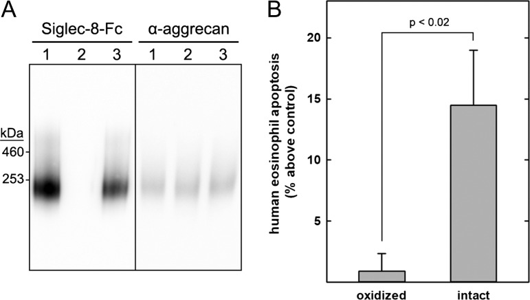
Fig. 10.
Purified human tracheal Siglec-8 ligand induces human eosinophil apoptosis. Siglec-8 ligands were extracted from human trachea and purified by size-exclusion chromatography followed by Siglec-8 affinity chromatography. A portion of purified S8-250K was treated with cold periodate to selectively oxidize the glycerol sidearm of its sialic acid followed by sodium borohydride reduction (negative control). An equal portion was incubated without periodate and treated with sodium borohydride (intact ligand). Oxidized and intact S8-250K were dialyzed against RPMI medium and added at equal concentrations to freshly isolated primary human eosinophils. After 24 h in culture, eosinophil apoptosis was quantified by flow cytometry. (A) Oxidized and intact S8-250 K were electrophoretically resolved on a composite agarose–acrylamide gel, blotted to PVDF membranes. Replicate blots were probed with Siglec-8-Fc to reveal Siglec-8 ligands or anti-aggrecan antibody (7D4). Lanes: (1) untreated S8-250K; (2) periodate oxidized and reduced S8-250K; (3) reduced S8-250K (intact ligand). (B) After isolation and overnight interleukin 5 (IL-5) priming, human eosinophils were incubated with equal portions of oxidized and intact S8-250K and apoptosis was assessed 18–24 h later. Results are expressed relative to untreated eosinophils, which had average apoptosis of 42 ± 5% (SEM). Data are displayed as mean and SEM of six replicates performed on three separate primary human eosinophil preparations.

