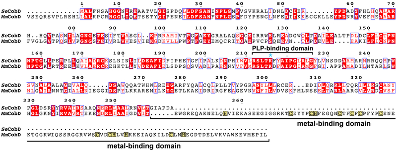
Figure 2.
Protein sequence alignment - Protein sequence alignment of CobD from S. enterica and M. mazei. Conserved residues are highlighted in red, and residues with similar properties are boxed in blue. The pyridoxal-5’-phosphate (PLP)-binding domain is bracketed and the active site lysine is marked with an asterisk. The cysteine-rich, putative metal-binding domain is indicated with brackets, and the cysteinyl and histidinyl residues in this region are boxed in yellow.

