



Abstract
Background:
Idiopathic pulmonary fibrosis (IPF) is an age-related and progressive interstitial lung disease. Up to 20% of cases of IPF cluster in families, genetic factors contribute significantly to the pathogenesis of the disease. This study aimed to explore the association between rare genetic variants and IPF in Chinese Han families.
Methods:
A Han family, comprising three IPF patients and five unaffected their first-degree relatives, and 100 ethnically matched control individuals from North China were enrolled in this study. Peripheral blood was collected, and genomic DNA was extracted. To elucidate if rare genetic variants are associated with the familial IPF, we performed whole-exome sequencing of affected members from a Chinese Han IPF family. Candidate rare variants were then confirmed by Sanger sequencing.
Results:
We identified a potentially damaging rare variant-a heterozygous mutation c.2146G>A in exon 6 of the gene encoding for telomerase reverse transcriptase (TERT), which results in an amino acid substitution (p.Ala716Thr). We confirmed the missense mutation by Sanger sequencing in all the affected family members but did not detect this mutation in 100 ethnically matched healthy controls. Patients carried this mutation were characterized by the frequently acute exacerbation of IPF phenotype, with poor prognosis. The mean time to death was 2.8 years after diagnosis.
Conclusion:
Using next-generation sequencing technology in familial IPF patients, we identified the heterozygous rare variant in TERT gene, and strengthened the importance of genetic variants in telomere-related pathogenesis in Chinese IPF patients.
Keywords: Genetics, Idiopathic Pulmonary Fibrosis, Telomerase Reverse Transcriptase, Telomere
摘要
背景:
特发性肺纤维化(IPF)是一种和年龄相关的进展性间质性肺疾病。多达20%的IPF患者呈现家族聚集性,遗传因素在IPF发病过程中起到重要作用。本研究的目的是探寻罕见遗传变异与中国汉族家族特发性肺纤维化的相关性。
方法:
收集了一个中国汉族IPF家系,包含3例IPF患者和5名非患病一级亲属。同时收集了100名与上述IPF患者性别、年龄相匹配的非患病的中国北方汉族人作为对照。所有受试者采集外周血提取DNA。为了研究罕见遗传变异是否与家族性IPF相关,我们对该家系的IPF患者进行了全外显子组测序。候选罕见变异位点采用Sanger测序证实。
结果:
我们在该家系发现了一个可能致病的罕见变异位点,该杂合变异c.2146G>A位于端粒逆转录酶基因(TERT)的第6外显子区,导致p.Ala716Thr错义突变。Sanger测序在家系中证实了该变异,在100名对照受试者中未检测到该变异。携带该突变的IPF患者表现为频繁急性加重表型,预后差,诊断后平均生存时间为2.8年。
结论:
通过对家族性IPF进行新一代测序技术分析,我们发现了与疾病相关的TERT基因的罕见变异,在中国IPF患者中进一步证实了遗传变异在端粒相关致病机制中的重要作用。
INTRODUCTION
Idiopathic pulmonary fibrosis (IPF) is an age-related and progressive interstitial lung disease, with a life expectancy of 3–5 years after diagnosis.[1] Accumulating evidence indicates that genetic factors, both common and rare variants, contribute significantly to the pathogenesis of IPF.[2] Genome-wide association studies have identified several common variants linked to IPF risk, for example, the most widely replicated variant rs35705950 located in the promoter region of the MUC5B gene.[3,4,5] Next generation sequencing (NGS) technologies have facilitated the identification of rare genetic variants. To date, rare genetic variants in multiple genes have been discovered in familial IPF, which mainly be subdivided into two categories as follows: genes related to surfactant protein metabolism and genes that maintain telomere length.[6] Germline defects in telomere maintenance are common in IPF, several genes in the telomere maintenance pathway have been implicated in IPF families, including those that affect telomerase catalytic activity (TERT, TERC), telomere biogenesis (DKC1, PARN, NAF1) or telomere end regulation (TINF2, RTEL1). Among them, rare coding variants in TERT gene which encodes the protein component of telomerase, are found up to 15% of familial IPF and show autosomal dominant transmission with age-dependent penetrance.[7,8] About 2–20% of cases of idiopathic interstitial pneumonia cluster in families,[9] leading to the potential to advance our understanding of the genetic basis of this disease; however, these mutations that have been implicated in pulmonary fibrosis account for only a small proportion of the population risk.
Recently, NGS studies have linked pathogenic rare variants in multiple new genes to the familial form of IPF. Here, we performed whole-exome sequencing (WES) on familial IPF patients, and first confirmed a rare missense variant in TERT among the Chinese Han population.
METHODS
Ethical approval
This study was approved by the Ethics Committee of the Beijing Chaoyang Hospital, Capital Medical University, China. All patients signed informed consent for genetic testing.
Subjects and specimens
A family, comprising three IPF patients and five unaffected their first-degree relatives, and 100 ethnically matched control individuals from North China were enrolled in this study. Clinical records of affected members were obtained, clinical evaluations including high-resolution computed tomography (HRCT) scan, and pulmonary function tests (PFT) were perform on the eight familial members. The diagnosis of IPF was established in accordance with the ATS/ERS/JRS/ALAT diagnostic criteria.[10] Eligible patients were at least 40 years of age and reported having symptoms of idiopathic interstitial pneumonia for at least 3 months. A HRCT scan was performed, and all the three IPF patients were consistent with a definitive usual interstitial pneumonia (UIP) pattern on HRCT (which is updated as “typical UIP” according to the Fleischner Society White Paper[11]), including the presence of bilateral, predominantly subpleural, basal reticular abnormalities, traction bronchiectasis, and honeycombing and the absence of additional features considered incompatible with a diagnosis of IPF. Familial pulmonary fibrosis was defined by the presence of two or more cases of definitive or probable idiopathic interstitial pneumonia within three generations of a family, with at least one case of idiopathic interstitial pneumonia established as a definitive or probable case of IPF. Patients with clinically significant exposure to known fibrogenic agents or other causes of interstitial lung disease were excluded from the study.
Peripheral blood was collected and genomic DNA was extracted using the QIAamp DNA blood mini Kit (Qiagen, Hilden, Germany) according to the manufacturer's instructions.
Whole-exome sequencing
To elucidate if rare genetic variants are associated with the familial IPF, we performed WES (Illumina HiSeq X Ten Analyzers, Illumina, San Diego, USA) of affected members from a Chinese IPF family. Variants were called using the Genome Analysis Tool Kit and were annotated using ANNOVAR (http://www.openbioinformatics.org/annovar/). Variants obtained from previous steps are then filtered with the minor allele frequency (AF) >1% in East Asian Population in the 1000 genomes database. Single-nucleotide variants were predicted damaging using the SIFT, Poly-Phen or the Mutation Taster software. Unique variants in telomere genes that were not found in the 1000 Genomes Project Database and the Exome Variant Server were prioritized for additional studies.
Sanger sequencing
Confirmatory sequencing of DNA samples for the TERT c.2146G>A mutation detected with WES method was performed by polymerase chain reaction (PCR) amplification of the specific region of exon 6. The primers used for were forward 5'-GGTGACCCTGTCACTGTTGAGG-3' and reverse 5'-GTGAACCTTACGTGGCTCTTG-3'. The conditions used for thermal cycling included an initial denaturation at 95°C for 5 min, 35 cycles at 95°C for 30 s, at 56°C for 30 s and at 72°C for 30s, and a final elongation at 72°C for 10 min. The PCR products were purified and then sequenced bidirectionally with the ABI 3700 DNA sequencer (Applied Biosystems, Foster City, CA, USA).
RESULTS
Clinical characteristics of idiopathic pulmonary fibrosis patients
The diagnosis of IPF was established by experienced clinical physicians and radiologists, according to the ATS diagnostic criteria.[10] Pedigree analysis revealed an autosomal dominant mode of inheritance as shown in Figure 1. All the three IPF patients were consistent with a definitive UIP pattern on HRCT scan. Moreover for the patient II2, the histopathological examination after his lung transplantation confirmed a pathological UIP pattern [Figure 2]. The proband presented with IPF at 51 years of age. All the patients did not have premature graying, blood count abnormalities, or any mucocutaneous features of dyskeratosis congenita.
Figure 1.
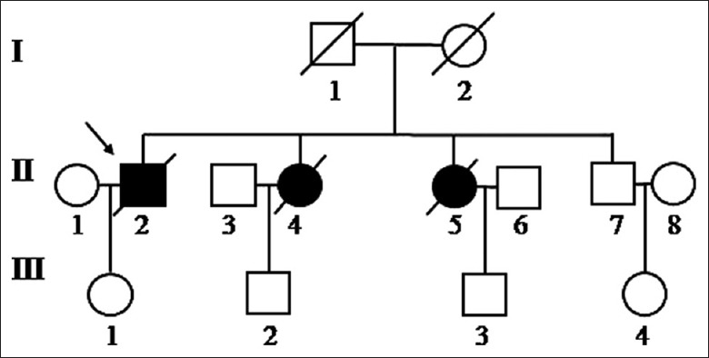
Pedigree of the Chinese family with IPF. Squares and circles indicate males and females, respectively; open symbols indicate unaffected individuals, filled symbols indicate affected individuals; diagonal indicates deceased individuals; arrow indicates the proband. IPF: Idiopathic pulmonary fibrosis.
Figure 2.
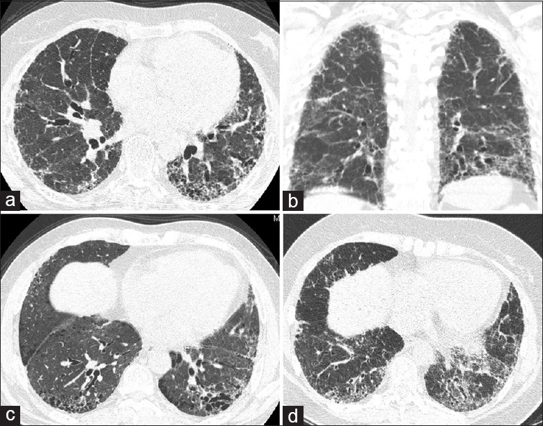
HRCT scans of three affected members in the FPF pedigree. (a and b) The CT scan of the proband, II2. (c and d) The CT scans of his younger sisters, II4 and II5, respectively. Reticular patter, honeycombing, and traction bronchiectasis were identified throughout both lungs in the CT sections, with subpleural and lower lobe predominance. HRCT: High-resolution computed tomography; FPF: Familial pulmonary fibrosis; CT: Computed tomography.
Table 1 shows the clinical features and outcomes of IPF patients. All patients were diagnosed IPF around their 50s, characterized by the frequently acute exacerbation of IPF phenotype, with poor prognosis. Patient II2 died 4.5 years after diagnosis, the 2nd day after he received bilateral lung transplantation, and II4 died after 1.5 years after diagnosis, II5 died after 2.5 years after diagnosis despite the oxygen therapy and oral N-acetylcysteine for them. The mean time to death was 2.8 years after diagnosis.
Table 1.
Demographic and clinical features of IPF patients
| Subject | Gender | Age at diagnosis | Smoking history | Outcome |
|---|---|---|---|---|
| II2 | Male | 51 | 30 packs/year | Died 4.5 years after diagnosis, the 2nd day after he received bilateral lung transplantation |
| II4 | Female | 51 | No | Died 1.5 years after the diagnosis |
| II5 | Female | 50 | No | Died 2.5 years after the diagnosis |
IPF: Idiopathic pulmonary fibrosis.
Table 2 shows the physiological evaluation of all the patients, including PFT, arterial blood gas, and cardiac ultrasound, 6 min walk test. All the three affected members had pulmonary hypertension, the pulmonary artery systolic pressure were assessed by the tricuspid incompetence with cardiac ultrasound.
Table 2.
Respiratory physiological examinations of IPF patients
| Subject | Pulmonary function test | Arterial blood gas (room air) | PASP (mmHg) Measured by cardiac ultrasound, TI | 6MWT (m) | ||||
|---|---|---|---|---|---|---|---|---|
| FVC (L) | FVC%pred (%) | DLCO (%) | pH | PaCO2 (mmHg) | PaO2 (mmHg) | |||
| II2 | 2.07 | 40.8 | 29.5 | 7.37 | 37 | 59 | 50 | N/A |
| II4 | 1.38 | 55.6 | 30.1 | 7.36 | 36 | 56 | 43 | 327 |
| II5 | 1.88 | 69.4 | 34.9 | 7.40 | 39 | 76 | 54 | 450 |
N/A: Not available; FVC: Forced vital capacity; FVC%pred: Ratio of FVC to predicted value for FVC; PASP: Pulmonary artery systolic pressure; 6MWT: 6 min walk test; TI: Tricuspid incompetence; IPF: Idiopathic pulmonary fibrosis; DLCO: Diffusing capacity of the lung for carbon monoxide.
Germline mutation of the telomerase reverse transcriptase gene
WES and analysis led to the identification of one variant in the TERT gene. Then, Sanger sequencing confirmed the heterozygous mutation in TERT, c.2146G>A, which causes a substitution of alanine for threonine at amino acid 716 in exon 6 [Figure 3]. This missense mutation is a single-nucleotide polymorphism (SNP) in dbSNP Build 147 (rs387907249) with no AF data according to the ExAC and 1000 genomes databases. By Sanger sequencing we did not detect this mutation in 100 ethnically matched healthy controls. This alanine is highly conserved in all vertebrate species that we have examined and is located within the putative oligomerization domain [Figure 4]. Furthermore, in silico analysis with the use of the SNPs3D database suggests that the A716T substitution is functionally deleterious.
Figure 3.
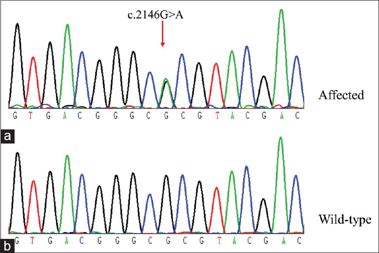
Sanger sequencing confirms TERT mutation. (a) The missense mutation c.2146G>A in exon 6 of the TERT gene (arrow) was found in the proband. (b) Wild-type sequence was from an unaffected member II7. TERT: Telomerase reverse transcriptase.
Figure 4.
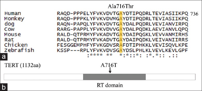
The sequence conservation and functional domain analysis of A716T substitution of TERT protein. (a) Partial sequence alignment of vertebrate TERT proteins. The shaded yellow region identifies the conserved alanine (A) mutated in the proband. Sequence numbering refers to the human TERT. (b) Location of the A716T substitution in TERT (arrow). Dark gray indicates the RT domain. RT: Reverse transcriptase; TERT: Telomerase reverse transcriptase.
DISCUSSION
Genetic variants, both common and rare, contribute to the genetic architecture of IPF. Compared with common variants, rare variants are considered to be highly penetrant, and generally have greater effect sizes as indicated by the high odds ratios for association of these rare variants with disease.[12] In addition, common variants are usually characterized by a single tagging SNP, but the rare variants often directly linked to disease-related genetic changes that are likely to alter the function of encoded proteins. Recently, increasing genetic studies of familial forms of IPF led to the discovery of such rare variants within telomere-related genes by using next-generation sequencing technologies to facilitate genetic discovery.[13,14,15,16,17] Thus, it is a powerful strategy to use exome sequencing of familial IPF patients to discover rare variants of large effect despite complexities such as clinical heterogeneity, reduced penetrance, and late-onset disease.
Here we report a rare variant in TERT associated with IPF in a Chinese Han family by above research strategy, which has never been reported in Chinese Han IPF families before. TERT encodes telomerase reverse transcriptase, and TERC encodes telomerase RNA, which are two major components of telomerase. Telomerase restores telomere length by adding telomeric DNA forming telomeres to the ends of linear chromosome. Germline mutations in the human TERT and TERC cause autosomal dominant dyskeratosis congenita, a rare hereditary disorder associated with premature death from aplastic anemia and pulmonary fibrosis.[18] TERT variants are the most frequently identified rare mutations related to pulmonary fibrosis, which were found in up to 15% of familial interstitial pneumonitis and in 1–3% sporadic cases.[8] This Ala716Thr variant of TERT was firstly reported in children with severe aplastic anemia and a family history of lung fibrosis.[19,20] Thus, it was considered that within a single family carried this mutation, older generations were more likely to affected by adult-onset pulmonary fibrosis, whereas bone marrow failure was the first presentation in subsequent generations at a younger age. However, in this Chinese Han IPF family, the next generation of the IPF patients (III1, III2, and III3) who carried this rare genetic variant of TERT are unaffected by neither aplastic anemia nor pulmonary fibrosis. This inconsistence may due to the limited number of cases, the ethnic differences and incomplete penetrance. More IPF families are required to clarify the association between phenotypes and genotypes in Chinese IPF patients.
In the present study, all patients carried this TERT mutation were characterized by the frequently acute exacerbation of IPF phenotype, with poor prognosis in both transplant and transplant-free conditions. Similar results had been reported previously that TERT mutations and/or short telomere lengths are associated with worse survival of IPF patients.[7,21] The mechanism through which TERT and other telomere-pathway genes contribute to lung fibrosis remains incompletely understood. Animal models of bleomycin-induced pulmonary fibrosis mice showed telomerase induction in lung fibroblasts associated with increased TERT expression, but without significant effect on telomere length. In contrast, TERT deficiency leads to lower myofibroblast differentiation and reduces lung fibrosis, which could be partially reversed by wild type bone marrow transplantation resulting in telomerase restoration in bleomycin induced lung fibrosis mice.[22] TRET, as an essential components of the enzyme telomerase, may cause haploinsufficiency, lead to short telomeres, and affect DNA damage responses. How these aging-related events lead to lung fibrosis and whether other mechanisms involved remain areas of active investigation.
Ethnic/racial differences were also found in the IPF population. A study in the United States including 251,058 cases of IPF patients using the National Center for Health Statistics database showed that 87.2% were non-Hispanic Whites, 5.1% were non-Hispanic African–American, and 2.2% were from other ethnic/racial groups. Compared with white decedents, African-Americans were significantly less likely to be coded with IPF, which were considered to be related to genetic differences.[23] While MUC5B promoter polymorphism rs35705950 is associated with IPF in independent cohorts, the AF and the strength of association are different between the Caucasian population and the Asian population.[24] Besides our present study, there are several other rare genetic variants have been reported to be associated with Chinese IPF patients. For example, six novel mutations in the TERT/TERC genes were identified in individuals diagnosed with sporadic IPF in the Chinese Han population.[25] Other rare variants in the genes encoding surfactant proteins, which maintain the alveolar stability and avert endoplasmic reticulum stress, was reported in a Chinese Han IPF family.[26] In generally, genetic research of IPF in various populations should be explored more active to reveal the whole picture and elucidate the mechanisms.
In conclusion, mutations in the telomerase pathway are the most frequently identified genetic cause of familial IPF. In this study, we confirmed a rare missense mutation in TERT associated with IPF in a Chinese Han family. More IPF families should be verified in the future. The mechanisms by which telomerase dysfunction and short telomeres lead to lung fibrosis still needs to be further understood.
Financial support and sponsorship
This study was supported by a grant from the National Natural Science Foundation of China (No. 81400045).
Conflicts of interest
There are no conflicts of interest.
Footnotes
Edited by: Yi Cui
REFERENCES
- 1.Wolters PJ, Collard HR, Jones KD. Pathogenesis of idiopathic pulmonary fibrosis. Annu Rev Pathol. 2014;9:157–79. doi: 10.1146/annurev-pathol-012513-104706. doi: 10.1146/annurev-pathol-012513-104706. [DOI] [PMC free article] [PubMed] [Google Scholar]
- 2.Schwartz DA. Idiopathic pulmonary fibrosis is a complex genetic disorder. Trans Am Clin Climatol Assoc. 2016;127:34–45. [PMC free article] [PubMed] [Google Scholar]
- 3.Fingerlin TE, Murphy E, Zhang W, Peljto AL, Brown KK, Steele MP, et al. Genome-wide association study identifies multiple susceptibility loci for pulmonary fibrosis. Nat Genet. 2013;45:613–20. doi: 10.1038/ng.2609. doi: 10.1038/ng.2609. [DOI] [PMC free article] [PubMed] [Google Scholar]
- 4.Noth I, Zhang Y, Ma SF, Flores C, Barber M, Huang Y, et al. Genetic variants associated with idiopathic pulmonary fibrosis susceptibility and mortality: A genome-wide association study. Lancet Respir Med. 2013;1:309–17. doi: 10.1016/S2213-2600(13)70045-6. doi: 10.1016/S2213-2600(13)70045-6. [DOI] [PMC free article] [PubMed] [Google Scholar]
- 5.Allen RJ, Porte J, Braybrooke R, Flores C, Fingerlin TE, Oldham JM, et al. Genetic variants associated with susceptibility to idiopathic pulmonary fibrosis in people of European ancestry: A genome-wide association study. Lancet Respir Med. 2017;5:869–80. doi: 10.1016/S2213-2600(17)30387-9. doi: 10.1016/S2213-2600(17)30387-9. [DOI] [PMC free article] [PubMed] [Google Scholar]
- 6.Kropski JA, Blackwell TS, Loyd JE. The genetic basis of idiopathic pulmonary fibrosis. Eur Respir J. 2015;45:1717–27. doi: 10.1183/09031936.00163814. doi: 10.1183/09031936.00163814. [DOI] [PMC free article] [PubMed] [Google Scholar]
- 7.Armanios MY, Chen JJ, Cogan JD, Alder JK, Ingersoll RG, Markin C, et al. Telomerase mutations in families with idiopathic pulmonary fibrosis. N Engl J Med. 2007;356:1317–26. doi: 10.1056/NEJMoa066157. doi: 10.1056/NEJMoa066157. [DOI] [PubMed] [Google Scholar]
- 8.Alder JK, Chen JJ, Lancaster L, Danoff S, Su SC, Cogan JD, et al. Short telomeres are a risk factor for idiopathic pulmonary fibrosis. Proc Natl Acad Sci U S A. 2008;105:13051–6. doi: 10.1073/pnas.0804280105. doi: 10.1073/pnas.0804280105. [DOI] [PMC free article] [PubMed] [Google Scholar]
- 9.García-Sancho C, Buendía-Roldán I, Fernández-Plata MR, Navarro C, Pérez-Padilla R, Vargas MH, et al. Familial pulmonary fibrosis is the strongest risk factor for idiopathic pulmonary fibrosis. Respir Med. 2011;105:1902–7. doi: 10.1016/j.rmed.2011.08.022. doi: 10.1016/j.rmed.2011.08.022. [DOI] [PubMed] [Google Scholar]
- 10.Raghu G, Collard HR, Egan JJ, Martinez FJ, Behr J, Brown KK, et al. An official ATS/ERS/JRS/ALAT statement: Idiopathic pulmonary fibrosis: Evidence-based guidelines for diagnosis and management. Am J Respir Crit Care Med. 2011;183:788–824. doi: 10.1164/rccm.2009-040GL. doi: 10.1164/rccm.2009-040GL. [DOI] [PMC free article] [PubMed] [Google Scholar]
- 11.Lynch DA, Sverzellati N, Travis WD, Brown KK, Colby TV, Galvin JR, et al. Diagnostic criteria for idiopathic pulmonary fibrosis: A Fleischner society white paper. Lancet Respir Med. 2018;6:138–53. doi: 10.1016/S2213-2600(17)30433-2. doi: 10.1016/S2213-2600(17)30433-2. [DOI] [PubMed] [Google Scholar]
- 12.Manolio TA, Collins FS, Cox NJ, Goldstein DB, Hindorff LA, Hunter DJ, et al. Finding the missing heritability of complex diseases. Nature. 2009;461:747–53. doi: 10.1038/nature08494. doi: 10.1038/nature08494. [DOI] [PMC free article] [PubMed] [Google Scholar]
- 13.Kannengiesser C, Borie R, Ménard C, Réocreux M, Nitschké P, Gazal S, et al. Heterozygous RTEL1 mutations are associated with familial pulmonary fibrosis. Eur Respir J. 2015;46:474–85. doi: 10.1183/09031936.00040115. doi: 10.1183/09031936.00040115. [DOI] [PubMed] [Google Scholar]
- 14.Alder JK, Stanley SE, Wagner CL, Hamilton M, Hanumanthu VS, Armanios M, et al. Exome sequencing identifies mutant TINF2 in a family with pulmonary fibrosis. Chest. 2015;147:1361–8. doi: 10.1378/chest.14-1947. doi: 10.1378/chest.14-1947. [DOI] [PMC free article] [PubMed] [Google Scholar]
- 15.Cogan JD, Kropski JA, Zhao M, Mitchell DB, Rives L, Markin C, et al. Rare variants in RTEL1 are associated with familial interstitial pneumonia. Am J Respir Crit Care Med. 2015;191:646–55. doi: 10.1164/rccm.201408-1510OC. doi: 10.1164/rccm.201408-1510OC. [DOI] [PMC free article] [PubMed] [Google Scholar]
- 16.Stuart BD, Choi J, Zaidi S, Xing C, Holohan B, Chen R, et al. Exome sequencing links mutations in PARN and RTEL1 with familial pulmonary fibrosis and telomere shortening. Nat Genet. 2015;47:512–7. doi: 10.1038/ng.3278. doi: 10.1038/ng.3278. [DOI] [PMC free article] [PubMed] [Google Scholar]
- 17.Kropski JA, Reiss S, Markin C, Brown KK, Schwartz DA, Schwarz MI, et al. Rare genetic variants in PARN are associated with pulmonary fibrosis in families. Am J Respir Crit Care Med. 2017;196:1481–4. doi: 10.1164/rccm.201703-0635LE. doi: 10.1164/rccm.201703-0635LE. [DOI] [PMC free article] [PubMed] [Google Scholar]
- 18.Armanios M, Blackburn EH. The telomere syndromes. Nat Rev Genet. 2012;13:693–704. doi: 10.1038/nrg3246. doi: 10.1038/nrg3246. [DOI] [PMC free article] [PubMed] [Google Scholar]
- 19.Du HY, Pumbo E, Ivanovich J, An P, Maziarz RT, Reiss UM, et al. TERC and TERT gene mutations in patients with bone marrow failure and the significance of telomere length measurements. Blood. 2009;113:309–16. doi: 10.1182/blood-2008-07-166421. doi: 10.1182/blood-2008-07-166421. [DOI] [PMC free article] [PubMed] [Google Scholar]
- 20.Parry EM, Alder JK, Qi X, Chen JJ, Armanios M. Syndrome complex of bone marrow failure and pulmonary fibrosis predicts germline defects in telomerase. Blood. 2011;117:5607–11. doi: 10.1182/blood-2010-11-322149. doi: 10.1182/blood-2010-11-322149. [DOI] [PMC free article] [PubMed] [Google Scholar]
- 21.Borie R, Tabèze L, Thabut G, Nunes H, Cottin V, Marchand-Adam S, et al. Prevalence and characteristics of TERT and TERC mutations in suspected genetic pulmonary fibrosis. Eur Respir J. 2016;48:1721–31. doi: 10.1183/13993003.02115-2015. doi: 10.1183/13993003.02115-2015. [DOI] [PubMed] [Google Scholar]
- 22.Liu T, Chung MJ, Ullenbruch M, Yu H, Jin H, Hu B, et al. Telomerase activity is required for bleomycin-induced pulmonary fibrosis in mice. J Clin Invest. 2007;117:3800–9. doi: 10.1172/JCI32369. doi: 10.1172/JCI32369. [DOI] [PMC free article] [PubMed] [Google Scholar]
- 23.Swigris JJ, Olson AL, Huie TJ, Fernandez-Perez ER, Solomon J, Sprunger D, et al. Ethnic and racial differences in the presence of idiopathic pulmonary fibrosis at death. Respir Med. 2012;106:588–93. doi: 10.1016/j.rmed.2012.01.002. doi: 10.1016/j.rmed.2012.01.002. [DOI] [PMC free article] [PubMed] [Google Scholar]
- 24.Zhu QQ, Zhang XL, Zhang SM, Tang SW, Min HY, Yi L, et al. Association between the MUC5B promoter polymorphism rs35705950 and idiopathic pulmonary fibrosis: A Meta-analysis and trial sequential analysis in Caucasian and Asian populations. Medicine (Baltimore) 2015;94:e1901. doi: 10.1097/MD.0000000000001901. doi: 10.1097/MD.0000000000001901. [DOI] [PMC free article] [PubMed] [Google Scholar]
- 25.Dai J, Cai H, Zhuang Y, Wu Y, Min H, Li J, et al. Telomerase gene mutations and telomere length shortening in patients with idiopathic pulmonary fibrosis in a Chinese population. Respirology. 2015;20:122–8. doi: 10.1111/resp.12422. doi: 10.1111/resp.12422. [DOI] [PubMed] [Google Scholar]
- 26.Zhang X, Jiang J, Chen WJ, Su LX, Xie LX. Genetic characterization of a Chinese family with familial idiopathic pulmonary fibrosis. Chin Med J. 2012;125:1945–51. doi: 10.3760/cma.j.issn.0366-6999.2012.11.020. [PubMed] [Google Scholar]