Capripoxviruses are etiological agents of important diseases in sheep, goats, and cattle. There are rare reports about viral protein function related to capripoxviruses. In the present study, we found that the 135 protein of GTPV plays an important role in inhibition of innate immunity and apoptosis in host cells. Use of the 135 gene as the insertion site to generate a vectored vaccine resulted in stronger adaptive immune responses than those obtained using the tk locus as the insertion site. As capripoxviruses are promising virus-vectored vaccines against many important diseases in small ruminants and cattle, the 135 gene may serve as an improved insertion site to generate recombinant capripoxvirus-vectored live dual vaccines.
KEYWORDS: GTPV 135, NF-κB pathway, apoptosis, viral vector
ABSTRACT
Goatpox virus (GTPV) is an important member of the Capripoxvirus genus of the Poxviridae. Capripoxviruses have large and complex DNA genomes encoding many unknown proteins that may contribute to virulence. We identified that the 135 open reading frame of GTPV is an early gene that encodes an ∼18-kDa protein that is nonessential for viral replication in cells. This protein functioned as an inhibitor of NF-κB activation and apoptosis and is similar to the N1L protein of vaccinia virus. In the natural host, sheep, deletion of the 135 gene from the GTPV live vaccine strain AV41 resulted in less attenuation than that induced by deletion of the tk gene, a well-defined nonessential gene in the poxvirus genome. Using the 135 gene as the insertion site, a recombinant AV41 strain expressing hemagglutinin of peste des petits ruminants virus (PPRV) was generated and elicited stronger neutralization antibody responses than those obtained using the traditional tk gene as the insertion site. These results suggest that the 135 gene of GTPV encodes an immunomodulatory protein to suppress host innate immunity and may serve as an optimized insertion site to generate capripoxvirus-vectored live dual vaccines.
IMPORTANCE Capripoxviruses are etiological agents of important diseases in sheep, goats, and cattle. There are rare reports about viral protein function related to capripoxviruses. In the present study, we found that the 135 protein of GTPV plays an important role in inhibition of innate immunity and apoptosis in host cells. Use of the 135 gene as the insertion site to generate a vectored vaccine resulted in stronger adaptive immune responses than those obtained using the tk locus as the insertion site. As capripoxviruses are promising virus-vectored vaccines against many important diseases in small ruminants and cattle, the 135 gene may serve as an improved insertion site to generate recombinant capripoxvirus-vectored live dual vaccines.
INTRODUCTION
Goatpox virus (GTPV) is a member of the Capripoxvirus genus of the Poxviridae, which includes etiological agents of important diseases in sheep and goats. The diseases caused by GTPV are endemic throughout Africa and Asia. They are categorized by the World Organisation for Animal Health (OIE) as notifiable due to their potential for rapid spread and substantial economic impact. Attenuated live vaccines have been developed and used in the field to prevent and control capripoxvirus infections (1). Different attenuated poxviruses have been used to express heterologous pathogen antigen genes to develop recombinant vaccines against important diseases. Many recombinant poxvirus-vectored vaccines have shown excellent safety profiles, significant immunogenicity, and protective efficacy (2–8). GTPV, which expresses antigens from important diseases, such as peste des petits ruminants virus (PPRV) infection, has been considered a promising vaccine for differentiating infected from vaccinated animals (DIVA vaccine) for a peste des petits ruminants (PPR) elimination campaign. The vaccine has several advantages, such as dual protection against PPR and goat pox, thermal stability, and a lower manufacturing cost. However, current GTPV-vectored vaccine candidates must be used at high doses and with repeated administration to achieve a high PPRV neutralization antibody (NA) seroconversion rate and protection from GTPV challenge (9, 10), which may hinder their practical application. An optimized GTPV-vectored vaccine to trigger strong and long-lasting immunity with high protective efficacy against selected diseases and goat pox itself is still sought.
Several factors may limit host immune responses to poxvirus-vectored vaccines. One is that use of the tk gene as an insertion site for foreign antigen genes usually results in attenuated replication of the recombinant virus in vivo. Another crucial factor is that poxviruses use a complex strategy to escape immune control by expressing immunomodulatory proteins (11, 12). It has been suggested that deletion of viral immunomodulatory genes in the poxvirus genome is a potential approach to improve the immunogenicity of poxvirus-vectored vaccines (13, 14). GTPV replicates in the cytoplasm of host cells and has an ∼145-kb, double-stranded DNA genome that comprises at least 147 putative genes (15), including replicative and structural genes that are conserved in the poxvirus family. So far, putative genes and their encoded proteins responsible for the virulence and host immunomodulation of GTPV are largely unknown (15). Bioinformatics analysis shows that the GTPV 135 gene encodes a protein that potentially plays roles similar to those of the N1L protein of vaccinia virus (VACV). N1L is an immunosuppressive protein and virulence factor that inhibits NF-κB signaling and apoptosis of host cells (16–18).
In this study, we demonstrate that the protein encoded by the GTPV 135 gene functions as an inhibitor of NF-κB and apoptosis. The 135 gene is not essential for GTPV replication in cells. Deletion of the 135 gene resulted in less attenuation than that with deletion of the tk gene in sheep. With the 135 gene used as a foreign gene insertion site, a recombinant GTPV strain expressing the PPRV H gene elicited significantly higher neutralization antibody responses to PPRV and GTPV than those obtained using the tk locus as the insertion site.
RESULTS
GTPV 135 protein is expressed early during infection and is nonessential for viral replication in cell culture.
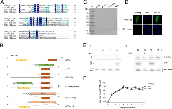
We sequenced the genome of GTPV vaccine strain AV41, and bioinformatics analysis showed that the 135 open reading frame (ORF) encodes a protein with 20% amino acid sequence identity to N1L of VACV strain WR, 42% identity to N1L of myxoma virus, and 20% identity to N1L of ectromelia virus (Fig. 1A).
FIG 1.
135 is an early gene and is nonessential for replication of GTPV in vitro. (A) Alignment of amino acid sequences of the 135 protein of GTPV (accession number AGZ95457.1) and the N1L proteins of VACV (accession number 2I39_A), myxoma virus (accession number NP_051859.1), and ectromelia virus (accession number NP_671537.1). Amino acids identical across all four viruses and three of the four viruses are highlighted in dark blue and light blue, respectively. (B) Schematic showing the genomes of wild-type and recombinant GTPV AV41 strains. Western blotting confirmed expression of the GTPV 135 protein (∼18 kDa) (C), and immunofluorescence assay indicated the subcellular localization of the 135 protein in LT cells 48 h after infection with AV41 and its r135-Flag recombinant (D). (E) The GTPV 135 gene is an early gene. LT cells were infected with r135Flag-GFP/tk at an MOI of 0.05 in the presence or absence of AraC, and expression of the 135 protein, GFP, and β-actin was detected by Western blotting. (F) The 135 gene is nonessential for GTPV replication. LT cells were infected with AV41, r135-Flag, or the Δ135 virus at an MOI of 0.05. Cells and medium were harvested at different times by freeze-thawing three times. After low-speed centrifugation, the supernatants were collected for viral titration in LT cells. Data are representative of independent experiments performed in triplicate.
To characterize the GTPV 135 protein, we first constructed three recombinants of GTPV AV41. The Δ135 strain was generated using the enhanced green fluorescent protein (GFP) gene under the control of the poxvirus late promoter p11 to replace the ORF of the 135 gene (Fig. 1B). The r135-Flag strain was generated by using a C-terminal Flag fusion with the 135 ORF to replace the ORF of the 135 gene (Fig. 1B). The r135Flag-GFP/tk strain was generated by inserting the GFP gene under the control of the poxvirus late promoter p11 into the tk gene locus of r135-Flag (Fig. 1B). The correct recombinants were selected based on PCR. These recombinants were confirmed by Western blotting with mouse polyclonal antibodies against GTPV 135. The results showed that the 135 protein had a molecular weight of ∼18 kDa and could be detected in LT cells infected by AV41 and the r135-Flag strain but not the Δ135 strain (Fig. 1C). To determine the subcellular localization of the 135 protein, LT cells were infected with r135-Flag and GTPV AV41 at a multiplicity of infection (MOI) of 0.05 for 48 h and were stained with a Flag monoclonal antibody. The 135 protein was distributed throughout the cytoplasm (Fig. 1D). Additionally, LT cells were seeded into a 10-cm plate and infected with GTPV AV41 at an MOI of 1 or were mock infected. Forty-eight hours after infection, the supernatant was collected and concentrated. Concentrated samples were tested by Western blotting, and no 135 protein was detected in the supernatant (data not shown). These results indicated that the 18-kDa protein expressed by the 135 ORF was distributed in the cytoplasm.
To clarify whether 135 is an early or late gene during GTPV infection, LT cells were infected with r135Flag-GFP/tk at an MOI of 0.05 in the absence or presence of arabinofuranosyl cytosine (AraC), an inhibitor of DNA replication and late gene expression. Expression of the 135-Flag protein and GFP in LT cells was detected by Western blotting with Flag and GFP monoclonal antibodies, respectively. In the absence of AraC in culture medium, 135 protein was detected 12 h after infection, while expression of GFP under the control of the p11 promoter was delayed until 24 h after infection (Fig. 1E, panel I). Forty-eight and 72 h after infection in the presence of AraC in culture medium, only the 135 protein was detected (Fig. 1E, panel II). These results indicate that 135 is an early gene during GTPV infection. To clarify whether the 135 gene is nonessential for GTPV replication in vitro, LT cells were inoculated with GTPV AV41, Δ135, and r135-Flag at an MOI of 0.05. The cell culture samples were collected every day for up to 8 days to determine virus titers. All three viruses reached peak titers 4 days after infection and showed no significant difference in growth titer at 8 days (Fig. 1F). This showed that deletion of the 135 gene did not affect the proliferation of GTPV in LT cells and suggested that 135 is a nonessential gene for replication of GTPV in vitro.
Deletion of 135 gene attenuates GTPV virulence in sheep.
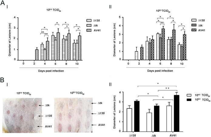
The 135 protein did not affect the proliferation of GTPV in vitro, which may not be in accordance with the situation in vivo. Therefore, we compared infections with the Δ135 and wild-type AV41 strains and a recombinant with a tk gene deletion (Δtk) in sheep. One-year-old sheep were inoculated with the Δ135, wild-type AV41, and Δtk strains at 104, 105, or 106 50% tissue culture infective doses (TCID50). Each virus at each dose was inoculated intradermally at four points in the thorax and abdomen. Sheep were observed daily for 2 weeks after inoculation. There were no lesions observed at sites inoculated with 104 TCID50 for all three viruses. For inoculations with 105 and 106 TCID50, mild edematous swelling was observed at all inoculation sites, and no secondary lesions were found throughout the observation period. The lesions caused by the Δ135 strain were smaller than those caused by wild-type AV41 but larger than those caused by the Δtk strain (Fig. 2A, panels I and II). For each virus, 105 TCID50 resulted in milder edematous swelling than that of lesions caused by 106 TCID50. Based on the most significant diameter of edematous swelling at inoculated sites 6 days after inoculation, there were significant differences in the sizes of the lesions caused by the Δ135 and Δtk strains and by the AV41 and Δ135 strains (Fig. 2B, panels I and II). These results suggest that the 135 gene contributes to virulence of GTPV in the natural host (sheep). However, deletion of the 135 gene impairs replication of GTPV in vivo less than deletion of the tk gene does.
FIG 2.
Deletion of the 135 gene results in less attenuation than that with deletion of the tk gene for GTPV in sheep. Sheep were inoculated intradermally with the AV41, Δ135, or Δtk virus. (A) Diameters of lesions at inoculation sites in sheep that received 105.0 TCID50 (I) and 106.0 TCID50 (II) were measured and recorded every day. (B) Representative lesions are shown for day 6 after inoculation (I), and the significance of differences in diameters between viruses was determined using the t test (II). *, P < 0.05; **, P < 0.01.
135 protein inhibits NF-κB and inflammatory signaling in cells.
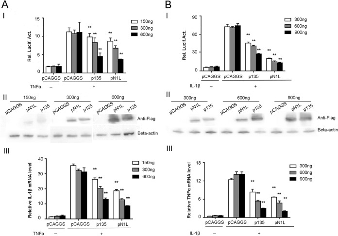
The VACV N1L protein can inhibit the NF-κB signaling pathway (16, 18). We speculated that the 135 protein may play a role similar to that of the N1L protein in host cells during GTPV infection. To test this hypothesis, we performed plasmid transfection and recombinant virus infection analysis in vitro to explore the biological function of the 135 protein. For transfection assays, a reporter plasmid containing the luciferase gene under the control of the NF-κB-specific promoter (pNF-κB-luc) and the internal control plasmid pTK-Renilla (pTK-luc) were cotransfected with plasmid expressing the Flag-tagged 135 protein (p135) or the VACV N1L protein (pN1L) into HEK 293T cells. The vector plasmid pCAGGS was also cotransfected with pNF-κB-luc and pTK-luc, as a control. Twenty-four hours after transfection, cells were treated with tumor necrosis factor alpha (TNF-α) and interleukin-1β (IL-1β) for 8 h before luciferase activity testing. Expression of the N1L protein of VACV and the 135 protein of GTPV was detected by Western blotting, and mRNA transcription of inflammatory cytokines was tested by quantitative reverse transcription-PCR (qRT-PCR). Expression of the 135 protein and the N1L protein resulted in significantly lower levels of relative luciferase activity than those observed with the control plasmid pCAGGS (Fig. 3A, panel I, and B, panel I). Western blotting indicated that the 135 and N1L proteins showed a pattern of dose-dependent inhibition (Fig. 3A, panel II, and B, panel II). qRT-PCR revealed that expression of the 135 and N1L proteins also resulted in significantly lower levels of transcription of the TNF-α and IL-1β genes (Fig. 3A, panel III, and B, panel III). These results suggest that the 135 protein significantly inhibits the NF-κB pathway that is activated by TNF-α or IL-1β, in a manner similar to that with the VACV N1L protein (16).
FIG 3.
Expression of the 135 protein inhibits NF-κB signaling pathway activation. HEK293T cells were cotransfected with p135, pN1L, and pCAGGS together with pNFκB-luc and pTK-Renilla. At 24 h, cells were treated with 100 ng/ml of TNF-α (AI) or IL-1β (BI) or left untreated for 8 h. The cell lysates were prepared for assays of NF-κB-inducible luciferase activity (AI and BI), and mRNAs were extracted for determination of TNF-α and IL-1β mRNA levels by qPCR (AIII and BIII). (AII and BII) Expression of the GTPV 135 protein and the VACV N1L protein was measured by Western blotting. Data are representative of independent experiments performed in triplicate. The significance of differences was determined by the t test. *, P < 0.05; **, P < 0.01.
GTPV with deletion of the 135 gene shows weakened inhibition of NF-κB and inflammatory signaling during infection.
To confirm the function of the 135 protein during GTPV infection, HEK293T cells were infected with wild-type AV41 and the Δ135 strain at MOIs of 0.01, 0.1, and 1.0. Twenty-four hours after infection, cells were cotransfected with pNF-κB-luc and pTK-luc. Forty-eight hours after infection, cells were treated with TNF-α and IL-1β for an additional 8 h before analysis of luciferase activity and quantification of IL-1β mRNA transcription by qRT-PCR. Compared to the Δ135 strain, GTPV AV41 showed significantly lower levels of luciferase activity (Fig. 4A) and IL-1β mRNA transcription (Fig. 4B) after infection and treatment with TNF-α and IL-1β. This result confirms that expression of the 135 protein inhibits the activation of NF-κB and the inflammatory signaling pathway during GTPV infection in cells.
FIG 4.
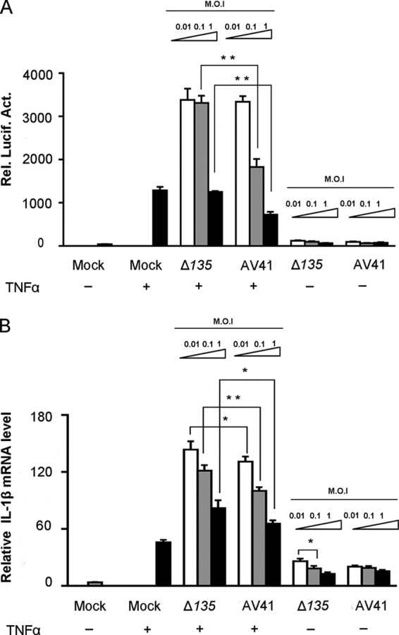
GTPVΔ135 is attenuated in inhibition of NF-κB activation. HEK293T cells were infected with AV41 or the Δ135 virus at MOIs of 0.01, 0.1, and 1. Twenty-four hours after infection, cells were cotransfected with pNFκB-luc and pTK-Renilla. At 24 h posttransfection, cells were treated with 100 ng/ml of TNF-α or left untreated for 8 h. Cell lysates were prepared for assays of NF-κB-inducible luciferase activity (A), and mRNA was extracted for determination of the IL-1β mRNA level by qPCR (B). Data are representative of independent experiments performed in triplicate. The significance of differences was determined by the t test. *, P < 0.05; **, P < 0.01.
135 protein inhibits apoptosis in HeLa cells.
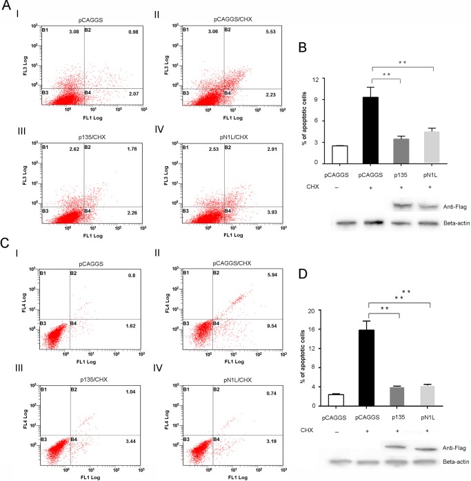
The N1L protein of VACV prevents apoptosis in host cells (16–18). To clarify if the 135 protein has a similar function, we performed transfection with p135, pN1L, or pCAGGS in HeLa cells. Twenty-four hours after transfection, cells were treated with 2 μM cycloheximide (CHX) and 5 ng/ml TNF-α for 12 h or were left untreated for 12 h before apoptosis assay by flow cytometry following staining with annexin V/propidium iodide (PI) or caspase 3/7 green. p135 and pN1L transfections caused significantly lower rates of apoptosis in HeLa cells, based on annexin V/PI (Fig. 5A and B) and caspase 3/7 green (Fig. 5C and D) staining. Western blotting confirmed that expression of the GTPV 135 protein and the VACV N1L protein was correlated with inhibition of apoptosis induced by CHX and TNF-α treatment in HeLa cells. The results suggest that the 135 protein functions as an inhibitor of apoptosis.
FIG 5.
Expression of the 135 protein inhibits apoptotic death of HeLa cells after treatment with CHX and TNF-α. HeLa cells were transfected with p135, pN1L, and pCAGGS and treated with 2 μM CHX and 5 ng/ml of TNF-α for 12 h. CHX-treated cells were harvested for annexin V/PI staining (A) or treatment with caspase 3/7 detection reagents and Sytox AADvanced Ready flow reagent (C), examined by flow cytometry, and analyzed with FlowJo, using biexponential scaling. The horizontal axis shows the quantity of cells stained with annexin V dye (A) or caspase 3/7 green dye (C). The vertical axis represents the quantity of cells stained by PI (A) or Sytox AADvanced (C). B2 represents the percentage of late apoptotic cells, and B4 represents the percentage of early apoptotic cells. (B and D) Total numbers of apoptotic cells were calculated by adding late and early apoptotic counts. Expression of the GTPV 135 and VACV N1L proteins was confirmed by Western blotting. The significance of differences was determined by the t test. *, P < 0.05; **, P < 0.01.
GTPV-vectored vaccine generated by use of the 135 gene as an insertion site elicits stronger adaptive immune responses than those obtained with the tk gene as an insertion site.
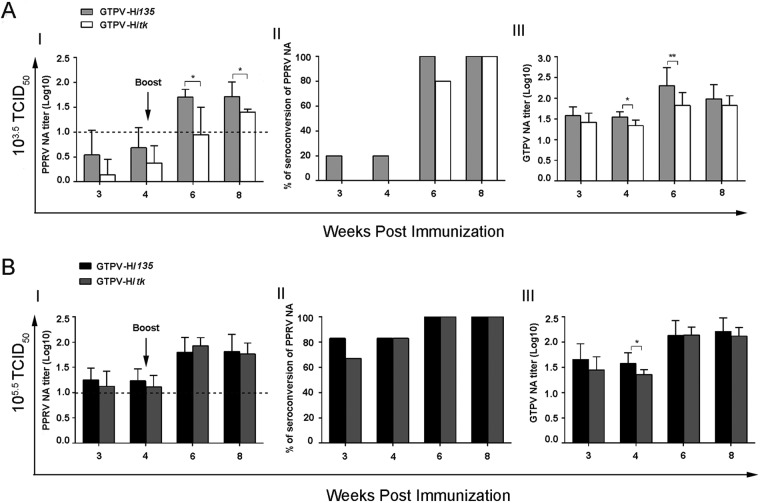
Poxvirus vectors that express heterologous antigens but lack different inhibitors of the central host cell signaling pathway enhance innate and adaptive immune responses (14, 19). The 135 gene is involved in inhibition of the signaling pathway of innate immunity and is not essential for replication in vitro and in vivo. We speculated that deletion of the 135 gene would be an alternative strategy to generate a GTPV-vectored vaccine with improved immunogenicity. Therefore, we constructed recombinant GTPV AV41 vaccine strains expressing the PPRV hemagglutinin protein (H) by using the 135 and tk genes as insertion sites (GTPV-H/135 and GTPV-H/tk, respectively) (Fig. 1B) and compared the immunogenicities of the strains in target animals. Sheep were immunized with GTPV-H/135 or GTPV-H/tk at 103.5 and 105.5 TCID50 and received a booster dose after 4 weeks. Serum samples were collected for measurement of PPRV- and GTPV-specific neutralization antibodies (NAs). For sheep that received 103.5 TCID50, GTPV-H/135 elicited higher titers of NAs to PPRV and GTPV than those seen with GTPV-H/tk (Fig. 6A). There were significant differences in NAs to PPRV between the GTPV-H/135 and GTPV-H/tk groups after booster immunization and significant differences in NAs to GTPV between the GTPV-H/135 and GTPV-H/tk groups after priming and booster immunizations (Fig. 6A, panels I and III). Animals with seroconversion of PPRV NA (titers of >10) were observed only in the GTPV-H/135-immunized group after the priming immunization (Fig. 6A, panel II). For sheep that received 105.5 TCID50, there were no significant differences in NAs to PPRV and GTPV between the GTPV-H/135 and GTPV-H/tk groups after immunization (Fig. 6B). These results suggest that GTPV-H/135 is more immunogenic than GTPV-H/tk for eliciting protective immunity against GTPV and PPRV.
FIG 6.
GTPV-H/135 is more immunogenic than GTPV-H/tk for eliciting PPRV and GTPV NAs. Groups of six sheep were vaccinated with GTPV-H/135 or GTPV-H/tk at 103.5 TCID50 (A) and 105.5 TCID50 (B) and were boosted 4 weeks after priming. Sera were collected to determine the titers of NAs to PPRV (AI, AII, BI, and BII) and GTPV (AIII and BIII). The titers are presented as the mean and standard error for each group. Dotted lines indicate the thresholds of seroconversion of PPRV NAs (AI and BI). The significance of differences was determined by the t test (*, P < 0.05; **, P < 0.01).
DISCUSSION
Capripoxviruses are important causative agents for many diseases of domestic and wild ruminants, including sheep pox, goat pox, and cattle lumpy skin disease (LSD). Goat pox and sheep pox have been endemic in many countries of Africa and Eurasia for a long time, while LSD has spread from Africa to southern and eastern Europe, the Middle East, and Central Asia in recent years; furthermore, Southeast and East Asia currently face an imminent threat (20, 21). Although capripoxviruses have a huge impact on domestic animal husbandry worldwide, there are few detailed studies of the molecular basis of their virulence and pathogenesis.
For host antiviral responses, acute inflammation and apoptosis induced by infection are major contributors to the innate immune response. Accordingly, viruses have evolved multiple mechanisms to subvert the strong barrier of host innate immunity (11, 12). Poxviruses, such as VACV, encode many proteins to evade host immunity by neutralizing complement factors, interferons, cytokines, and chemokines or by inhibiting apoptosis and signaling pathways to produce interferons and proinflammatory cytokines and chemokines (11, 12). All these factors may contribute to the virulence of poxviruses. The N1L protein is an intracellular virulence factor that belongs to a family of VACV Bcl2-like proteins, and it inhibits both proinflammatory and proapoptotic signaling by using independent domains of the protein (17, 18, 22). Although apoptosis represents a powerful mechanism for eliminating virus-infected cells (23), abolishing the antiapoptotic activity of the N1L protein does not affect the virulence of VACV in mice (18). The ability of the N1L protein to inhibit inflammatory NF-κB signaling is a major contributor to virulence (18). The present study revealed that the GTPV 135 protein is an early intracellular protein with a molecular weight of ∼18 kDa and is completely nonessential for viral replication in vitro. The 135 protein showed significant inhibition of NF-κB activation and apoptosis in cells. These results were consistent with studies of the VACV N1L protein and suggested that 135 is an important virulence gene for GTPV, which was confirmed in our study by use of the Δ135 strain, a recombinant AV41 strain with a 135 gene deletion. Sheep inoculated with the Δ135 strain developed milder lesions than those in sheep inoculated with wild-type AV41. Whether the 135 protein inhibits NF-κB activation and apoptosis by using different independent domains and whether inhibition of NF-κB activation contributes to virulence more than apoptosis does remain to be investigated.
Our results showed that recombinant GTPV AV41 expressing PPRV H, using the 135 gene as the insertion site, induced a stronger adaptive immune response than that seen with GTPV-H/tk. These results were inconsistent with previous studies. It has been reported that poxvirus vectors that express heterologous antigens and lack immunomodulator genes usually induce enhanced innate and adaptive immune responses. VACV with deletion of the A41L gene induced a stronger virus-specific CD8+ T-cell response in mice (14). VACV with deletion of the N1L gene or an N1L mutant that abolished inhibition of NF-κB resulted in increased central and memory CD8+ T-cell populations and CD8+ T-cell cytotoxicity and lower virus titers in organs after challenge (19). A pathogenic LSD virus (LSDV) strain attenuated by removal of a putative virulence factor gene (IL-10-like gene) conferred on goats and sheep a solid protective immunity against virulent GTPV and sheeppox virus (SPPV) challenges (24). This modified LSDV may be a candidate vaccine against capripoxviruses in goats, sheep, and cattle. A study based on a modified vaccinia virus Ankara (MVA) vector expressing human immunodeficiency virus type 1 (HIV-1) antigens revealed that deletion of the C6L and K7R genes enhanced the magnitude and quality of HIV-1-specific CD4+ and CD8+ T-cell responses in mice, as well as Env antibody levels (25, 26). Other studies with MVA-vectored vaccines lacking the A41L and B16R or F1L gene and expressing HIV-1 antigens showed similarly enhanced adoptive immune responses (27, 28).
The function of the 135 protein involves the inhibition of NF-κB signaling and apoptosis and may contribute to the enhancement of specific immune responses by a GTPV vector with destruction of the 135 gene. NF-κB is considered crucial for B- and T-cell development (29, 30), participates in secondary lymphoid organogenesis after cytokine receptor activation (31, 32), supports the activities of different CD4+ T-cell subtypes (33), and is crucial for survival of activated CD8+ T cells (34). It is also important for costimulatory receptor functions, such as those of CD28 (35). Apoptosis may also facilitate antigen presentation to T cells through major histocompatibility complex class I (MHC-I) and CD1, at least for infection with Mycobacterium tuberculosis (36). Moreover, use of the 135 gene as an insertion site while keeping the tk gene intact may favor replication of poxvirus in vivo, which facilitates viral antigen expression and presentation. However, details about the mechanism remain to be investigated.
Berhe et al. and Diallo et al. generated recombinant capripoxviruses expressing the F and H glycoproteins of PPRV, respectively, and both recombinant viruses provided good protection from challenge with PPRV, even with low vaccination doses and without detectable PPRV-specific NAs (37, 38). GTPV AV41 expressing PPRV H and F, using tk as an insertion site, showed that the recombinant viruses elicited dose-dependent immune responses and long-lasting protective antibody duration against PPRV in goats and sheep. Moreover, repeat vaccination with GTPV-vectored PPRV vaccines successfully overcame the interference from preexisting immunity in animals that received GTPV prevaccination (9). In the present study, AV41 expressing PPRV H showed a dose-dependent antibody response pattern that was similar to that in previous studies with recombinants generated using tk as an insertion site (9, 10). For NA responses to PPRV, GTPV-H/135 induced higher titers than those observed with GTPV-H/tk at the lower dose of 103.5 TCID50 after priming, although there was no significant difference between the two recombinants at the higher dose of 105.5 TCID50. GTPV-H/135 elicited significantly more NAs to GTPV than those obtained with GTPV-H/tk at the higher dose after priming and at the lower dose after priming and boosting. There was no significant difference in NAs to GTPV between the higher and lower doses for GTPV-H/135. According to the OIE manual of diagnostic tests and vaccines for terrestrial animals (39), a titer of NA to PPRV of ≥1:10 indicates a protective efficacy that is valid for vaccine quality control (39). Our results showed that titers of NAs to PPRV were 1:51 to 1:83 after booster vaccination, suggesting that GTPV-H/135 is a promising PPR DIVA vaccine candidate.
AV41 at a TCID50 of 103.5 is a typical dose in the field, and a single vaccination usually provides good protection against GTPV. For the GTPV-vectored PPRV vaccine generated using tk as an insertion locus, single vaccination with a higher dose (105.0 TCID50) or two vaccinations with a lower dose (104.0 TCID50) provided efficient protection against virulent GTPV challenge in goats (9). However, two vaccinations with a dose of 103.0 TCID50 provided only partial protection against virulent SPPV challenge in sheep (10). Therefore, GTPV-H/tk must depend on a higher dose to obtain efficient protection against GTPV and SPPV, which implies a higher cost for vaccine manufacture and hinders application in developing countries. Previous studies suggested that both live attenuated and inactivated capripoxvirus vaccines induced specific NAs in goats and sheep. After vaccination with live and killed vaccines, most goats and sheep showed neutralization antibody titers of <100 and received protection from challenge with a virulent capripoxvirus (24, 40). In this study, sheep vaccinated with GTPV-H/135 showed titers of neutralization antibodies to GTPV ranging from 1.8 to 2.3 log10, which indicated that this recombinant has potency to serve as a dual live vectored vaccine against PPRV and capripoxviruses in goats and sheep. Our study demonstrates that GTPV-H/135 is an improved candidate vaccine against PPRV and GTPV, with advantages over GTPV-H/tk in terms of immunogenicity.
Based on data from the UN Food and Agriculture Organization (FAO), PPR causes losses of up to $2.1 billion annually. In 2016, FAO and OIE launched an ambitious global campaign to eradicate PPR worldwide by 2030. That would be the second important animal disease to be eradicated, after rinderpest. A safe and efficacious PPR and goat pox dual vaccine with DIVA function, low cost, and thermal stability, such as GTPV-H/135, would be an attractive tool to use to realize this goal. Using the 135 gene as an insertion site may be an improved strategy to generate recombinant vectored vaccines not only for GTPV but also for other important capripoxviruses, including SPPV and LSDV.
MATERIALS AND METHODS
Cells and viruses.
LT cells and chicken embryo fibroblasts (CEFs) were prepared as described previously (9, 41) and maintained in modified Eagle's minimal essential medium (MEM) containing 10% fetal bovine serum (FBS), 100 U/ml penicillin, and 100 μg/ml streptomycin at 37°C in a 5% CO2 humidified atmosphere. HEK-293T, HeLa, and Vero cells were maintained in complete Dulbecco's modified Eagle's medium containing 10% FBS. GTPV AV41 and its recombinant viruses, the avian poxvirus vaccine strain, and PPRV vaccine strain Nigeria/75/1 were propagated and titrated in LT cells, CEFs, and Vero cells, respectively, as previously described (9, 10).
Plasmid construction.
Two primer pairs (5′-CACGAATTCATGGGAAATAAAAAAAATGATG-3′/5′-TCAGGTACCTTACTTGTCATCGTCATCCTTGTAATCCATTATTTTACTTTTAATGTAG-3′ and 5′-TCGAATTCATGAGGACTCTACTTATTAGATATATTC-3′/5′-TAGGTACCTTACTTGTCATCGTCATCCTTGTAATCTTTTTCACCATATAGATCAATCATTAG-3′) were used to amplify the 135 gene of GTPV (GenBank accession number KC951854.1) and the N1L gene of VACV (GenBank accession number LT966077.1) from genomic DNAs of AV41 and VACV (prepared in our laboratory), respectively. The restriction sites are indicated in italics, and Flag tag sequences (underlined) were fused to the C-terminal regions of both ORFs. The amplified 135 gene and N1L gene were cloned into pCAGGS (Addgene, Cambridge, MA, USA) and the resulting plasmids named p135 and pN1L, respectively.
Recombinant viruses.
To generate recombinant GTPV AV41 with deletion of the 135 ORF, an upstream arm (681 bp) and a downstream arm (554 bp) that flanked the ORF of the GTPV 135 gene were amplified using the primer pairs 5′-ATCCCGGGATGAAAAATATACATATGTTATTG-3′/5′-TAGCGGCCGCATTTTATTTATAATATTTTATAC-3′ and 5′-TCGCGGCCGCTGTAAAACATAAAATAAATAAC-3′/5′-CCGCGGTGAAATACATCCATATGGTTTATTG-3′, respectively. XmaI and NotI restriction sites were introduced at the two termini of the upstream arm, and NotI and SacII restriction sites were introduced at the two termini of the downstream arm. An expression cassette containing the ORF of the EGFP gene under the control of the poxvirus p11 promoter was amplified from pTk-gig-PPRV-H (9) by use of the primer pair 5′-ACGCGGCCGCTTACTTGTACAGCTCGTCC-3′/5′-TAGCGGCCGCAGCGATGCTACGCTAGTCACAATC-3′, and NotI sites were introduced at both termini. The resultant DNA fragments were assembled in the order upstream arm-p11-enhanced GFP (EGFP)-downstream arm in pBluescript II SK(+) (Clontech, Mountain View, CA, USA) to construct the recombinant plasmid pΔ135. To generate recombinant virus, LT cells were infected with GTPV AV41 and transfected with pΔ135 by use of calcium phosphate (Invitrogen, Carlsbad, CA, USA). The AV41 mutant clone with deletion of the 135 gene was selected and purified based on expression of EGFP. The resultant virus was named Δ135. To generate a recombinant GTPV AV41 strain expressing a Flag-tagged 135 protein, the recombinant pr135 was constructed by replacing the p11 and EGFP ORF sequences in pΔ135 with the sequence of a C-terminally Flag-tagged 135 ORF. LT cells were infected with the avian poxvirus vaccine strain at an MOI of 5 for 2 h and then cotransfected with pr135 and the genomic DNA of the Δ135 virus after treatment with NotI, for which there are no sites within the genome of wild-type AV41. The rescued recombinant virus was designated r135-Flag. To generate a recombinant AV41 clone expressing the Flag-tagged 135 protein at the 135 locus and EGFP at the tk locus, the recombinant pEGFP/tk was constructed by replacing the sequence between the tk upstream arm and the tk downstream arm with the ORF for EGFP under the control of p11. LT cells were transfected with pEGFP/tk and infected with r135-Flag. The recombinant virus was selected and purified based on the expression of EGFP. The resultant virus was designated r135-Flag/GFP. A recombinant AV41 strain expressing the H protein of PPRV under the control of the poxvirus H6 promoter at the 135 locus, GTPV-H/135, was generated following a strategy similar to that for r135-Flag described above. To generate recombinant AV41 expressing the PPRV H protein under the control of the H6 promoter at the tk locus, a recombinant AV41 strain expressing EGFP was constructed, in whose genome the ORF of the tk gene was replaced by a sequence containing p11 and the ORF for EGFP, with NotI sites at both termini. The resultant virus was designated Δtk and then was used to construct the target recombinant GTPV-H/tk, following a strategy similar to that for r135-Flag and GTPV-H/135. The recombinant viruses were screened and confirmed by PCR, sequencing, and Western blotting. The primer pairs used for PCR were as follows: 5′-ATCCCGGGATGAAAAATATACATATGTTATTG-3′/5′-CCGCGGTGAAATACATCCATATGGTTTATTG-3′ and 5′-GAATCACCAGAGCCGATAACATATATAG-3′/5′-CCAGATTGGACAGAAGTAAGGAATGTT-3′.
Western blotting and immunofluorescence assay.
For Western blotting, HEK293 and HeLa cells transfected with p135 or LT cells infected with AV41 or its recombinants were lysed with cell lysis buffer (50 mM Tris-HCl, pH 7.4, 150 mM NaCl, 1% Triton X-100, 5 mM MgCl2, 1 mM EDTA, and 10% glycerol) containing 1 mM phenylmethylsulfonyl fluoride and protease inhibitor cocktail (Roche Diagnostics, Indianapolis, IN, USA). Equal amounts of cell lysates were resolved by 10 to 12% SDS-PAGE and transferred to polyvinylidene difluoride membranes (EMD Millipore, Billerica, MA, USA). After incubation with anti-Flag, anti-GFP, and anti-β-actin monoclonal antibodies (Sigma-Aldrich, St. Louis, MO, USA) or a mouse anti-135 polyclonal antibody that was prepared by immunizing mice with a synthesized polypeptide corresponding to the 135 protein (KEYIRWRGNSEKNIC) and adjuvant, the blots were developed by incubation with a horseradish peroxidase (HRP)-conjugated goat anti-mouse IgG(H+L) secondary antibody (Genscript, Piscataway, NJ, USA). Finally, the membranes were visualized using enhanced chemiluminescence (ECL; GE Healthcare, Little Chalfont, Bucks, United Kingdom).
For indirect immunofluorescence assay, LT cells were infected with AV41 or its recombinants at an MOI of 0.05. Forty-eight hours after infection, cells were fixed with 3% paraformaldehyde and stained with mouse anti-Flag monoclonal antibody as the primary antibody and fluorescein isothiocyanate (FITC)-conjugated goat anti-mouse IgG as the secondary antibody. Cell nuclei were stained with DAPI (4′,6-diamidino-2-phenylindole; Invitrogen, Eugene, OR, USA). Stained cells were analyzed with a Leica TCS SP5 laser scanning confocal microscope (Leica, Mannheim, Germany).
Luciferase assays and qRT-PCR.
The pNFκB-luc and pTK-Renilla plasmids were supplied by Promega (Madison, WI, USA). HEK293T cells in 48-well plates were transfected with different concentrations of p135 and pN1L or infected with AV41 and the Δ135 virus at different MOIs, in combination with 100 ng/well pNF-κB-Luc and 5 ng/well pTK-Renilla as internal controls. In each experiment, the total amount of plasmid DNA in each sample was kept constant by supplementation with the corresponding empty parental expression vector. Twenty-four hours after transfection, cells were stimulated with 100 ng/ml of IL-1β or TNF-α for 8 h. After that, the firefly and Renilla luciferase activities were measured using a dual-luciferase reporter assay system (Promega).
To quantitate mRNA transcription for human TNF-α and IL-β, HEK293T cells were transfected with p135 and pN1L or infected with AV41 or the Δ135 virus at different MOIs for 24 h and then stimulated with 100 ng/ml of IL-1β or TNF-α for 8 h. Total cellular mRNA was extracted using TRIzol reagent (Invitrogen). Reverse transcription was carried out using Primescript RT master mix (TaKaRa Bio, Tokyo, Japan). qRT-PCR was performed using an Agilent-Stratagene Mx real-time qPCR system according to SYBR Premix DimerEraser instructions (TaKaRa Bio). The amount of mRNA for the target gene was normalized to the amount of β-actin mRNA in the same sample. The primer pairs 5′-GCTGAGGAACAAGCACCG-3′/5′-CCTCTCACATACTGACCCACG-3′, 5′-CGGCCACATTTGGTTCTAAGA-3′/5′-AGGGAAGCGGTTGCTCAT-3′, and 5′-CCTTCCTGGGCATGGAGTCCTG-3′/5′-GGAGCAATGATCTTGATCTTC-3′ were used to target the human TNF-α, IL-1β, and β-actin mRNAs, respectively.
Flow cytometry.
HeLa cells were transfected with p135, pN1L, or pCAGGS. Twenty-four hours after transfection, the cells were treated with 2 μM CHX and 5 ng/ml TNF-α (both from Sigma-Aldrich, St. Louis, MO) for 12 h. After that, HeLa cells were dyed by use of an Alexa Fluor 488-annexin V/dead cell apoptosis kit (Invitrogen, Carlsbad, CA, USA) for 15 min at room temperature or a Cell Event caspase-3/7 green flow cytometry assay kit (Invitrogen, Carlsbad, CA, USA) for 45 min at room temperature. Fluorescence emission was detected at 530 and 575 nm and at 530 and 650 nm, respectively, by using a Cytomics FC 500 instrument (Beckman Coulter, Los Angeles, CA, USA) and a 488-nm excitation wavelength.
Animal experiments.
The animal experimental protocols in this study were approved by the Harbin Veterinary Research Institutional Animal Care Committee. Four 1-year-old sheep naive to GTPV vaccination and serologically negative in the GTPV neutralization test were used for virulence assay. Each animal was simultaneously inoculated with GTPV AV41 and the Δ135 and Δtk viruses at 104.0, 105.0, or 106.0 TCID50, as well as with phosphate-buffered saline (PBS) as a control, in the thorax and abdomen. Each virus was inoculated intradermally in four replicates (0.1 ml per inoculum). The skin lesions at inoculation sites were observed, and the diameters of the lesions were measured every day for 2 weeks after inoculation. The titers for the challenge virus to induce obvious macules/papules were calculated from the inocula.
For immunization studies, we used four groups of six sheep aged 6 months to 2 years that were serologically negative for GTPV and PPRV. Each group of sheep was inoculated intradermally with GTPV-H/135 or GTPV-H/tk diluted in serum-free MEM to 103.5 or 105.5 TCID50 (0.1 ml per inoculum) and received a booster vaccination 4 weeks after priming. Sera were collected for detection of NAs. The NA titers against PPRV and GTPV were determined by following the protocols recommended by the OIE (39), with minor modifications of the previously described method (9).
Accession number(s).
The sequence of GTPV vaccine strain AV41 was submitted to GenBank under accession number MH381810.
ACKNOWLEDGMENTS
This work was sponsored by the National Natural Science Fund of China (grant 31602074), the International S&T Cooperation Program of China (grant 2015DFA31300), and the National Key R&D Program of China (grant 2017YFD0501800).
REFERENCES
- 1.Babiuk S, Bowden TR, Boyle DB, Wallace DB, Kitching RP. 2008. Capripoxviruses: an emerging worldwide threat to sheep, goats and cattle. Transbound Emerg Dis 55:263–272. doi: 10.1111/j.1865-1682.2008.01043.x. [DOI] [PubMed] [Google Scholar]
- 2.Lazaro-Frias A, Gomez-Medina S, Sanchez-Sampedro L, Ljungberg K, Ustav M, Liljestrom P, Munoz-Fontela C, Esteban M, Garcia-Arriaza J. 2018. Distinct immunogenicity and efficacy of poxvirus-based vaccine candidates against Ebola virus expressing GP and VP40 proteins. J Virol 92:e00363-18. doi: 10.1128/JVI.00363-18. [DOI] [PMC free article] [PubMed] [Google Scholar]
- 3.Saunders KO, Santra S, Parks R, Yates NL, Sutherland LL, Scearce RM, Balachandran H, Bradley T, Goodman D, Eaton A, Stanfield-Oakley SA, Tartaglia J, Phogat S, Pantaleo G, Esteban M, Gomez CE, Perdiguero B, Jacobs B, Kibler K, Korber B, Montefiori DC, Ferrrari G, Vandergrift N, Liao HX, Tomaras GD, Haynes BF. 2018. Immunogenicity of NYVAC prime-protein boost human immunodeficiency virus type 1 envelope vaccination and simian-human immunodeficiency virus challenge of nonhuman primates. J Virol 92:e02035-17. doi: 10.1128/JVI.02035-17. [DOI] [PMC free article] [PubMed] [Google Scholar]
- 4.Haagmans BL, van den Brand JM, Raj VS, Volz A, Wohlsein P, Smits SL, Schipper D, Bestebroer TM, Okba N, Fux R, Bensaid A, Solanes Foz D, Kuiken T, Baumgartner W, Segales J, Sutter G, Osterhaus AD. 2016. An orthopoxvirus-based vaccine reduces virus excretion after MERS-CoV infection in dromedary camels. Science 351:77–81. doi: 10.1126/science.aad1283. [DOI] [PubMed] [Google Scholar]
- 5.Teigler JE, Phogat S, Franchini G, Hirsch VM, Michael NL, Barouch DH. 2014. The canarypox virus vector ALVAC induces distinct cytokine responses compared to the vaccinia virus-based vectors MVA and NYVAC in rhesus monkeys. J Virol 88:1809–1814. doi: 10.1128/JVI.02386-13. [DOI] [PMC free article] [PubMed] [Google Scholar]
- 6.Kyriakis CS, De Vleeschauwer A, Barbe F, Bublot M, Van Reeth K. 2009. Safety, immunogenicity and efficacy of poxvirus-based vector vaccines expressing the haemagglutinin gene of a highly pathogenic H5N1 avian influenza virus in pigs. Vaccine 27:2258–2264. doi: 10.1016/j.vaccine.2009.02.006. [DOI] [PubMed] [Google Scholar]
- 7.Guthrie AJ, Quan M, Lourens CW, Audonnet JC, Minke JM, Yao J, He L, Nordgren R, Gardner IA, Maclachlan NJ. 2009. Protective immunization of horses with a recombinant canarypox virus vectored vaccine co-expressing genes encoding the outer capsid proteins of African horse sickness virus. Vaccine 27:4434–4438. doi: 10.1016/j.vaccine.2009.05.044. [DOI] [PubMed] [Google Scholar]
- 8.Boone JD, Balasuriya UB, Karaca K, Audonnet JC, Yao J, He L, Nordgren R, Monaco F, Savini G, Gardner IA, Maclachlan NJ. 2007. Recombinant canarypox virus vaccine co-expressing genes encoding the VP2 and VP5 outer capsid proteins of bluetongue virus induces high level protection in sheep. Vaccine 25:672–678. doi: 10.1016/j.vaccine.2006.08.025. [DOI] [PubMed] [Google Scholar]
- 9.Chen W, Hu S, Qu L, Hu Q, Zhang Q, Zhi H, Huang K, Bu Z. 2010. A goat poxvirus-vectored peste-des-petits-ruminants vaccine induces long-lasting neutralization antibody to high levels in goats and sheep. Vaccine 28:4742–4750. doi: 10.1016/j.vaccine.2010.04.102. [DOI] [PubMed] [Google Scholar]
- 10.Fakri F, Bamouh Z, Ghzal F, Baha W, Tadlaoui K, Fihri OF, Chen W, Bu Z, Elharrak M. 2018. Comparative evaluation of three capripoxvirus-vectored peste des petits ruminants vaccines. Virology 514:211–215. doi: 10.1016/j.virol.2017.11.015. [DOI] [PubMed] [Google Scholar]
- 11.Smith GL, Benfield CT, Maluquer de Motes C, Mazzon M, Ember SW, Ferguson BJ, Sumner RP. 2013. Vaccinia virus immune evasion: mechanisms, virulence and immunogenicity. J Gen Virol 94:2367–2392. doi: 10.1099/vir.0.055921-0. [DOI] [PubMed] [Google Scholar]
- 12.Seet BT, Johnston JB, Brunetti CR, Barrett JW, Everett H, Cameron C, Sypula J, Nazarian SH, Lucas A, McFadden G. 2003. Poxviruses and immune evasion. Annu Rev Immunol 21:377–423. doi: 10.1146/annurev.immunol.21.120601.141049. [DOI] [PubMed] [Google Scholar]
- 13.Garcia-Arriaza J, Gomez CE, Sorzano CO, Esteban M. 2014. Deletion of the vaccinia virus N2L gene encoding an inhibitor of IRF3 improves the immunogenicity of modified vaccinia virus Ankara expressing HIV-1 antigens. J Virol 88:3392–3410. doi: 10.1128/JVI.02723-13. [DOI] [PMC free article] [PubMed] [Google Scholar]
- 14.Clark RH, Kenyon JC, Bartlett NW, Tscharke DC, Smith GL. 2006. Deletion of gene A41L enhances vaccinia virus immunogenicity and vaccine efficacy. J Gen Virol 87:29–38. doi: 10.1099/vir.0.81417-0. [DOI] [PubMed] [Google Scholar]
- 15.Zeng X, Chi X, Li W, Hao W, Li M, Huang X, Huang Y, Rock DL, Luo S, Wang S. 2014. Complete genome sequence analysis of goatpox virus isolated from China shows high variation. Vet Microbiol 173:38–49. doi: 10.1016/j.vetmic.2014.07.013. [DOI] [PubMed] [Google Scholar]
- 16.DiPerna G, Stack J, Bowie AG, Boyd A, Kotwal G, Zhang Z, Arvikar S, Latz E, Fitzgerald KA, Marshall WL. 2004. Poxvirus protein N1L targets the I-kappaB kinase complex, inhibits signaling to NF-kappaB by the tumor necrosis factor superfamily of receptors, and inhibits NF-kappaB and IRF3 signaling by Toll-like receptors. J Biol Chem 279:36570–36578. doi: 10.1074/jbc.M400567200. [DOI] [PubMed] [Google Scholar]
- 17.Cooray S, Bahar MW, Abrescia NG, McVey CE, Bartlett NW, Chen RA, Stuart DI, Grimes JM, Smith GL. 2007. Functional and structural studies of the vaccinia virus virulence factor N1 reveal a Bcl-2-like anti-apoptotic protein. J Gen Virol 88:1656–1666. doi: 10.1099/vir.0.82772-0. [DOI] [PMC free article] [PubMed] [Google Scholar]
- 18.Maluquer de Motes C, Cooray S, Ren H, Almeida GM, McGourty K, Bahar MW, Stuart DI, Grimes JM, Graham SC, Smith GL. 2011. Inhibition of apoptosis and NF-kappaB activation by vaccinia protein N1 occur via distinct binding surfaces and make different contributions to virulence. PLoS Pathog 7:e1002430. doi: 10.1371/journal.ppat.1002430. [DOI] [PMC free article] [PubMed] [Google Scholar]
- 19.Ren H, Ferguson BJ, Maluquer de Motes C, Sumner RP, Harman LE, Smith GL. 2015. Enhancement of CD8(+) T-cell memory by removal of a vaccinia virus nuclear factor-kappaB inhibitor. Immunology 145:34–49. doi: 10.1111/imm.12422. [DOI] [PMC free article] [PubMed] [Google Scholar]
- 20.Kahana-Sutin E, Klement E, Lensky I, Gottlieb Y. 2017. High relative abundance of the stable fly Stomoxys calcitrans is associated with lumpy skin disease outbreaks in Israeli dairy farms. Med Vet Entomol 31:150–160. doi: 10.1111/mve.12217. [DOI] [PubMed] [Google Scholar]
- 21.Agianniotaki EI, Tasioudi KE, Chaintoutis SC, Iliadou P, Mangana-Vougiouka O, Kirtzalidou A, Alexandropoulos T, Sachpatzidis A, Plevraki E, Dovas CI, Chondrokouki E. 2017. Lumpy skin disease outbreaks in Greece during 2015–16, implementation of emergency immunization and genetic differentiation between field isolates and vaccine virus strains. Vet Microbiol 201:78–84. doi: 10.1016/j.vetmic.2016.12.037. [DOI] [PubMed] [Google Scholar]
- 22.Aoyagi M, Zhai D, Jin C, Aleshin AE, Stec B, Reed JC, Liddington RC. 2007. Vaccinia virus N1L protein resembles a B cell lymphoma-2 (Bcl-2) family protein. Protein Sci 16:118–124. doi: 10.1110/ps.062454707. [DOI] [PMC free article] [PubMed] [Google Scholar]
- 23.Tait SW, Green DR. 2010. Mitochondria and cell death: outer membrane permeabilization and beyond. Nat Rev Mol Cell Biol 11:621–632. doi: 10.1038/nrm2952. [DOI] [PubMed] [Google Scholar]
- 24.Boshra H, Truong T, Nfon C, Bowden TR, Gerdts V, Tikoo S, Babiuk LA, Kara P, Mather A, Wallace DB, Babiuk S. 2015. A lumpy skin disease virus deficient of an IL-10 gene homologue provides protective immunity against virulent capripoxvirus challenge in sheep and goats. Antiviral Res 123:39–49. doi: 10.1016/j.antiviral.2015.08.016. [DOI] [PubMed] [Google Scholar]
- 25.Garcia-Arriaza J, Najera JL, Gomez CE, Tewabe N, Sorzano CO, Calandra T, Roger T, Esteban M. 2011. A candidate HIV/AIDS vaccine (MVA-B) lacking vaccinia virus gene C6L enhances memory HIV-1-specific T-cell responses. PLoS One 6:e24244. doi: 10.1371/journal.pone.0024244. [DOI] [PMC free article] [PubMed] [Google Scholar]
- 26.Garcia-Arriaza J, Arnaez P, Gomez CE, Sorzano CO, Esteban M. 2013. Improving adaptive and memory immune responses of an HIV/AIDS vaccine candidate MVA-B by deletion of vaccinia virus genes (C6L and K7R) blocking interferon signaling pathways. PLoS One 8:e66894. doi: 10.1371/journal.pone.0066894. [DOI] [PMC free article] [PubMed] [Google Scholar]
- 27.Perdiguero B, Gomez CE, Najera JL, Sorzano CO, Delaloye J, Gonzalez-Sanz R, Jimenez V, Roger T, Calandra T, Pantaleo G, Esteban M. 2012. Deletion of the viral anti-apoptotic gene F1L in the HIV/AIDS vaccine candidate MVA-C enhances immune responses against HIV-1 antigens. PLoS One 7:e48524. doi: 10.1371/journal.pone.0048524. [DOI] [PMC free article] [PubMed] [Google Scholar]
- 28.Garcia-Arriaza J, Najera JL, Gomez CE, Sorzano CO, Esteban M. 2010. Immunogenic profiling in mice of a HIV/AIDS vaccine candidate (MVA-B) expressing four HIV-1 antigens and potentiation by specific gene deletions. PLoS One 5:e12395. doi: 10.1371/journal.pone.0012395. [DOI] [PMC free article] [PubMed] [Google Scholar]
- 29.Feng B, Cheng S, Pear WS, Liou HC. 2004. NF-κB inhibitor blocks B cell development at two checkpoints. Med Immunol 3:1. doi: 10.1186/1476-9433-3-1. [DOI] [PMC free article] [PubMed] [Google Scholar]
- 30.Gerondakis S, Fulford TS, Messina NL, Grumont RJ. 2014. NF-kappaB control of T cell development. Nat Immunol 15:15–25. doi: 10.1038/ni.2785. [DOI] [PubMed] [Google Scholar]
- 31.Alcamo E, Hacohen N, Schulte LC, Rennert PD, Hynes RO, Baltimore D. 2002. Requirement for the NF-kappaB family member RelA in the development of secondary lymphoid organs. J Exp Med 195:233–244. doi: 10.1084/jem.20011885. [DOI] [PMC free article] [PubMed] [Google Scholar]
- 32.Hayden MS, Ghosh S. 2011. NF-kappaB in immunobiology. Cell Res 21:223–244. doi: 10.1038/cr.2011.13. [DOI] [PMC free article] [PubMed] [Google Scholar]
- 33.Oh H, Ghosh S. 2013. NF-kappaB: roles and regulation in different CD4(+) T-cell subsets. Immunol Rev 252:41–51. doi: 10.1111/imr.12033. [DOI] [PMC free article] [PubMed] [Google Scholar]
- 34.Mondor I, Schmitt-Verhulst AM, Guerder S. 2005. RelA regulates the survival of activated effector CD8 T cells. Cell Death Differ 12:1398–1406. doi: 10.1038/sj.cdd.4401673. [DOI] [PubMed] [Google Scholar]
- 35.Schmitz ML, Krappmann D. 2006. Controlling NF-kappaB activation in T cells by costimulatory receptors. Cell Death Differ 13:834–842. doi: 10.1038/sj.cdd.4401845. [DOI] [PubMed] [Google Scholar]
- 36.Schaible UE, Winau F, Sieling PA, Fischer K, Collins HL, Hagens K, Modlin RL, Brinkmann V, Kaufmann SH. 2003. Apoptosis facilitates antigen presentation to T lymphocytes through MHC-I and CD1 in tuberculosis. Nat Med 9:1039–1046. doi: 10.1038/nm906. [DOI] [PubMed] [Google Scholar]
- 37.Diallo A, Minet C, Berhe G, Le Goff C, Black DN, Fleming M, Barrett T, Grillet C, Libeau G. 2002. Goat immune response to capripox vaccine expressing the hemagglutinin protein of peste des petits ruminants. Ann N Y Acad Sci 969:88–91. doi: 10.1111/j.1749-6632.2002.tb04356.x. [DOI] [PubMed] [Google Scholar]
- 38.Berhe G, Minet C, Le Goff C, Barrett T, Ngangnou A, Grillet C, Libeau G, Fleming M, Black DN, Diallo A. 2003. Development of a dual recombinant vaccine to protect small ruminants against peste-des-petits-ruminants virus and capripoxvirus infections. J Virol 77:1571–1577. doi: 10.1128/JVI.77.2.1571-1577.2003. [DOI] [PMC free article] [PubMed] [Google Scholar]
- 39.World Organisation for Animal Health. 2012. Manual of diagnostic tests and vaccines for terrestrial animals. OIE, Paris, France. [Google Scholar]
- 40.Boumart Z, Daouam S, Belkourati I, Rafi L, Tuppurainen E, Tadlaoui KO, El Harrak M. 2016. Comparative innocuity and efficacy of live and inactivated sheeppox vaccines. BMC Vet Res 12:133. doi: 10.1186/s12917-016-0754-0. [DOI] [PMC free article] [PubMed] [Google Scholar]
- 41.Ge J, Deng G, Wen Z, Tian G, Wang Y, Shi J, Wang X, Li Y, Hu S, Jiang Y, Yang C, Yu K, Bu Z, Chen H. 2007. Newcastle disease virus-based live attenuated vaccine completely protects chickens and mice from lethal challenge of homologous and heterologous H5N1 avian influenza viruses. J Virol 81:150–158. doi: 10.1128/JVI.01514-06. [DOI] [PMC free article] [PubMed] [Google Scholar]


