Abstract
Background
Tenofovir disoproxil fumarate (TDF) is relatively safe, although renal toxicity has been reported. In Nigeria, there is insufficient data on renal toxicity among patients on TDF. This study assesses TDF-associated tubular dysfunction among human immunodeficiency virus (HIV) patients at a hospital in Nigeria.
Methods
In this cohort study, 104 adult HIV patients were recruited with a simple random technique from the outpatient clinic. Biochemical indices of renal function were estimated from serum and urine at the 16th and 24th week after an initial assessment at baseline.
Results
There were no significant differences in baseline proteinuria or glycosuria between TDF and non-TDF groups. Mean baseline urine and serum parameters did not differ significantly between the two groups (P > 0.05). In the TDF group, all urine parameters differed significantly between baseline and 24th week values (P < 0.001). After 16 weeks, mean urine phosphate and urine uric acid increased significantly (P < 0.05) by 2.97 mg/dL and 50.9 mg/dL, respectively, in the TDF group. The rise in mean urine glucose from baseline to the 24th week was more marked in the TDF than the non-TDF group (0.25 vs. 0.07 mmol/L). Higher mean differences in urine albumin were also recorded in the TDF group from baseline to the 24th week.
Conclusion
Indicators of tubular dysfunction were markedly higher among patients on the TDF-based treatment regimen. Biomarkers of tubular dysfunction could be useful for detecting pre-symptomatic nephrotoxicity before marked reduction of glomerular filtration rate in HIV patients on TDF.
Keywords: Chronic renal insufficiency, Proximal renal tubular dysfunction, Renal Fanconi syndrome
Introduction
The fact that antiretroviral drugs improve the health and well-being of individuals living with human immunodeficiency virus infection and acquired immune deficiency syndrome (HIV/AIDS) has been clearly established and proven [1]. The first and foremost goal of antiretroviral therapy (ART) is to achieve improved immune function with a significantly reduced viral load while at the same time ensuring minimal toxicity. However, associated drug-induced toxicity remains an issue of great concern [2,3]. Every antiretroviral regimen has one or more adverse effects, which vary from mild and manageable to intolerable and require medical intervention [4].
Tenofovir is usually available as tenofovir disoproxil fumarate (TDF) or tenofovir alafenamide. TDF, an acyclic phosphate, is an orally administered pro-drug of tenofovir. It is the first nucleoside analogue of reverse transcriptase inhibitor approved for treatment of HIV [5–8].
Tenofovir is eliminated from the body through the glomerular filtration of the kidney, with 20% to 30% transported actively into the proximal renal tubular cells by organic anion transporter 1. Tenofovir is generally considered safe, although renal toxicity has been reported following its administration [9–13]. As reported by Woodward et al [14], tenofovir can cause proximal renal tubulopathy, such as Fanconi syndrome and other nephrotoxicity-related syndromes. These are characterized by calcium and phosphorus dysregulation with accompanying bone disease, diabetes insipidus, and reduction in glomerular activity [15,16]. Several authors have outlined the risk factors associated with tenofovir, including baseline-impaired renal function due to nephrotoxic drugs and reduced body weight [17–19]. Others have reported that in a number of cases, tubular dysfunction is reversible when tenofovir is withdrawn [12,13].
In Nigeria, limited information is available on the rate at which adverse drug reactions occur among patients on ART. Data are insufficient on various environmental, medication, and patient factors that predispose a patient on ART to adverse effects. It is worth noting that rates of adverse drug reactions observed in clinical trials are usually not a true reflection of rates observed in clinical practice. A number of factors need to be evaluated to better understand the pattern of adverse drug events in a population. This observational study, therefore, adopted a prospective cohort design to establish the extent to which tenofovir is associated with renal toxicity, especially tubular dysfunction, among HIV patients at the University of Port Harcourt Teaching Hospital (UPTH), Rivers State in Nigeria.
Methods
Study subjects
This was a prospective cohort study conducted among HIV-infected patients attending the medical outpatient clinic of UPTH. There were two groups: a TDF group, consisting of newly-diagnosed HIV patients on a tenofovir-based ART regimen, and a non-TDF group consisting of newly diagnosed HIV patients on ART not containing tenofovir. All HIV-positive patients aged 18 to 50 years newly commenced on ARTs were recruited, however, those who were severely ill (too ill to stand or walk without support), hypertensive, diabetic, or with any other comorbidity were excluded. Those who received any herbal, traditional, or complementary medicine capable of altering liver or kidney function within 2 weeks prior to the commencement of the study, were pregnant or planning to get pregnant within 4 months of the study, or were unable or unwilling to give informed consent were also excluded.
The essence of the study was verbally explained to patients attending the HIV clinics at UPTH. Exclusion criteria were discussed verbally, and with permission, reference was made to patients’ medical records where available. Current and past medical history, including drug history, adherence, adverse effects and intercurrent ailments, were also obtained. Blood and urine samples were obtained for determination of parameters at baseline.
A pre-treatment run-in visit was scheduled to identify potential non-adherent patients and those unwilling to participate in the study. At the end of a two-week run-in period, during which patients were counseled on strategies to overcome problems with non-adherence, adherence was ascertained by means of recall questions and pill count. Only those who had > 90% adherence were included in the study.
Four visits (screening, end of run-in period/commencement, 16 weeks and 24 weeks) were scheduled for study participants. At all visits, patient history, physical examination and adherence to medications were assessed and recorded. Recently passed urine and venous blood samples were also collected for measurement of biochemical parameters.
Random urine samples were taken from participants for urinalysis and measurement of biomarkers of tubular function (uric acid [mg/dL], normal: 16–100; albumin [mg/dL], normal: 0.2–1.9; creatinine [mg/dL], normal: 40–300; glucose [mmol/L], normal: 0; phosphate [mg/dL], normal: 2.5–4.5; and total protein [mg/dL], normal: 0–20). Presence of glucose in urine was regarded as glycosuria while uric aciduria, phosphaturia or proteinuria was reported to be present if the normal range was exceeded. Serum creatinine (μmol/L) was measured for estimated glomerular filtration rate (eGFR), and two-hour post-prandial serum glucose (mmol/L) was also measured. GFR was determined from a simplified 4-variable Modification of Diet in Renal Disease equation, where GFR = 1.86 × serum creatinine−1.154 × age−0.203 × 1.72 (if black) × 0.745 (if female) [20]. Hence, patients with eGFR < 60 mL/min/1.73 m2, which persisted to the 16th and/or 24th week were categorized as those with chronic kidney disease. Renal impairment was GFR < 90 mL/min/1.73 m2, while an abnormal urine albumin-creatinine ratio (UACR) was regarded as UACR > 30 mg/g.
A follow-up of study participants at 8 weeks, 16 weeks and 24 weeks was done to ascertain drug adherence and presence of side effects, and for drug refill.
Sampling technique
A total of 104 patients were recruited over four months. An average of 12 new HIV-positive patients was enrolled into the HIV Testing Services program each week, making a total of at least 48 new cases per month. From the clinic register, six new HIV positive patients who met the selection criteria and willingly gave consent to participate were selected per week using a simple random technique of balloting until sample size was attained.
Statistical analysis
All collected data were analyzed with the Statistical Package for Social Sciences (SPSS) version 22.0 (IBM Co., Armonk, NY, USA). Continuous data were abridged as means ± standard deviation, while categorical data were expressed in proportions. Associations between categorical variables were tested using chi-square and corrected with Fisher’s exact and likelihood ratio chi-square tests where more than 20% of the cells had expected values less than five. Student t test and a one-way ANOVA of repeated measures were employed to test the effect of TDF use on urine parameters at baseline, 16 weeks and 24 weeks with a two-sided significance level set at P < 0.05.
Ethical approval
Ethical clearance was obtained from the Research Ethics Committee of UPTH, as part of a larger study. Written informed consent was obtained from all volunteer subjects recruited for the study. Utmost confidentiality was maintained throughout the study. Participation in the study was entirely voluntary and participants were at liberty to withdraw from the study at any time without any penalty. A copy of the filled informed consent form was given to the patient and the others were retained in the patients’ medical notes and the study master file.
Results
About half (48.1%) of the patients were in the modal age group (25–34 years), and the age distribution was almost similar in both groups (P = 0.263). The mean age of the patients was 34.1 ± 8.2 years; the mean ages for the two groups were almost comparable (P = 0.125). Similarly, sex distribution was about the same for the groups (P = 0.226). The mean serum glucose was within normal limits throughout the study. While no patient in the non-TDF group had hematuria, a higher proportion of TDF patients had proteinuria (8.8% vs. 6.4%) and glycosuria (7.0% vs. 4.3%). However, the association between outcome of urinalysis and use of TDF was not significant (P = 0.279; Table 1).
Table 1.
Age and sex distribution among treatment groups
| Variable | TDF group (n = 57) | Non-TDF group (n = 47) | Total (n = 104) | P value |
|---|---|---|---|---|
| Age (yr) | 35.3 ± 7.5 | 32.7 ± 8.9 | 34.1 ± 8.3 | 0.125 |
| Age group (yr) | 0.263 | |||
| 15–24 | 3 (5.3) | 8 (17.0) | 11 (10.6) | |
| 25–34 | 28 (49.1) | 22 (46.8) | 50 (48.1) | |
| 35–44 | 16 (28.1) | 10 (21.3) | 26 (25.0) | |
| 45–54 | 10 (17.5) | 7 (14.9) | 17 (16.3) | |
| Sex | 0.226 | |||
| Male | 19 (33.3) | 11 (23.4) | 30 (28.8) | |
| Female | 38 (66.7) | 36 (76.6) | 74 (71.2) | |
| Baseline urinalysis | 0.279 | |||
| Normal | 46 (80.7) | 42 (89.4) | 88 (84.6) | |
| Proteinuria | 5 (8.8) | 3 (6.4) | 8 (7.7) | |
| Glycosuria | 4 (7.0) | 2 (4.3) | 6 (5.8) | |
| Hematuria | 2 (3.5) | 0 (0.0) | 2 (1.9) |
Data are presented as number (%) or mean ± standard deviation.
TDF, tenofovir disoproxil fumarate.
Definition: proteinuria, ≥ 15 mg/dL; glycosuria, ≥ 100 mg/dL; hematuria, ≥ 5–10 erythrocyte/μL.
There was no statistically significant difference in mean values of urine glucose and phosphate between the two treatment groups at baseline (P = 0.171 and P = 0.465, respectively). However, their mean concentrations at 24 weeks were significantly higher among TDF group (P < 0.001 and P = 0.007, respectively). The differences in loss of albumin, total protein, creatinine and uric acid in urine were not statistically significant between the TDF and non-TDF groups from baseline to the 24th week (Table 2). Mean values of all urine parameters increased significantly among the TDF group before treatment and 24 weeks after treatment (P < 0.001). However, among the non-TDF group differences in mean values of total protein and creatinine in urine did not change significantly from baseline to the 24th week (P > 0.05; Table 3).
Table 2.
Mean differences in urine parameters
| Variable | TDF (n = 57) | Non-TDF (n = 47) | 95% CI of mean difference | P value | |
|---|---|---|---|---|---|
| Glucose (mmol/L) | Baseline | 0.68 ± 0.16 | 0.64 ± 0.13 | −0.02 to 0.10 | 0.171 |
| 16th wk | 0.76 ± 0.41 | 0.78 ± 0.33 | −0.17 to 0.13 | 0.788 | |
| 24th wk | 0.93 ± 0.3 | 0.71 ± 0.19 | 0.12 to 0.32 | < 0.001 | |
| Phosphate (mg/dL) | Baseline | 10.6 ± 4.7 | 9.8 ± 6.8 | −1.41 to 3.06 | 0.465 |
| 16th wk | 13.6 ± 5.9 | 10.8 ± 4.5 | 0.71 to 4.86 | 0.009 | |
| 24th wk | 18.9 ± 4.1 | 16.3 ± 5.4 | 0.70 to 4.38 | 0.007 | |
| Albumin (mg/dL) | Baseline | 0.79 ± 0.17 | 0.81 ± 0.41 | −0.14 to 0.10 | 0.738 |
| 16th wk | 1.43 ± 0.57 | 1.32 ± 0.75 | −0.15 to 0.37 | 0.398 | |
| 24th wk | 2.22 ± 1.59 | 1.99 ± 1.67 | −0.41 to 0.86 | 0.475 | |
| Creatinine (mg/dL) | Baseline | 352 ± 113 | 378 ± 125 | −72.85 to 19.93 | 0.260 |
| 16th wk | 395 ± 170 | 392 ± 135 | −57.46 to 63.60 | 0.920 | |
| 24th wk | 417 ± 107 | 412 ± 147 | −45.03 to 53.87 | 0.860 | |
| Uric acid (mg/dL) | Baseline | 147 ± 44 | 150 ± 138 | −24.73 to 35.09 | 0.732 |
| 16th wk | 197 ± 80 | 192 ± 72 | −11.45 to 87.13 | 0.131 | |
| 24th wk | 289 ± 136 | 251 ± 112 | −72.85 to 19.93 | 0.260 | |
| Total protein (mg/dL) | Baseline | 7.06 ± 2.36 | 6.87 ±1.78 | −0.64 to 1.02 | 0.650 |
| 16th wk | 9.60 ± 6.60 | 8.75 ± 3.67 | −1.29 to 3.00 | 0.433 | |
| 24th wk | 9.09 ±3.33 | 9.43 ± 3.41 | −1.66 to 0.97 | 0.609 | |
Data are presented as mean ± standard deviation.
CI, confidence interval; TDF, tenofovir disoproxil fumarate.
Table 3.
Paired differences in urine parameters at baseline and 24th week
| Variable | Baseline | 24th wk | P value |
|---|---|---|---|
| TDF group (n = 57) | |||
| Phosphate (mg/dL) | 10.6 ± 4.7 | 18.9 ± 4.1 | < 0.001 |
| Glucose (mmol/L) | 0.68 ± 0.16 | 0.93 ± 0.30 | 0.009 |
| Uric acid (mg/dL) | 147 ± 44 | 289 ± 136 | < 0.001 |
| Albumin (g/dL) | 0.79 ± 0.17 | 2.22 ± 1.59 | < 0.001 |
| Creatinine (mg/dL) | 352 ± 113 | 412 ± 147 | 0.654 |
| Total protein (g/dL) | 7.06 ± 2.36 | 9.09 ± 3.33 | < 0.001 |
| Non-TDF group (n = 47) | |||
| Phosphate (mg/dL) | 9.8 ± 6.8 | 16.31 ± 5.37 | < 0.001 |
| Glucose (mmol/L) | 0.64 ± 0.13 | 0.71 ± 0.19 | 0.040 |
| Uric acid (mg/dL) | 150 ± 138 | 251 ± 112 | < 0.001 |
| Albumin (g/dL) | 0.81 ± 0.41 | 1.99 ± 1.67 | < 0.001 |
| Creatinine (mg/dL) | 378 ± 125 | 412 ± 147 | 0.230 |
| Total protein (g/dL) | 6.87 ± 1.78 | 9.43 ± 3.41 | 0.083 |
Data are presented as mean ± standard deviation.
TDF, tenofovir disoproxil fumarate.
With analysis of variance for repeated for measures, the use of TDF-based ARTs had significant effects on all urine parameters except creatinine (P = 0.093), while the use of non-TDF-based ARTs only affected urine albumin and uric acid (P = 0.001 and P = 0.031, respectively). After 16 weeks of treatment with TDF-based ART, mean urine phosphate increased significantly (P = 0.003) by 2.97 mg/dL whereas the non-TDF group had a mean rise of only 1.02 mg/dL, which was not statistically significant (P = 0.394). The rise in urine phosphate from baseline to 24 weeks was higher among the TDF than the non-TDF group (8.22 vs. 6.51 mg/dL). Similarly, uric acid rose precipitously by 50.90 mg/dL from baseline to 16 weeks after treatment with TDF-based ART; however, the rise among the non-TDF for the same period was not significant (P = 0.065). The subsequent increase in uric acid between the 16th and 24th weeks (91.64 vs. 59.01 mg/dL) and the mean difference between baseline and 24th week values (142.58 vs. 101.55 mg/dL) were higher among the TDF group.
The rise in mean urine glucose from baseline to the 24th week was more marked among the TDF than non-TDF group (0.25 vs. 0.07 mmol/L). Higher increases in mean differences in urine albumin were recorded among the TDF than non-TDF group (0.64, 0.79, 1.43 vs. 0.51, 0.67, 1.18 mg/dL (Table 4). Serum creatinine and glucose, as well as GFR, were virtually comparable across the two groups, and their mean differences were not statistically significant (P > 0.05; Table 5).
Table 4.
ANOVA of repeated measures with pair-wise comparisons for urine parameters
| Variable | Baseline vs. 16th wk | 16th vs. 24th wk | Baseline vs. 24th wk | F | P value | |||
|---|---|---|---|---|---|---|---|---|
|
|
|
|
||||||
| Mean diff. | P value | Mean diff. | P value | Mean diff. | P | |||
| TDF group | ||||||||
| Phosphate | 2.97 | 0.003 | 5.25 | < 0.001 | 8.22 | < 0.001 | 30.246 | < 0.001 |
| Glucose | 0.08 | 0.173 | 0.17 | 0.013 | 0.25 | < 0.001 | 4.269 | 0.017 |
| Uric acid | 50.90 | < 0.001 | 91.67 | < 0.001 | 142.58 | < 0.001 | 20.837 | < 0.001 |
| Albumin | 0.64 | < 0.001 | 0.79 | 0.001 | 1.43 | < 0.001 | 16.003 | < 0.001 |
| Creatinine | 44.59 | 0.110 | 21.31 | 0.425 | 65.90 | < 0.001 | 2.443 | 0.093 |
| Total protein | 2.54 | 0.007 | −0.51 | 0.603 | 2.03 | < 0.001 | 6.904 | 0.002 |
| Non-TDF group | ||||||||
| Phosphate | 1.02 | 0.394 | 5.49 | < 0.001 | 6.51 | < 0.001 | 8.143 | 0.062 |
| Glucose | 0.14 | 0.008 | −0.07 | 0.211 | 0.07 | 0.040 | 3.775 | 0.051 |
| Uric acid | 43.54 | 0.065 | 59.01 | 0.003 | 101.55 | < 0.001 | 10.494 | 0.031 |
| Albumin | 0.51 | < 0.001 | 0.67 | 0.014 | 1.18 | < 0.001 | 25.281 | 0.001 |
| Creatinine | 14.04 | 0.601 | 9.97 | 0.493 | 34.01 | 0.230 | 1.083 | 0.342 |
| Total protein | 1.88 | 0.002 | −0.68 | 0.355 | 2.56 | 0.083 | 3.147 | 0.047 |
Mean diff., difference between means; TDF, tenofovir disoproxil fumarate.
Table 5.
Serum parameters and glomerular filtration rate (GFR) for both groups
| Variable | TDF group (n = 57) | Non-TDF group (n = 47) | P value* |
|---|---|---|---|
| At baseline | |||
| Serum creatinine (μmol/L) | 99.4 ± 9.8 | 97.4 ± 8.9 | 0.298 |
| GFR (mL/min/1.73 m2) | 105 ± 47 | 110 ± 54 | 0.656 |
| Serum glucose (mmol/L) | 5.19 ± 1.00 | 5.18 ± 0.85 | 0.957 |
| At 16th wk | |||
| Serum creatinine (μmol/L) | 136 ± 23 | 129 ± 18 | 0.109 |
| GFR (mL/min/1.73 m2) | 78.6 ± 28.7 | 83.5 ± 27.2 | 0.383 |
| Serum glucose (mmol/L) | 5.69 ± 0.97 | 6.05 ± 1.07 | 0.072 |
| At 24th wk | |||
| Serum creatinine (μmol/L) | 154 ± 55 | 136 ± 58 | 0.083 |
| GFR (mL/min/1.73 m2) | 64.7 ± 26.2 | 73.2 ± 22.3 | 0.083 |
| Serum glucose | 5.87 ± 0.86 | 5.61 ± 0.74 | 0.108 |
Data are presented as mean ± standard deviation.
TDF, tenofovir disoproxil fumarate.
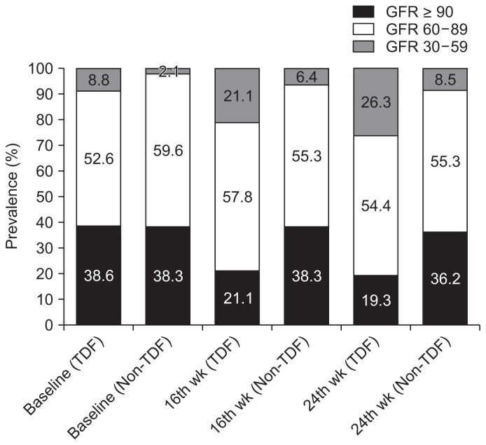
About three-fifths (61.4%) of the patients on tenofovir had impaired renal function at baseline. Over a third (38.3%) of the non-TDF group had normal GFRs at 16 weeks, although slightly over a fifth (21.3%) of the TDF group were already at the stage of chronic kidney disease. The difference in proportions of patients with various categories of GFR was statistically significant between the groups (P = 0.038). The categories of GFRs differed significantly between the TDF and non-TDF groups (P = 0.027) as over a quarter (26.3%) of the TDF group had GFRs reflective of chronic kidney disease by the 24th week (Fig. 1).
Figure 1. Prevalence of renal impairment among tenofovir diso-proxil fumarate (TDF) and non-TDF groups.
Renal impairment was defined as glomerular filtration rate (GFR) < 90 mL/min/1.73 m2; CKD as GFR < 60 mL/min/1.73 m2 persisting from baseline to the 16th and/or the 24th week. P for association between GFR and TDF use at baseline, the 16th and the 24th week were 0.254, 0.038, and 0.027, respectively.
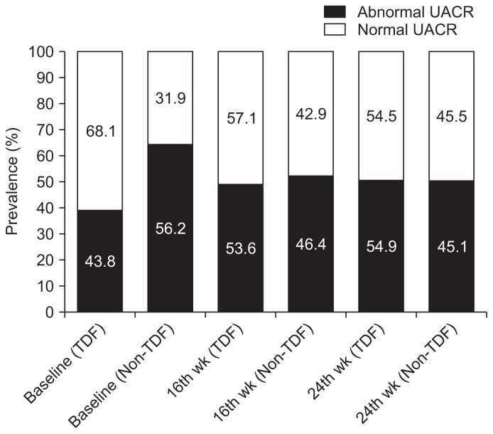
A greater proportion of the patients who had abnormal UACR at baseline were those on non-TDF ARTs (56.2% vs. 43.8%; P = 0.013). The proportion of patients with abnormal UACR among the TDF group increased progressively from less than half (43.8%) to almost three-fifths (54.9%) at 24 weeks; however, the difference in prevalence of abnormal UACR between the two groups from 16 weeks to the end of the follow-up period was not significant (P = 0.773 and 0.971, respectively; Fig. 2).
Figure 2. Prevalence of abnormal urine albumin-creatinine ratio (UACR) among tenofovir disoproxil fumarate (TDF) and non-TDF groups.
Abnormal UACR was defined as of > 30 mg/g. Blue bar present percentage of patients with normal value. Red bar presents percentage of patients with abnormal value. P for difference in proportions between TDF and non-TDF groups at baseline, the 16th and the 24th week were 0.013, 0.773, and 0.971, respectively.
Discussion
The advent of antiretroviral agents for the effective treatment of HIV is one of the best things that have ever happened to people living with HIV/AIDS in the twenty-first century; however, some of these drugs have adverse effects, particularly on the kidneys and possibly other vital organs. Evidence suggests that tenofovir has devastating effects on renal tubular and glomerular function [20,21]. This study sought to highlight some of the effects that tenofovir may have on renal function by comparison with a group of HIV-infected patients not on a tenofovir-based regimen in order to identify the connection between renal dysfunction and tenofovir use among HIV-positive patients.
There was a female predominance in this study. Females are more prone to HIV infection, especially via sexual transmission, and this could explain the higher female to male ratio. Conversely, male predominance was reported from similar studies conducted in Spain and the USA [22,23]. High-risk sexual behavior and differing sexual or gender preferences may account for this variation.
The age and sex distributions of the patients were considerably similar in the two groups. The modal age group was 25 to 34 years and the mean age was 34.1 ± 8.2 years. Similarly, a study conducted in Zambia reported an average age of 32 years for patients treated with tenofovir [24]. Because the modal age in this study represents a young population constituting a major part of the ‘non-dependent’ economically-productive age group, it is unpromising that young people bear the burden of HIV/AIDS. There is no known cure for this pandemic, and anti-retroviral drugs have many adverse effects. Thus, the quality of life of HIV patients is, beyond doubt, not optimal. The burden of ingesting pills daily, compounded with stigmatization, further reduces the measure of satisfaction they derive from life.
Dipstick urinalysis at baseline revealed that about one-tenth of the patients had proteinuria. Although no significant association between proteinuria and the use of TDF was recognized at the initial assessment, about 1 in 20 of the TDF group also had hematuria. These findings of hematuria and proteinuria are suspicious of pre-therapy abnormalities and may be substantiated by the high level of urinary proteinuria observed at baseline for all HIV-positive patients. However, they are not confirmatory of pre-treatment renal abnormality. A study conducted in England also reported a high prevalence of pre-therapy subclinical proteinuria among HIV-positive patients [21].
Persistently-increasing proteinuria observed in the weeks following baseline assessment is probably due to damage to the kidneys by the administration of tenofovir in this study and is corroborated by the finding from a previous study in the USA which reported repeated measures of proteinuria among subjects who had tenofovir [22].
There was persistent glycosuria in spite of normal blood glucose among these patients, a finding that may point to proximal tubular damage as there was progressive excretion of glucose in urine. Similarly, there was significantly increased excretion of phosphate, albumin, uric acid and protein after initiation of ART. However, use of tenofovir-based ART seemed to have had a greater impact on renal function, as phosphaturia was significantly higher among the tenofovir group. Persistent excretion of phosphate in urine could reduce bone density in tenofovir users and increase their susceptibility to pathological fractures. This finding is supported by a similar report from Denmark where the use of TDF has been associated with phosphate wastage in urine and subsequent bone demineralisation [25].
Although urinary bicarbonate and amino acids were not assessed in this study, the observation of persistent renal glycosuria and phosphaturia may also be early warning signs of progression to Fanconi syndrome in tenofovir users [26], who may be susceptible to developing partial Fanconi syndrome as suggested by a 2014 review [27]. Similar findings of partial Fanconi syndrome among patients on TDF have also been documented in previous studies [14,28].
There was a sustained rise in prevalence of chronic kidney disease among all patients. This upsurge was significantly more marked among the tenofovir-exposed group than the controls, particularly at the 16th and 24th weeks. Evidence from previous studies supports the association of tenofovir use with progressive decline in kidney function [22,26,29,30]. Unfortunately, this renal abnormality may worsen with continued treatment as recent studies have reported that renal dysfunction due to tenofovir toxicity is not totally revocable even if treatment is discontinued, and progress to the symptomatic stage is inevitable without appropriate intervention [24,31]. It is, however, noteworthy that none of the patients in this study had eGFR < 30 mL/min/1.73 m2 and mean GFR was comparable between the two treatment groups, suggesting more of a tubular than a glomerular dysfunction. Similarly, a previous study reported that progressive decline in eGFR among tenofovir users is likely to be associated with tubular rather glomerular dysfunction [32]. Thus, biomarkers of tubular dysfunction will be instrumental in the recognition of pre-symptomatic nephrotoxicity before the marked reduction in GFR becomes obvious in HIV patients on TDF.
Nonetheless, the prevalence of abnormal UACR was high among all patients at baseline. It was, therefore, almost impossible to exclude a pre-treatment renal condition, although the baseline renal abnormality in this study may have been due to HIV-associated nephropathy as the virus also targets the kidneys in HIV patients without treatment [33].
Similarly, there was a gradual decrease in the proportion of patients with normal UACR, and this trend was more pronounced in the tenofovir group than the control group. The above findings all demonstrate likely ongoing damage to the kidneys with continued administration of TDF-based ART.
Surprisingly, the non-TDF patients had a higher prevalence of abnormal UACR at baseline. This finding suggests pre-treatment nephropathy which improved progressively from baseline to the 24th week. Although a vivid explanation for this finding is not readily available, it is possible that incremental rate of creatinine excretion exceeded albumin excretion in urine after initiating non-TDF-based ART.
Generally, most baseline serum and urine biomarkers of renal function in this study were not significantly different between the two groups, indicating a comparable pre-treatment renal status among all patients. This makes the effect of tenofovir demonstrated in this study plausible. However, selection bias was inevitable since participants were drawn from one tertiary hospital. An additional limitation is that this study reported the prevalence of renal tubular dysfunction as patients with prior subclinical biochemical derangement were not originally excluded; thus, further studies to elicit incidence of tubular dysfunction are required in the future.
In conclusion, this study demonstrated a progressive rise in the prevalence of renal dysfunction among HIV-positive patients following administration of tenofovir-based ART. However, pre-treatment renal dysfunction could not be completely excluded from all HIV-positive patients in this study. Indicators of tubular, rather than glomerular, dysfunction were found to be markedly higher among those patients on tenofovir-based ART. Therefore, biomarkers of tubular dysfunction will be valuable in detecting pre-symptomatic nephrotoxicity before a marked reduction in GFR becomes obvious in HIV patients on TDF.
Footnotes
Conflicts of interest
All authors have no conflicts of interest to declare.
References
- 1.Weller IV, Williams IG. ABC of AIDS: antiretroviral drugs. BMJ. 2001;322:1410–1412. doi: 10.1136/bmj.322.7299.1410. [DOI] [PMC free article] [PubMed] [Google Scholar]
- 2.Knobel H, Guelar A, Montero M, et al. Risk of side effects associated with the use of nevirapine in treatment-naïve patients, with respect to gender and CD4 cell count. HIV Med. 2008;9:14–18. doi: 10.1111/j.1468-1293.2008.00513.x. [DOI] [PubMed] [Google Scholar]
- 3.Caron M, Auclairt M, Vissian A, Vigouroux C, Capeau J. Contribution of mitochondrial dysfunction and oxidative stress to cellular premature senescence induced by antiretroviral thymidine analogues. Antivir Ther. 2008;13:27–38. [PubMed] [Google Scholar]
- 4.O’Brien ME, Clark RA, Besch CL, Myers L, Kissinger P. Patterns and correlates of discontinuation of the initial HAART regimen in an urban outpatient cohort. J Acquir Immune Defic Syndr. 2003;34:407–414. doi: 10.1097/00126334-200312010-00008. [DOI] [PubMed] [Google Scholar]
- 5.Gallant JE, DeJesus E, Arribas JR, et al. Tenofovir DF, emtricitabine, and efavirenz vs. zidovudine, lamivudine, and efavirenz for HIV. N Engl J Med. 2006;354:251–260. doi: 10.1056/NEJMoa051871. [DOI] [PubMed] [Google Scholar]
- 6.Gallant JE, Staszewski S, Pozniak AL, et al. Efficacy and safety of tenofovir DF vs stavudine in combination therapy in antiretroviral-naive patients: a 3-year randomized trial. JAMA. 2004;292:191–201. doi: 10.1001/jama.292.2.191. [DOI] [PubMed] [Google Scholar]
- 7.Nelson MR, Katlama C, Montaner JS, et al. The safety of tenofovir disoproxil fumarate for the treatment of HIV infection in adults: the first 4 years. AIDS. 2007;21:1273–1281. doi: 10.1097/QAD.0b013e3280b07b33. [DOI] [PubMed] [Google Scholar]
- 8.Izzedine H, Launay-Vacher V, Deray G. Antiviral drug-induced nephrotoxicity. Am J Kidney Dis. 2005;45:804–817. doi: 10.1053/j.ajkd.2005.02.010. [DOI] [PubMed] [Google Scholar]
- 9.Rollot F, Nazal EM, Chauvelot-Moachon L, et al. Tenofovir-related Fanconi syndrome with nephrogenic diabetes insipidus in a patient with acquired immunodeficiency syndrome: the role of lopinavir-ritonavir-didanosine. Clin Infect Dis. 2003;37:e174–e176. doi: 10.1086/379829. [DOI] [PubMed] [Google Scholar]
- 10.Barrios A, García-Benayas T, González-Lahoz J, Soriano V. Tenofovir-related nephrotoxicity in HIV-infected patients. AIDS. 2004;18:960–963. doi: 10.1097/00002030-200404090-00019. [DOI] [PubMed] [Google Scholar]
- 11.Rifkin BS, Perazella MA. Tenofovir-associated nephrotoxicity: Fanconi syndrome and renal failure. Am J Med. 2004;117:282–284. doi: 10.1016/j.amjmed.2004.03.025. [DOI] [PubMed] [Google Scholar]
- 12.Peyriere H, Reyenes J, Rouanet I, et al. Renal tubular dysfunction associated with tenofovir therapy: report of 7 cases. J Acquir Immune Defic Syndr. 2004;35:269–273. doi: 10.1097/00126334-200403010-00007. [DOI] [PubMed] [Google Scholar]
- 13.Zimmermann AE, Pizzoferrato T, Bedford J, Morris A, Hoffman R, Braden G. Tenofovir-associated acute and chronic kidney disease : a case of multiple drug interactions. Clin Infect Dis. 2006;42:283–290. doi: 10.1086/499048. [DOI] [PubMed] [Google Scholar]
- 14.Woodward CL, Hall AM, Williams IG, et al. Tenofovir-associated renal and bone toxicity. HIV Med. 2009;10:482–487. doi: 10.1111/j.1468-1293.2009.00716.x. [DOI] [PubMed] [Google Scholar]
- 15.Créput C, Gonzalez-Canali G, Hill G, et al. Renal lesions in HIV-1-positive patient treated with tenofovir. AIDS. 2003;17:935–937. doi: 10.1097/00002030-200304110-00026. [DOI] [PubMed] [Google Scholar]
- 16.Szczech LA. Renal dysfunction and tenofovir toxicity in HIV-infected patients. Top HIV Med. 2008;16:122–126. [PubMed] [Google Scholar]
- 17.Karras A, Lafaurie M, Furco A, et al. Tenofovir-related nephrotoxicity in human immunodeficiency virus-infected patients: three cases of renal failure, Fanconi syndrome, and nephrogenic diabetes insipidus. Clin Infect Dis. 2003;36:1070–1073. doi: 10.1086/368314. [DOI] [PubMed] [Google Scholar]
- 18.Antoniou T, Raboud J, Chirhin S, et al. Incidence of and risk factors for tenofovir-induced nephrotoxicity: a retrospective cohort study. HIV Med. 2005;6:284–290. doi: 10.1111/j.1468-1293.2005.00308.x. [DOI] [PubMed] [Google Scholar]
- 19.Gupta Samir K. Editorial comment: tenofovir-related nephrotoxicity: who’s at risk? AIDS Read. 2007;17:102–103. [PubMed] [Google Scholar]
- 20.Levey AS, Bosch JP, Lewis JB, et al. A more accurate method to estimate glomerular rate from serum creatinine: a new prediction equation. Modification of Diet in Renal Disease Study Group. Ann Intern Med. 1999;130:461–470. doi: 10.7326/0003-4819-130-6-199903160-00002. [DOI] [PubMed] [Google Scholar]
- 21.Kohler JJ, Hosseini SH, Hoying-Brandt A, et al. Tenofovir renal toxicity targets mitochondria of renal proximal tubules. Lab Invest. 2009;89:513–519. doi: 10.1038/labinvest.2009.14. [DOI] [PMC free article] [PubMed] [Google Scholar]
- 22.Hall AM, Hendry BM, Nitsch D, Connolly JO. Tenofovir-associated kidney toxicity in HIV-infected patients: a review of the evidence. Am J Kidney Dis. 2011;57:773–780. doi: 10.1053/j.ajkd.2011.01.022. [DOI] [PubMed] [Google Scholar]
- 23.Scherzer R, Estrella M, Li Y, et al. Association of tenofovir exposure with kidney disease risk in HIV infection. AIDS. 2012;26:867–875. doi: 10.1097/QAD.0b013e328351f68f. [DOI] [PMC free article] [PubMed] [Google Scholar]
- 24.Rodríguez-Nóvoa S, Labarga P, Soriano V, et al. Predictors of kidney tubular dysfunction in HIV-infected patients treated with tenofovir: a pharmacogenetic study. Clin Infect Dis. 2009;48:e108–e116. doi: 10.1086/598507. [DOI] [PubMed] [Google Scholar]
- 25.Mulenga L, Musonda P, Mwango A, et al. Effect of baseline renal function on tenofovir-containing antiretroviral therapy outcomes in Zambia. Clin Infect Dis. 2014;58:1473–1480. doi: 10.1093/cid/ciu117. [DOI] [PMC free article] [PubMed] [Google Scholar]
- 26.Rasmussen TA, Jensen D, Tolstrup M, et al. Comparison of bone and renal effects in HIV-infected adults switching to abacavir or tenofovir based therapy in a randomized trial. PLoS One. 2012;7:e32445. doi: 10.1371/journal.pone.0032445. [DOI] [PMC free article] [PubMed] [Google Scholar]
- 27.Waheed S, Attia D, Estrella MM, et al. Proximal tubular dysfunction and kidney injury associated with tenofovir in HIV patients: a case series. Clin Kidney J. 2015;8:420–425. doi: 10.1093/ckj/sfv041. [DOI] [PMC free article] [PubMed] [Google Scholar]
- 28.Hall AM, Bass P, Unwin RJ. Drug-induced renal Fanconi syndrome. QJM. 2014;107:261–269. doi: 10.1093/qjmed/hct258. [DOI] [PubMed] [Google Scholar]
- 29.Patel KK, Patel AK, Ranjan RR, Patel AR, Patel JK. Tenofovir-associated renal dysfunction in clinical practice: an observational cohort from western India. Indian J Sex Transm Dis. 2010;31:30–34. doi: 10.4103/0253-7184.68998. [DOI] [PMC free article] [PubMed] [Google Scholar]
- 30.Martin M, Vanichseni S, Suntharasamai P, et al. Renal function of participants in the Bangkok tenofovir study--Thailand, 2005–2012. Clin Infect Dis. 2014;59:716–724. doi: 10.1093/cid/ciu355. [DOI] [PMC free article] [PubMed] [Google Scholar]
- 31.Jose S, Hamzah L, Campbell LJ, et al. UK Collaborative HIV Cohort Study Steering Committee. Incomplete reversibility of estimated glomerular filtration rate decline following tenofovir disoproxil fumarate exposure. J Infect Dis. 2014;210:363–373. doi: 10.1093/infdis/jiu107. [DOI] [PMC free article] [PubMed] [Google Scholar]
- 32.Vrouenraets SM, Fux CA, Wit FW, et al. Persistent decline in estimated but not measured glomerular filtration rate on tenofovir may reflect tubular rather than glomerular toxicity. AIDS. 2011;25:2149–2155. doi: 10.1097/QAD.0b013e32834bba87. [DOI] [PubMed] [Google Scholar]
- 33.Estrella MM, Fine DM, Atta MG. Recent developments in HIV-related kidney disease. HIV Ther. 2010;4:589–603. doi: 10.2217/hiv.10.42. [DOI] [PMC free article] [PubMed] [Google Scholar]


