History
A 49-year-old, nonsmoking female was admitted to our clinic on 20 June 2001 with a one-year history of progressive dyspnea and dry cough. She denied sputum production, hemoptysis, night sweats and fever. At the time of her evaluation, she had been taking antituberculous drugs for two months. She had been noted to have abnormal shadows on a chest radiograph in 1990, but she had not been treated for it due to her good clinical status. She was treated for tuberculosis for about one year in 1993. Her family history was negative for any lung disease.
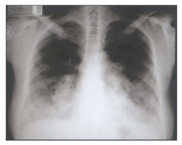
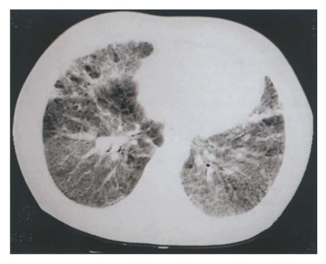
Vital signs were within normal limits. Examination of the chest indicated bibasilar end-inspiratory fine rales. Extremities showed digital clubbing of grade V with slight cyanosis. Although the patient appeared well at rest, she became dyspneic after walking about twenty to thirty meters. Laboratory findings are shown in the table. Plain chest x-ray and thorax CT are shown in Figures 1 and 2, respectively.
Figure 1.
Posteroanterior chest roentgenogram.
Figure 2.
Thorax CT scan.
(see answer on page 68)
Laboratory findings
| Hematology | |
| White blood cell count | 6500/mm3 with normal differential |
| Hemoglobin | 16.3 d/L |
| Hematocrit | 47.3% |
| Erythrocyte sedimentation rate | 50 mm/h |
| Serum chemistry profile | |
| Calcium | 8.3 mg/dL |
| Total protein | 9.1 g/dL |
| Albumin | 3.33 g/dL |
| Globulin | 5.77 g/dL |
| Resting arterial blood gas (ABG) | |
| pH | 7.44 |
| PaO2 | 61.5 mm Hg |
| PaCO2 | 35.8 mm Hg |
| Repeat ABG immediately after exercise | |
| pH | 7.36 |
| PaO2 | 46.4 mm Hg |
| PaCO2 | 35.1 mm Hg |
| Serum immune electrophoresis | |
| IgG | 29.8 g/L (N:5.5–19) |
| IgM | 2.12 g/L (N:0.6–2.7) |
| IgA | 7.18 g/L (N:0.6–3.3) |
| Pulmonary function tests | |
| Forced vital capacity (FVC) | 1.96 L (66.3% predicted) |
| Forced expiratory volume in one second (FEV1) | 1.64 L (66.7% predicted) |
| FEV1/FVC | 102.5% predicted* |
demonstrated restrictive disturbance


