Proximal femoral focal deficiency (PFFD) is a rare congenital malformation with an approximate incidence of 0.2/10 000 live births.1 The abnormality results in shortening and altered function of the involved lower extremity and can occur rarely as an isolated defect or in association with other skeletal malformations. This case report of a newborn with isolated unilateral PFFD and a well-developed acetabulum, which is unusual and diagnosed antenatally only twice in the literature.
Case
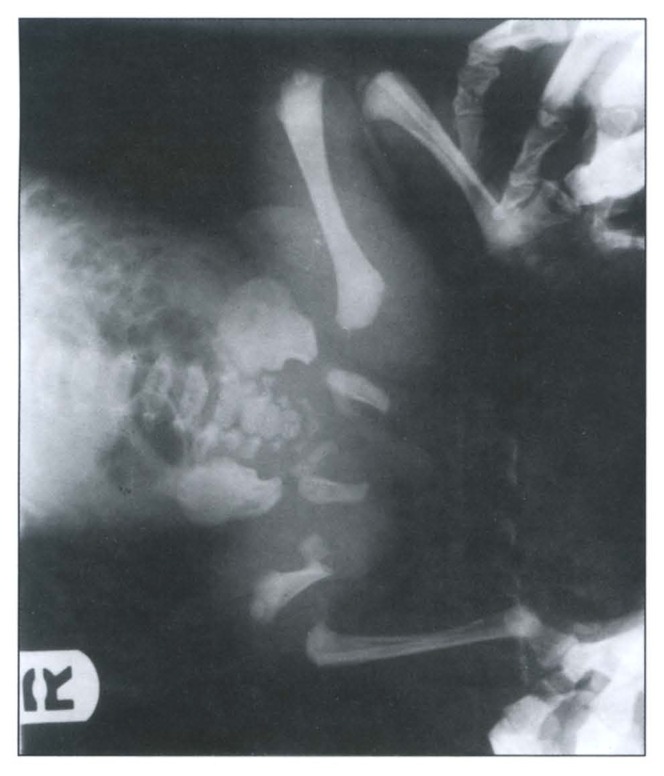
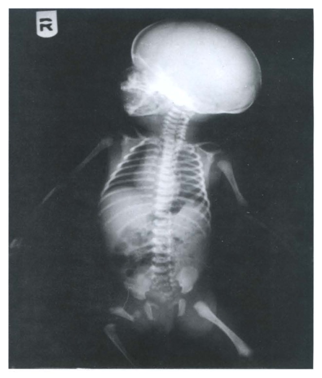
An 18-year-old primigravida was seen at the antenatal booking clinic at 25 weeks of gestation. Her routine antenatal screening was normal. She had an ultrasound examination that showed a normal fetus with breech presentation. All measurements were reported as normal. A repeat ultrasound scan was done at 38 weeks of gestation for weight estimation because of the breech presentation, and it was also reported to be normal. She delivered at 39 weeks of pregnancy by emergency cesarean section for fetal distress in labor. The female newborn weighed 2.4 kg with an Apgar score of 8 at 1 minute and 9 at 5 minutes. An extremely short right thigh was noticed in the newborn with the femoral shaft felt on the lateral side of the right thigh, but it was severely angulated. No other associated anomalies were noticed. Radiological survey of the long bones showed severe PFFD (Figure 1). A good acetabular development and a good space between that and the rest of the femur was noted (Figure 2). Other long bones were symmetrical in their measurements. Pediatric orthopedic consultation was obtained, and a long term follow-up plan was made.
Figure 1.
A skeletal survey of the long bones shows the fetus with proximal femoral focal deficiency.
Figure 2.
An x-ray of the affected limb showing the acetabulum and the space between it and the femur.
Discussion
Mid-trimester ultrasound screening for fetal malformations of all pregnancies is well-accepted routine antenatal care. Despite this fact, prenatal diagnosis of PFFD has been reported only twice in the literature. Once it was described by ultrasound in an otherwise unremarkable pregnancy,2 and the second time in a diabetic pregnant woman.3 Reasons for missing the diagnosis by ultrasound include inappropriate measurement of the femur length, shadowing the end of one bone by another bony structure, such as the iliac wing, the other femur or by the presence of a twin. In our case, the persistent breech presentation was the main reason for missing the diagnosis.
Most cases of PFFD are sporadic. Although some have reported a history of exposure to environmental factors such as drugs, viral infections, radiation, and focal ischemia, as well as trauma between the fourth and eighth week of gestation,1,4 others found no evidence of a specific environmental cause,5 as in our case, and there is still controversy over the etiology. Familial recurrence is quite unusual and chromosomal studies have failed to show any abnormality, but a postulated somatic mutation in the early embryo has been suggested.5
The short limb used to be managed by either amputation or prosthesis in the past. Surgical correction used to result in a significantly short limb,3 but the use of the Ilizarov technique in the correction of such deformities and extended limb lengthening has an excellent outcome,6 especially in cases similar to our patient as the acetabulum was not severely undeveloped.
Although a routine mid-trimester scan for anomalies should include viewing all long bones, we recommend keeping the image of the two fetal femurs, especially in cases of maternal diabetes. In cases of persistent breech presentation, a repeat scan with a full bladder is recommended if the image of the two femurs cannot be obtained in the first scan as this may help to reduce the incidence of misdiagnosis antenatally.
References
- 1.Hamanishi C. Congenital short femur, clinical, genetic and epidemiological comparison of the naturally occurring condition with that caused by Thalidomide. J Bone Joint Surg. 1980;62:307–320. doi: 10.1302/0301-620X.62B3.7410462. [DOI] [PubMed] [Google Scholar]
- 2.Jeanty P, Kleinman G. Proximal Femoral Focal deficiency. J Ultrasound Med. 1989;8:639–642. doi: 10.7863/jum.1989.8.11.639. [DOI] [PubMed] [Google Scholar]
- 3.Hadi HA, Wade A. Prenatal diagnosis of unilateral proximal femoral focal deficiency in diabetic pregnancy: case report. Am J Perinat. 1993;10(4):285–287. doi: 10.1055/s-2007-994741. [DOI] [PubMed] [Google Scholar]
- 4.Ashkenazy M, Lurie S, Ben-ltzhak I, Appelman Z, Caspi B. Unilateral congenital short femur: case report. Prenat Diag. 1990;10:67–70. doi: 10.1002/pd.1970100110. [DOI] [PubMed] [Google Scholar]
- 5.Lenz W, Zygulska M, Horst J. FFU complex: An analysis of 491 cases. Hum Genet. 1993;91:347–356. doi: 10.1007/BF00217355. [DOI] [PubMed] [Google Scholar]
- 6.Bell DF, Boyer MI, Armstrong PF. The use of the ilizarov technique in the correction of limb deformities associated with skeletal dysplasia. J Pediatr Orthop. 1992;12(3):283–290. doi: 10.1097/01241398-199205000-00003. [DOI] [PubMed] [Google Scholar]


