Abstract
BACKGROUND
Although the majority of cerebrospinal (CSF) fistulas in the anterior skull base are traumatic in nature, the minority is non-traumatic or primary. Non-traumatic CSF leak can be a diagnostic and treatment challenge.
PATIENTS AND METHODS
We describe the diagnosis, modified methods of localization, and surgical repair of a series of nine patients who presented with non-traumatic CSF rhinorrhea and were managed between July 2000 and October 2002.
RESULTS
Eight patients were managed via an endoscopic approach and one patient through an intracranial approach. The RI/T2-FLAIR test was used for localization of the site of the leak. The test confirmed the site of CSF leak in 6 patients. Successful repair of CSF rhinorrhea was achieved in 7 of 8 patients with a single endoscopic procedure; one patient required two procedures after a re-leak 18 months following the first repair.
CONCLUSION
Non-traumatic CSF rhinorrhea is a relatively rare condition and occurs secondary to different etiologies. Among multiple techniques available for localization, MRI/FLAIR is effective, but requires further evaluation and polishing. In the absence of a large skull base lesion or tumor, endoscopic repair of CSF fistula carries a high success rate with a high margin of safety and low morbidity rate.
Keywords: Cerebrospinal fluid, rhinorrhea, encephalocele, endoscopic surgery
Cerebrospinal fluid (CSF) leak occurs as a result of an abnormal communication between the subarachnoid space and a pneumatized area in the skull base that includes the sinonasal tract or the middle ear/mastoid region. This communication or fistula must involve a breech of the arachnoid and dura matter, the bone of skull base, and the underlying mucosa.1 CSF rhinorrhea can be classified according to the etiology into traumatic and non-traumatic. 2 Traumatic fistulae are subdivided into accidental, which compose the majority (up to 80%), and iatrogenic or post-surgical (16%). The non-traumatic or primary CSF fistula accounts for less than 4% of all CSF fistulae. The non-traumatic CSF fistula can be further classified into high pressure or normal pressure.
Because of the serious potential complications of CSF rhinorrhea, (e.g. meningitis, brain abscess, pneumocephalus) prompt management and repair of all CSF rhinorrhea should be attempted. Different approaches have been described for the repair of sinonasal CSF fistula, including the intracranial approach,3 which has a high failure rate (up to 70%).3,4 Multiple extracranial approaches have been used for repair of CSF leak.5–10 Mattox and Kennedy were the first to explain the details of endoscopic repair, reporting an initial success rate of 88% with a final success rate of 100%.11
Careful review of the literature reveals a minor emphasis on non-traumatic CSF leak. Most of the published studies discuss CSF leaks of traumatic origin as it is considered the most common. The purpose of this study was to review our experience at our institution with presentation and management of non-traumatic CSF rhinorrhea. In addition, a modification of new diagnostic methodology and endoscopic techniques used for the repair of different pathologies are discussed.
Patients and Methods
We reviewed the charts of all patients who presented to our department with CSF rhinorrhea. Patients with non-traumatic CSF leak were included in our retrospective study, while those with CSF rhinorrhea secondary to trauma were excluded. Nine patients presenting with CSF rhinorrhea had no previous history of trauma, or nasal or neurosurgical procedures (Table 1). The presence of CSF was confirmed in all patients by a positive β2-transferrin analysis The most common presenting symptom was clear rhinorrhea. Other symptoms included headache, nasal congestion and post-nasal discharge. Two patients presented with meningitis.
Table 1.
Nine patients who presented with non-traumatic CSF rhinorrhea
| Patient number | Age | Sex | Location | Cause of the leak | Size | Surgical technique | Grafting |
|---|---|---|---|---|---|---|---|
| 1 | 47 | F | Lt sphenoid | Encephalocele | 5 mm | Transnasal endoscopic | Overlay |
| 2 | 25 | M | Lt Sphenoid | Meningocele | 3 mm | Transnasal endoscopic | Overlay |
| 3 | 42 | F | Rt sphenoid | Spontaneous defect | 3 mm | Transnasal endoscopic | Overlay |
| 4 | 62 | M | Rt sphenoid | Encephalocele | 3 mm | Transnasal endoscopic | Overlay |
| 5 | 42 | F | Lt sphenoid | Spontaneous defect | 2 mm | Transnasal endoscopic | Overlay |
| 6 | 52 | F | Rt cribriform | Spontaneous defect | 4 mm | Transnasal endoscopic | Underlay |
| 7 | 30 | M | Rt cribriform | Spontaneous defect | 3–4 mm | Transnasal endoscopic | Underlay |
| 8 | 38 | F | Rt ethmoid | Encephalocele | 1.5 cm | Rt frontal craniotomy | Underlay |
| 9 | 43 | F | Rt ethmoid | Meningocele | 5 mm | Transnasal endoscopic | Underlay |
Thin-cut (2 mm) coronal CT scans of the paranasal sinuses were obtained for all patients. Additional radiological tests were done to help identify the nature of the defect and localize the site of the defect in patients undergoing exploration and repair. Combined thin-section (2 mm) coronal T2 weighted MRI and corresponding thin-section fluid attenuated inversion recovery (MRI/T2-FLAIR) was done to confirm the site of the leak and the pathology causing the defect. All patients underwent endoscopic nasal examination using a rigid nasal scope of ‘0°’ and ‘30°’ to try to identify the site of the lesion and rule out other pathological lesions within the nasal cavity.
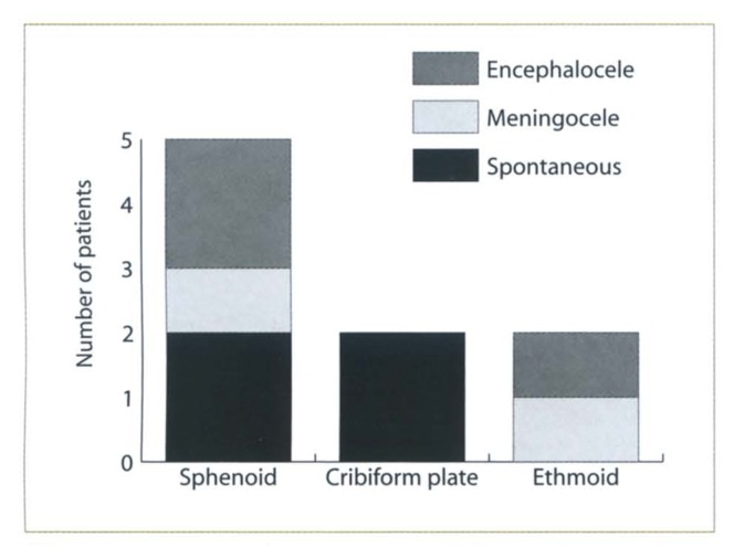
The site of CSF leak included the sphenoid sinus, cribriform plates and ethmoid sinuses (Table 1). Meningocele and encephalocele were the most commonly associated pathology encountered in the sphenoid (3 patients) and ethmoid sinuses (2 patients) (Figure 1). Spontaneous leak secondary to bony defect was encountered in the sphenoid sinus (2 patients) and the cribriform plate (2 patients).
Figure 1.
Distribution of the patients according to the site and the etiology of the lesions.
Operative techniques
Sphenoid fistula. Adequate exposure to the site of the leak was obtained by endoscopic sphenoidotomy performed from medial to middle turbinate. The sphenoid ostium was enlarged medially, inferiorly and latteraly at the same time, which provided fair support to the graft anteriorly. Once the site of the leak was identified, the mucosa of the sphenoid sinus was denuded using otologic microsurgical instruments like the Rosen knife. In the presence of encephalocele, its origin was cauterized using a bipolar cautery to reduce its size and maintain hemostasis control. It is essential to divide all the intranasal-intracranial connections of the cephalocele to allow it to recede intracranially, being extra cautious not to create intracranial bleeding. A fascia lata graft harvested from the thigh and trimmed into an appropriate size was applied to the site of the leak followed by a fat graft to obliterate the sinus and maintain pressure to the facial graft. In the case of a larger defect (>5 mm), cartilaginous graft can be used to seal the defect. Fibrin glue was applied to the graft using a large bore needle to seal the space between the graft and the defect. The sphenoidotomy opening and the anterior wall of sphenoid were packed with gel foam and the nasal cavity packed with a non-absorbable pack like a Sofratol pack for 3 to 5 days.
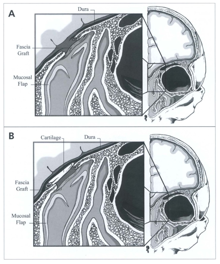
Fovea ethmoidalis and cribriform plate defect. The fistulae in the fovea ethmoidalis and the cribriform plate were repaired in a manner similar to the sphenoid sinus. Having identified the site of the fistula, the mucosa surrounding the defect was dissected as a pedicled flap for about 3 to 5 mm from the fistula site (Figure 2a). The size of the lesion determined the type of grafting technique used. In small defects (<3 mm), an overlay grafting technique was applied using fascia lata, which was further stabilized using fibrin glue. On the other hand, in larger defects, dissection of the dura from the surrounding bone of the skull base was done and a piece of cartilage graft of appropriate size was interpositioned between the dura and the bony edges in an underlay fashion (Figure 2b). This was supported by a facial graft applied using an overlay technique to achieve complete sealing of the fistula. Fibrin glue and Gel foam pack were applied in a similar fashion.
Figure 2.
(A) Overlay technique for small defect repair (<3mm); (B) Underlay technique for large defect repair (>3mm).
Results
Nine cases, 3 males and 6 females, were identified as having non-traumatic CSF rhinorrhea. Their ages ranged from 25 to 68 years. The follow-up period ranged between 6 to 24 months with an average of 15 months. The fistula defect was in the sphenoid sinus in five patients, secondary to cephalocele in three patients (two encephaloceles and one meningiocele), and a spontaneous defect in two patients. Two patients had CSF rhinorrhea secondary to one encephalocele and one meningocele in the ethmoid sinuses and two patients had a spontaneous defect in the cribriform plate. Prior to surgical repair, all patients had received conservative medical management that had failed.
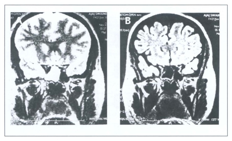
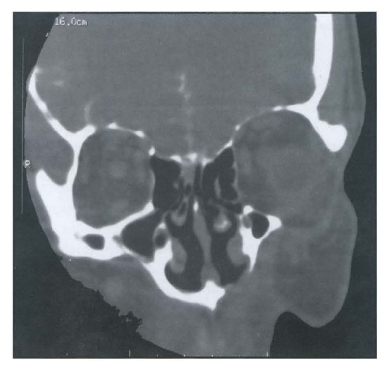
The clear fluid collected from the nose was confirmed to be CSF by positive β2-transferrin electrophoresis in all nine patients. CT studies were positive in five patients where a bony defect could be identified. On the other hand, MRI/T2-FLAIR tests done in seven patients confirmed the presence, and localized the CSF leak in six patients. A positive MRI result was documented when the fluid collection within the paranasal sinus had a CSF signal pattern that was hyperintense on T2 and equally suppressed to CSF on FLAIR (Figure 3). In three patients, MRI/T2-FLAIR localized the CSF fistula when the leak was not active during the study. In one patient, CT scan and MRI failed to locate the site of the lesion. In this particular patient, CT cisternography was done, which was accurate in localizing the site of the leak in the right cribriform plate (Figure 4).
Figure 3.
MRI/FLAIR imaging of non-traumatic CSF rhinorrhea for sphenoid lesions: (A) MRI T2-weighted image of spontaneous CSF rhinorrhea of the right sphenoid sinus; (B) FLAIR imaging of the same patient, notice the nulling signal of the CSF.
Figure 4.
CT-cisternography revealing CSF agress from right cribriform area.
One patient with a large ethmoid encephalocele (>2 cm defect) was elected for an intracranial subfrontal approach. Since an endonasal approach would be less efficient for dealing with intracranial bleeding secondary to injury to blood vessels that accompanied the encephalocele, this approach was elected. An endonasal endoscopic approach was used in eight patients to repair the CSF fistulae. Seven patients had successful cessation of the rhinorrhea after a single procedure. One patient who presented with a left sphenoid encephalocele had a recurrence 18 months after the first procedure. The recurrence could have happened due to incomplete resection of the origin of the basal encephalocele where it extended through the lateral wall in a laterally pneumatized recess of the sphenoid sinus. In the second procedure, which was done endoscopically, the lateral wall was exposed sufficiently and the origin of the encephalocele was approached and cauterized.
No major complication occurred secondary to surgical management. However, minor complications occurred in three patients, including headache and nasal congestion that were temporary and improved with symptomatic treatment.
Discussion
Non-traumatic CSF rhinorrhea can be spontaneous, caused by a bony defect or a cephalocele (encephalocele or meningiocele) protruding into the nasal cavity. However, the causes of spontaneous CSF rhinorrhea are variable and not very well understood. Some studies suggest a congenital defect, while others believe it is due to a small encephalocele that erodes the bone and ruptures.12,13 Spontaneous CSF leak might also occur secondary to focal atrophy of the olfactory nerve in the region of the cribriform plate. Additionally, defective development of the bony skull base could allow the arachnoid and brain tissue to protrude through the nose.14 Since the communication between a sterile intracranial compartment and a non-sterile sinonasal cavity can lead to life threatening complications (e.g., meningitis, pneumocephalus, or brain abscess,15–17 prompt diagnosis and management is demanded.
All patients in our study were subjected to conservative medical treatment that included bedrest in a semi-setting position for 10 days to 2 weeks, and avoidance of straining with the use of laxative and antitussive medication when needed. In the absence of obvious infection, the role of antibiotics is controversial. All nine patients with non-traumatic CSF rhinorrhea failed to stop leaking by conservative measures alone. Two patients responded to conservative treatment temporarily, but the leak restarted shortly after cessation of the conservative measure. Our experience with conservative treatment is consistent with the Mayo Clinic study where all 28 patients with spontaneous CSF rhinorrhea ended up closing surgically.18
Chemical analysis of the collected samples of the rhinorrhea is important to confirm the nature of the CSF fluid. An adequate amount of CSF was collected for β2-tansferrin analysis in all patients. All collected samples were positive for β2-tansferrin, which is considered a reliable specific marker for CSF. β2-tansferrin is also present in the perilymph of the inner ear and in the vitreous humor of the eye.
Radiological investigations play a pivotal role in identifying the underlying etiology of CSF rhinorrhea and in detecting the site, side, and size of the leaking fistula. With thin cuts of coronal sections, a CT scan was helpful in detecting bony dehiscence, which is not an uncommon finding in the skull base. Other CT findings in CSF leak cases include deviation of christa galli, causative intracranial lesions (e.g. encephalocele), complications of CSF leak (e.g. pneumocephalus), or fluid in the extracranial compartment adjacent to the fistula site.15 High resolution CT scan has an overall sensitivity of 70% in detecting bony dehiscence.19 However, the rate of detection is much lower in cases of spontaneous CSF rhinorrhea with less than 2 mm skull base defects, due to partial volume averaging, resulting in false positive and false negative results.20 MRI is a valuable non-invasive test for detecting and localizing CSF leak. Modification of MRI technique by using both T2 MRI images with fluid attenuated inversion recovery (FLAIR) imaging was very helpful in localizing CSF rhinorrhea. A FLAIR sequence is also capable of differentiating skull base cystic lesions of a CSF nature (arachnoid cyst, meningocele) from non-CSF lesions with similar signal intensity on T1, T2 sequence (e.g. epidermoid, inflammatory sinus changes).21,22
CT cisternography is another valuable radiographic study that is considered the procedure of choice in detecting CSF leak.24–26 It requires administration of a low osmolar non-ionic iodine contrast agent into the subarachnoid space with a subsequent search for agent egress through the fistula site.15 The sensitivity can reach up to 90% in active leaks. However, this technique is invasive and the sensitivity can be as low as 40% in cases of intermittent or inactive leaks.27
Surgical management of CSF rhinorrhea can be achieved by an intracranial or extracranial approach. The decision depends on the size and pathology causing the leak. Intracranial tumors or a large encephalocele eroding into the sinonasal cavity are difficult to manage by the endonasal approach, especially if complications, e.g. intracranial bleeding, should occur. However, management of CSF rhinorrhea by an intracranial approach carries morbidity and failure rates between 20 to 40%.18,28 On the other hand, an endoscopic approach is less morbid and has a success rate of 90 to 100%.11 In the present study, only one of nine patients was elected for an intracranial approach because of a large encephalocele protruding through the ethmoid sinus. In the other eight patients, an endoscopic approach was used to seal the CSF fistulae and treat associated pathologies. The overall success rate was comparable to other studies where eight of nine patients required a single procedure (one intracranial and seven endoscopic). One patient who suffered from left sphenoid encephalocele presented 18 months after the first procedure with CSF re-leak. The patient underwent a second endoscopic procedure for sealing of the CSF leak and remained leak-free up to his last follow-up visit, which was 14 months following the last procedure. The defect in this patient was due to an encephalocele that was protruding through a pneumatized lateral extension of the sphenoid sinus. The frequency of such lateral pneumatization of the sphenoid sinus is about 28% in normal skulls.29
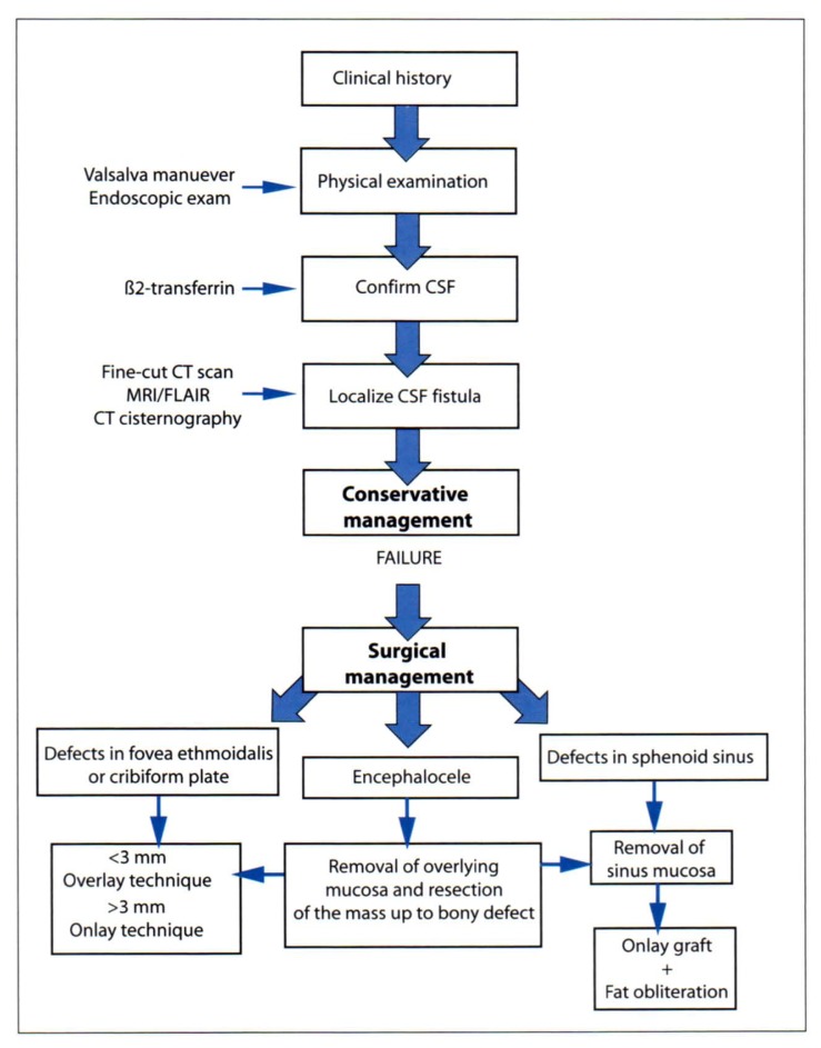
Adjunctive techniques that can be used to increase the success rate of CSF fistula repair include perioperative antibiotics, and complete bedrest with elevation of the head. The indications for postoperative lumbar drain are still controversial. Diagnosis and management of non-traumatic CSF rhinorrhea is shown in Figure 5.
Figure 5.
Diagnosis and management of non-trumatic CSF rhinorrhea. F.E., Fovea ethmoidalis; C.F.P. cribriform plate.
Conclusion
Non-traumatic CSF rhinorrhea is a relatively rare condition occurring secondary to different etiologies. Because of potential risky complications, prompt diagnosis and management should be attempted. A multidisciplinary team, composed of an otolaryngologist, radiologist, and neurosurgeon, should be involved in the diagnosis and repair of the CSF fistula. Precise preoperative localization is essential for successful repair. Among multiple techniques available for localization, MRI/FLAIR technique is effective but requires further evaluation and polishing. In the absence of a large skull base lesion or tumor, endoscopic repair of a CSF fistula carries a high success rate with a high margin of safety and low morbidity rate.
Acknowledgement
We would like to thank Dr. Jarah Al-Tubaikh for his fine work in producing the illustrations of surgical procedure.
References
- 1.Zapalac JS, Marple BF, Schwade ND. Skull base cerebrospinal fluid fistulas: A comprehensive diagnostic algorithm. Otolaryngol Head Neck Surg. 2002;126:669–676. doi: 10.1067/mhn.2002.125755. [DOI] [PubMed] [Google Scholar]
- 2.Ommaya AK, DiChiro G, Baldwin M, et al. Non-traumatic cerebrospinal fluid rhinorrhea. J Neurol Neurosurg Psychiatry. 1968;31:214–255. doi: 10.1136/jnnp.31.3.214. [DOI] [PMC free article] [PubMed] [Google Scholar]
- 3.Dandy WD. Pneumocephalus (intracranial pneumocele or aerocele) Arch Surg. 1926;12:949–982. [Google Scholar]
- 4.Park JI, Strezlow VV, Freidman WH. Current management of cerebrospinal fluid rhinorrhea. Laryngoscope. 1983;93:1294–1300. doi: 10.1002/lary.1983.93.10.1294. [DOI] [PubMed] [Google Scholar]
- 5.Dohlman G. Spontaneous cerebrospinal rhinorrhea. Acta Otolaryngol Suppl (Stockh) 1948;67:20–23. doi: 10.3109/00016484809129635. [DOI] [PubMed] [Google Scholar]
- 6.Hirsch O. Successful closure of cerebrospinal fluid rhinorrhea by endonasal surgery. Arch Otolaryngol. 1952;56:1–13. doi: 10.1001/archotol.1952.00710020018001. [DOI] [PubMed] [Google Scholar]
- 7.Vrabec DP, Hallberg OE. Cerebrospinal fluid rhinorrhea. Arch Otolaryngol. 1964;80:218–229. doi: 10.1001/archotol.1964.00750040224022. [DOI] [PubMed] [Google Scholar]
- 8.Wigand ME. Transnasal ethmoidectomy under endoscopic control. Rhinology. 1981;19:7–15. [PubMed] [Google Scholar]
- 9.Stankiewicz JA. Complications of endoscopic intranasal ethmoidectomy: an update. Laryngoscope. 1989;99:686–690. doi: 10.1288/00005537-198907000-00004. [DOI] [PubMed] [Google Scholar]
- 10.Papay FA, Maggiano H, Dominquez S, et al. Rigid endoscopic repair of paranasal sinus cerebrospinal fluid fistula. Laryngoscope. 1989;99:1195–1201. doi: 10.1288/00005537-198911000-00018. [DOI] [PubMed] [Google Scholar]
- 11.Mattox DE, Kennedy DW. Endoscopic management of cerebrospinal fluid leaks and cephaloceles. Laryngoscope. 1990;100:857–862. doi: 10.1288/00005537-199008000-00012. [DOI] [PubMed] [Google Scholar]
- 12.Ommaya AK. Spinal fluid fistulae. Clin Neurosurg. 1976;23:363–392. doi: 10.1093/neurosurgery/23.cn_suppl_1.363. [DOI] [PubMed] [Google Scholar]
- 13.Anderson WM, Schwartz GA, Gommon GD. Chronic spontaneous cerebrospinal rhinorrhea. Arch Intern Med. 1961;107:723–731. doi: 10.1001/archinte.1961.03620050089009. [DOI] [PubMed] [Google Scholar]
- 14.Casiano RR, Jassir D. Endoscopic cerebrospinal fluid rhinorrhea repair: Is lumbar drain necessary? Otolaryngol Head Neck Surg. 1999;121:745–750. doi: 10.1053/hn.1999.v121.a98754. [DOI] [PubMed] [Google Scholar]
- 15.Wax MK, Ramadan HH, Ortiz O, et al. Contemporary management of cerebrospinal fluid rhinorrhea. Otolaryngol Head Neck Surg. 1997;116:442–449. doi: 10.1016/S0194-59989770292-4. [DOI] [PubMed] [Google Scholar]
- 16.Calvert CA, Cairns H. Discussion on injuries of the frontal and ethmoid sinuses. Proc R Soc Med. 1942;35:805–810. [PMC free article] [PubMed] [Google Scholar]
- 17.Munro D. The modern treatment of craniocerebral injuries with special reference to the maximum permissible mortality and morbidity. N Engl J Med. 1935;213:893–906. [PMC free article] [PubMed] [Google Scholar]
- 18.Hubbard JL, McDonald TJ, Pearson BW, Laws ER. Spontaneous cerebrospinal fluid rhinorrhea: evolving concepts in diagnosis and surgical management based on the Mayo Clinic experience from 1970 through 1981. Neurosurgery. 1985;16:314–321. doi: 10.1227/00006123-198503000-00006. [DOI] [PubMed] [Google Scholar]
- 19.Stone JA, Castillo M, Neelon B, Mukherji SK. Evaluation of CSF Leaks: High resolution CT compared with contrast enhanced CT and radionuclide cisternography. Am J Neuroradiol. 1999;20(4):706–712. [PMC free article] [PubMed] [Google Scholar]
- 20.Lund VJ, Savy L, Llyod G, Howard D. Optimum imaging and diagnosis of cerebrospinal fluid rhinorrhoea. J Laryngol Otol. 2000;114:988–992. doi: 10.1258/0022215001904572. [DOI] [PubMed] [Google Scholar]
- 21.Gacek RR, Gacek MR, Tart R. Adult spontaneous cerebrospinal fluid otorhoea: diagnosis and management. Am J Otolaryngol. 1999;20:770–776. [PubMed] [Google Scholar]
- 22.Bradely WG, Bydder GM. Mosby Year Book. 1st edition. 1997. Advanced MR Imaging Techniques; pp. 143–162. [Google Scholar]
- 23.Moser FG, Panush D, Rubin JS, Honigsberg RM, Spragen S, Eisig SB. Incidental paranasal sinus abnormalities on MRI of the brain. Clin Radiol. 1991;43:252–254. doi: 10.1016/s0009-9260(05)80249-1. [DOI] [PubMed] [Google Scholar]
- 24.Johnson DBS, Toland BJ, O’Dwyer AJ. Magnetic resonance imaging in the evaluation of cerebrospinal fluid fistulae. Clin Radiol. 1996;51:837–841. doi: 10.1016/s0009-9260(96)80079-1. [DOI] [PubMed] [Google Scholar]
- 25.Shetty PG, Shroff MM, Sahani DV, et al. Evaluation of high-resolution CT and MR cisternography in the diagnosis of cerebrospinal fluid fistula. Am J Neuroradiol. 1998;19:633–639. [PMC free article] [PubMed] [Google Scholar]
- 26.Gammal TE, Sobol W, Wadlington VR, et al. cerebrospinal fluid fistula: detection with MR cisternography. Am J Neuroradiol. 1998;19:627–631. [PMC free article] [PubMed] [Google Scholar]
- 27.EL Jamel MS, Pidgeon CN, Toland J, et al. MR cisternographyand the localization of CSF fistulae. Br J Neurosurg. 1994;8:433–437. doi: 10.3109/02688699408995111. [DOI] [PubMed] [Google Scholar]
- 28.Aarabi B, Leibrock LG. Neurosurgical approaches to cerebrospinal fluid rhinorrhea. Ear Nose Throat J. 1992;71:300–305. [PubMed] [Google Scholar]
- 29.Morely TP, Wortzman G. The importance of the lateral extentionof sphenoid sinus in post-traumatic cerebrospinal rhinorrhea and meningitis. J Neurosurg. 1965;22:326–332. doi: 10.3171/jns.1965.22.4.0326. [DOI] [PubMed] [Google Scholar]


