Abdominal pain causes 5% to 10% of the emergency department (ED) admissions.1–5 One-third of these problems are related to surgery. Most of the diagnoses are nonspecific abdominal pain (34%–52%) or pain with unidentified origin.3,6–8 One of the important tasks of the ED physician is to determine life threatening causes of abdominal pain among the many abdominal pain admissions as well as institute due treatment. Emergency physicians should rule out serious pathological conditions.3,9 Some etiologies do not threaten life but need further investigation.
Spontaneous rectus sheath hematoma is one of these causes of abdominal pain. Although rare, it is a serious etiology that underlies acute-onset abdominal pain and prompts admission to ED. Spontaneous rectus sheath hematoma was diagnosed in four patients admitted to our institution with severe abdominal pain in a two-month period. This report highlights the specific features of the patients and reviews the literature on this entity.
Patient 1
A 54-year-old male patient with abdominal pain was admitted to ED. He had been suffering from cough, nasal secretion and sputum for 4 days. Three days before a sudden pain in the left subcostal area had begun after a prominent severe cough. The pain was exacerbated by coughing, breathing and moving. The pain was characterized as sharp and was more prominent on the left lower quadrant. The patient was on colchicine and indomethacin for 15 years for gout disease, but had not taken his drugs for the last 10 days. He denied having nausea or vomiting, or any change in urinary output. Passage of bowel gases and stool was as usual. The bowel sounds were not altered. However, the pain increased gradually over 3 days. His pulse rate was 80 beats/minute and rhythmic, his blood pressure was 115/85 mm Hg, his respiratory rate was 18 breaths/minute, his temperature was 37.8°C, and arterial oxygen saturation was 97%.
Examinations of the head, ear, nose, and throat were unremarkable. Breath sounds were equal on both hemithoraces and rales were heard on the left lung extending to the middle. No rubs were audible. On cardiovascular system examination there were no pathological heart sounds. Abdominal percussion revealed dullness and stiffness in the left upper and lower quadrants. Abdominal palpation of the left quadrant revealed a painful mass which lay from the left upper to lower quadrant. Genital examination was normal.
On laboratory examination, the whole blood count revealed white blood cell (WBC) 10 300 /mm3, hemoglobin was 14.3 mg/dL, platelet was 212 000/mm3, partial thromboplastin time (PTT) was 26.2 sec and international normalized ratio (INR) was 1.02, and bleeding time was within normal limits. Electrolytes, hepatic aminotransferase levels, troponin T, myoglobin and creatine kinase-MB (CK-MB) were all within normal ranges. Urinalysis was normal. Chest x-ray revealed pneumonic infiltration in the left lower lobe of the lung and there was no pneumothorax. Sputum stain revealed gram-positive diplococcal microorganisms.
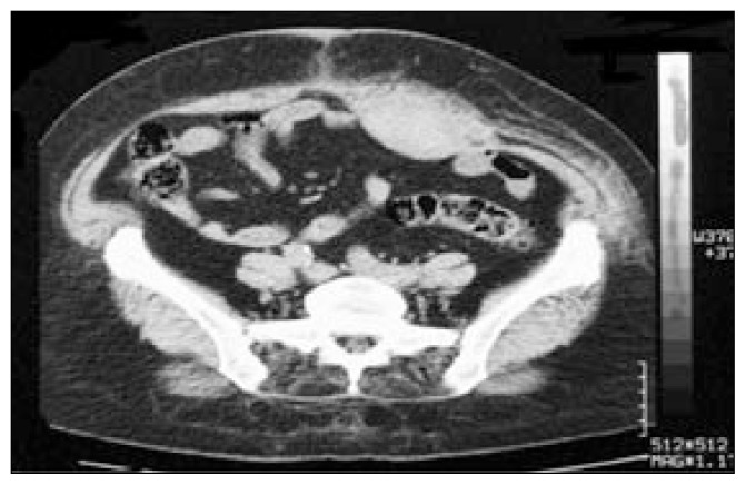
Abdominal ultrasonography (USG) was normal except for a suspect round hypoechoic image from the left upper to lower quadrants, and thus a thoracoabdominal computed tomography (CT) was obtained. Thoracoabdominal CT revealed an area of pneumonic consolidation on the left lung lower lobe and atelectasis, minimal pleural effusion and a hematoma of 8×27cm on the anterior wall of the abdomen and on the left side in the rectus abdominis muscle extending to the pubis (Figure 1). There was no acute pathology of the abdomen.
Figure 1.
CT scan of rectus sheath hematoma (patient 1).
He was followed up for 48 hours in ED. After surgical consultation he was discharged with antibiotic therapy for acute left lower lobe pneumonia. Daily control of the hematoma was performed, and the hematoma resolved spontaneously in 10 days.
Patient 2
A 70-year-old female was admitted to ED because of abdominal pain on the left lower quadrant. The pain had begun the day before and eventually was confined to the left lower quadrant. The pain lasted for the whole day and increased gradually, so she was brought to our ED. She had no trauma history. On presentation she had nausea and pain. She denied having experienced vomiting, or any change in urinary output. Stool passage was as usual. The bowel sounds were not altered. The pain increased gradually within 24 hours. She had aortic stenosis, hypertension and chronic atrial fibrillation and was on acetylsalicylate, warfarin and antihypertensive drugs (beta blockers, angiotensin-converting enzyme inhibitors).
Her pulse was 123 beats/minute and arrhythmic, her blood pressure was 145/85 mm Hg, her respiratory rate was 16 breaths/minute, her temperature was 36.8°C and arterial oxygen saturation was 96%. Examination of the head and ears, nose, and throat were unremarkable. Breath sounds were equal on both hemithoraces. No rales or rubs were heard. On examination of the cardiovascular system, there was a 2/6 systolic ejection murmur in the aortic area. Abdominal percussion revealed dullness and stiffness on the left lower quadrants. Abdominal palpation revealed a palpable tender mass in the lower left quadrant. There were ecchymoses on the suprapubic area. Genital examination was normal.
On laboratory examinations, the whole blood count showed WBC 5800/mm3, hemoglobin 11.5 mg/dL, platelets 168 000/mm3, PTT 34.9 seconds, INR 1.19, and bleeding time was within normal limits. Electrolytes, hepatic aminotransferase levels, troponin T, myoglobin and CK-MB were all within normal ranges. ECG disclosed atrial fibrillation and there was no ST-T wave change. Chest x-ray and urinalysis were normal.
Abdominal USG was normal except for a suspect round hypoechoic image of 7×3.5 cm which lay from the left lower quadrant to midline. Thoracoabdominal CT revealed a hematoma of 6×3.5 centimeters on the anterior wall of the abdomen and on the left side in the rectus abdominis muscle extending to the pubis (Figure 2). There was no acute pathology of the abdomen. The patient was followed up in the ED for seven days. Tachyarrhythmia was medically treated. Her vital signs and clinical condition were stabilized. After surgical consultation she was discharged. Daily control of the hematoma was performed, and the hematoma resolved gradually in 10 days.
Figure 2.
CT scan of rectus sheath hematoma (patient 2).
Patient 3
A 68-year-old female was admitted to ED because of abdominal pain in the left lower quadrant. The pain began suddenly, after two days of coughing and increased gradually, so she was brought to ED. She had no trauma history. She had nausea and abdominal pain on admission. She denied having experienced vomiting, or any change in urinary output. Passage of bowel gases and stool was as usual. The bowel sounds were not altered. But the pain increased gradually over a two-day period. She had been using drugs for chronic obstructive pulmonary disease (inhaled beta blockers).
Her pulse was 104 beats/min and rhythmic, blood pressure was 115/80 mm Hg, respiratory rate was 16 breaths/min, temperature was 36.8°C and arterial oxygen saturation was 95%. Examination of the head and ENT were unremarkable. Breath sounds were equal on both hemithoraces. No rales or rubs were noted. There were no pathological heart sounds. Abdominal percussion revealed dullness and stiffness in the left lower quadrant. Abdominal palpation revealed a palpable tender mass in the left lower quadrant and midline. Genital examination was normal.
The whole blood count was WBC 4400/mm3, hemoglobin 9.9 mg/dL, platelets 239 000/mm3, PTT 26.1 sec and INR 1.01; bleeding time was within normal limits. Electrolytes, hepatic aminotransferase levels, troponin T, myoglobin and CK-MB were all within normal ranges. ECG revealed atrial fibrillation, and there were no ST-T wave changes. Chest x-ray and urinalysis were normal.
Abdominal USG was normal except from a suspect round hypoechoic image of 7×15 cm which layed from the left lower quadrant to midline and thus a thoracoabdominal CT was obtained. Thoracoabdominal CT revealed a hematoma of 6×16-cm size on the anterior wall of abdomen, on the left side in the rectus abdominis muscle extending to the pubis (Figure 3). There was no acute pathology of the abdomen.
Figure 3.
CT scan of rectus sheath hematoma (patient 3).
The patient was admitted to the general surgery ward because of severe bleeding, decreased hemoglobin levels and tachycardia. After transfusion with 2 units of packed red blood cells, hemoglobin level rose to 11.9 g/dL, which was followed by improved vital signs. Daily control of hematoma was performed. Two days later the patient was discharged without any complications. The hematoma resolved spontaneously in 10 days.
Patient 4
A 62-year-old female was admitted to ED because of abdominal pain. She presented for right-sided abdominal pain that had begun after a sudden cough attack the day before. She had no trauma history and denied having experienced nausea and vomiting, and any change in urinary output. Passage of bowel gases and stool was as usual. The bowel sounds were not altered, but the pain increased gradually within a day. She had no chronic disease.
Her pulse was 92 beats/min and rhythmic, blood pressure was 122/80 mmHg, respiratory rate 16 breaths/min, temperature was 36.9 °C and arterial oxygen saturation was 97%. Examination of the head and ENT were unremarkable. Breath sounds were equal in both hemithoraces. No rales or rubs were heard. There were no pathological heart sounds. Percussion revealed dullness and stiffness in the right middle abdomen. Abdominal palpation of the right quadrant revealed a palpable tender mass, which lay from the right upper quadrant to the lower quadrant. ECG revealed a normal sinus rhythm and no ST-T wave change.
The whole blood count showed a WBC of 4000/mm3, hemoglobin 14.1 mg/dL, platelet 232 000/mm3, PTT 25.3 sec, INR 1.21 and bleeding time was within normal limits. Electrolytes, hepatic aminotransferase levels, BUN, creatinine, troponin T, myoglobin and CK-MB were all within normal ranges. Chest x-ray and urinalysis were normal.
Abdominal USG was normal except for a suspicious round hypoechoic image, which lay from the right upper to lower quadrants. Thoracoabdominal CT revealed a suspect image, 7×5 cm adjusted, with a hematoma in the right abdominis rectus muscle (Figure 4), but there was no pathological change in the abdomen. The patient was followed up for 48 hours in the ED with no complication. He was discharged with surgery department consultation. Outpatient follow up was undertaken for ten days with no complication, and the hematoma resolved spontaneously in 10 days.
Figure 4.
CT scan of rectus sheath hematoma (patient 4).
Discussion
Abdominal pain results from problems in the gastrointestinal tract (dyspepsia, gastritis, acute gastroenteritis, colon disorders, small bowel obstruction and appendicitis), gallbladder (acute cholecystitis and biliary colic), urinary tract (infection and ureteral colic), pancreas (acute pancreatitis) and from diseases of the pelvis and adjacent organs (pelvic inflammatory disease) (13–22%).3,9 Rectus sheath hematoma is generally not considered a reason for abdominal pain and its incidence as a cause of abdominal pain is unknown.10 The annual patient volume of our ED is nearly 30 000. In a time period of two months, four cases of rectus sheath hematoma were diagnosed among the acute surgical pathologies admitted.
A detailed history and physical examination is often enough to determine diagnosis and treatment,4 but sometimes a history and physical examination alone are not enough for accurate diagnosis and we may need to add radiographic and laboratory tests.3,9
Superior and inferior epigastric vessels run along the posterior border of the muscle within the sheath along its entire course.10–13 Tearing of these vessels or rupture of the rectus abdominis muscle causes rectus sheath hematoma.13 In the literature, risk factors for sheath hematoma include spontaneous or simple trauma, fast and immediate position changes, use of anticoagulant therapy,13,15 recent surgery, asthma exacerbation16 or coughing paroxysm,14 injections17 and pregnancy.13 Older patients may have more than one risk factor.13,14 In the four present cases, the mean age was 63.5 years. The 70-year-old female was on oral anticoagulant medications. Except for this patient, the main triggering factor was severe cough.
Severe ipsilateral tenderness is usually not present on the opposite side, as was the case in our patients. There can be ecchymoses on the abdominal wall or flank area, as in the reported case on warfarin therapy. If there is substantial blood in the rectus sheath hematoma, hypotension and tachycardia can be seen. Rectus sheath hematoma is self-limiting but it can be fatal in older patients.18 These findings should alert the emergency physician. Although rectus sheath hematoma is a non-surgical cause of abdominal pain, the localization can result in misdiagnosis of acute surgical diseases like abdominal wall tumors and hernia,19 appendicitis,20 periappendiceal abscess,21 biliary, diverticular,13,14 gynecologic11 diseases, and acute splenic22 or urinary tract diseases.
Accurate diagnosis and differential diagnosis should be made carefully to avoid unnecessary surgery and to get early appropriate treatment. So early diagnosis will help avoid usage of expensive diagnostic tests and unnecessary hospitalizations.10
In the diagnosis of rectus sheath hematoma abdominal USG, CT, and magnetic resonance imaging can be used. In high-risk patients, appropriate history, risk factors and physical examination are enough with only the help of abdominal USG. If these diagnostic modalities do not suffice, then CT can be used to rule out other significant causes.10 Rectus sheath hematoma can be managed non-operatively with bedrest and analgesics. It is rarely necessary to ligate epigastric vessels, to embolise bleeding vessels transvasculary13 or to evacuate hematoma.4,24 Deaths are rarely reported.18
Three of four patients had been hospitalized in the ED for two days before discharge from the hospital without any complication. One patient was hospitalized for seven days because of decreases in hemoglobin levels and was eventually discharged from the hospital with no other complication.
Emergency physicians should establish a differential diagnosis of potentially fatal acute abdomen among other etiologies for abdominal pain. Spontaneous hematoma of the rectus abdominis muscle should not be overlooked in the adult patient who presents with acute-onset abdominal pain. The diagnosis, whether clinical or based on imaging findings, needs accurate anatomopathologic knowledge of anterior abdominal wall. Once the diagnosis has been confirmed by abdominal USG or CT, treatment should be carried out conservatively. Surgery should be reserved for the complicated patient.
References
- 1.Brewer RJ, et al. Abdominal pain analysis of 100 cases. Am J Surg. 1976;131:219–220. doi: 10.1016/0002-9610(76)90101-x. [DOI] [PubMed] [Google Scholar]
- 2.Janzon L, Ryden CI, Zederfeldt B. Acute abdomen in the surgical emergency room. Who is taken care of when for what? Acta Chir Scand. 1982;148(2):141–8. [PubMed] [Google Scholar]
- 3.Alexander TT, Raymond HL. Acute Abdominal Pain. In: Rosen P, Barkin R, Danzl DF, editors. Emergency Medicine: Concepts and Clinical Practice. 4th ed. St. Louis: Mosby; 1998. pp. 1888–1903. [Google Scholar]
- 4.Stone R. Acute abdominal pain. Lippincott’s Prim Care Pract. 1998;2(4):341–57. [PubMed] [Google Scholar]
- 5.Sanson TG, O’Keefe KKP. Evaluation of abdominal pain in the elderly. Emerg Med Clin North Am. 1996;19:615–627. doi: 10.1016/s0733-8627(05)70270-4. [DOI] [PubMed] [Google Scholar]
- 6.de Dombal FT. The OMGE acute abdominal pain survey. Progress report, 1986. Scand J Gastroenterol Suppl. 1988;144:35–42. [PubMed] [Google Scholar]
- 7.Adelman A, Metcalf L. Abdominal pain in a university family practice setting. J Fam Pract. 1983 Jun;16(6):1107–11. [PubMed] [Google Scholar]
- 8.de Dombal FT. Computer diagnosis of acute abdominal pain in the elderly. Br Med J. 1972;2:9–12. doi: 10.1136/bmj.2.5804.9. [DOI] [PMC free article] [PubMed] [Google Scholar]
- 9.Gallagher EJ. Acute Abdominal Pain. In: Tintinalli JE, Kelen GD, Stapczynski JS, editors. Emergency Medicine: A Comprehensive Study Guide. 5th ed. North Carolina: McGraw-Hill; 1999. pp. 497–505. [Google Scholar]
- 10.Edlow JA, Juang P, Margulies S, et al. Rectus sheath hematoma. Ann Emerg Med. 1999;34:671–675. doi: 10.1016/s0196-0644(99)70172-1. [DOI] [PubMed] [Google Scholar]
- 11.Gocke JE, MacCarty RL, Foulk WT. Rectus sheath hematoma: Diagnosis by computed tomography scanning. Mayo Clin Proc. 1981;56:757–761. [PubMed] [Google Scholar]
- 12.Fukuda T, Sakamoto I, Kohzaki S, et al. Spontaneous rectus sheath hematoma: Clinical and radiologic features. Abdom Imaging. 1996:2158–61. doi: 10.1007/s002619900010. [DOI] [PubMed] [Google Scholar]
- 13.Zainea G, Jordan F. Rectus sheath hematomas: Their pathogenesis, diagnosis and management. Am Surg. 1988;54:630–633. [PubMed] [Google Scholar]
- 14.Titone C, Lipsius M, Krakauer J. “Spontaneous” hematoma of the rectus abdominis muscle: critical review of 50 cases with emphasis on early diagnosis and treatment. Surgery. 1972 Oct;72(4):568–72. [PubMed] [Google Scholar]
- 15.Scott W, Fishman E, Siegelman S. Anticoagulants and abdominal pain. JAMA. 1984;252:2053–2056. [PubMed] [Google Scholar]
- 16.Lee TM, Greenberger PA, Nahrwold DL, et al. Rectus sheath hematoma complicating an exacerbation of asthma. J Allergy Clin Immunol. 1986;78:290–292. doi: 10.1016/s0091-6749(86)80078-1. [DOI] [PubMed] [Google Scholar]
- 17.Webb K, Hadzima S. Hematoma of the rectus abdominis muscle: A complication of subcutaneous heparin therapy. South Med J. 1987;80:911–912. doi: 10.1097/00007611-198707000-00026. [DOI] [PubMed] [Google Scholar]
- 18.Ducatman BS, Ludwig J, Hurt RD. Fatal rectus sheath hematoma. JAMA. 1983;249:924–925. [PubMed] [Google Scholar]
- 19.Pandolfo I, Blandino A, Gaeta M, et al. CT findings in palpable lesions of the anterior abdominal wall. J Comput Assist Tomogr. 1986;10:629–633. doi: 10.1097/00004728-198607000-00016. [DOI] [PubMed] [Google Scholar]
- 20.Lohle PN, Puylaert JB, Coerkamp EG, et al. Nonpalpable rectus sheath hematoma clinically masquerading as appendicitis: US and CT diagnosis. Abdom Imaging. 1995;20:152–154. doi: 10.1007/BF00201526. [DOI] [PubMed] [Google Scholar]
- 21.Bober SE, Cohen HL, Setzen G, et al. Rectus sheath hematoma simulating periappendiceal abscess. J Ultrasound Med. 1992;11:179–180. doi: 10.7863/jum.1992.11.4.179. [DOI] [PubMed] [Google Scholar]
- 22.Noseda A, Bellens R, Van Gansbeke D, et al. Rectus sheath hematoma mimicking acute splenic disease. Am J Gastroenterol. 1983;78:566–568. [PubMed] [Google Scholar]
- 23.Levy JM, Gordon HW, Pitha NR, et al. Gelfoam embolisation for control of bleeding from rectus sheath hematoma. AJR Am J Roentgenol. 1980;135:1283–1284. doi: 10.2214/ajr.135.6.1283. [DOI] [PubMed] [Google Scholar]
- 24.Cervantes J, Sanchez-Cortazer J, Ponte RJ, et al. Ultrasound diagnosis of rectus sheath hematoma. Ann Surg. 1983;49:542–545. [PubMed] [Google Scholar]


