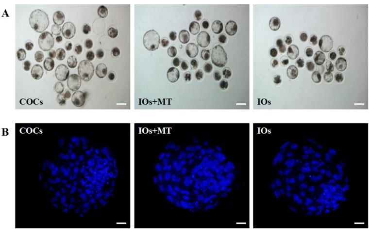
Figure 7.
Effects of melatonin on IVF embryo developmental potential and cell number of blastocysts. (A) Epifluorescence photomicrographs of in vitro-produced bovine blastocysts. COCs, cumulus–oocyte complexes; IOs, inferior bovine oocytes; MT, melatonin; scale bar = 100 μm; (B) Nuclear staining of bovine blastocyst after IVF in different groups. Scale bar = 20 μm.

