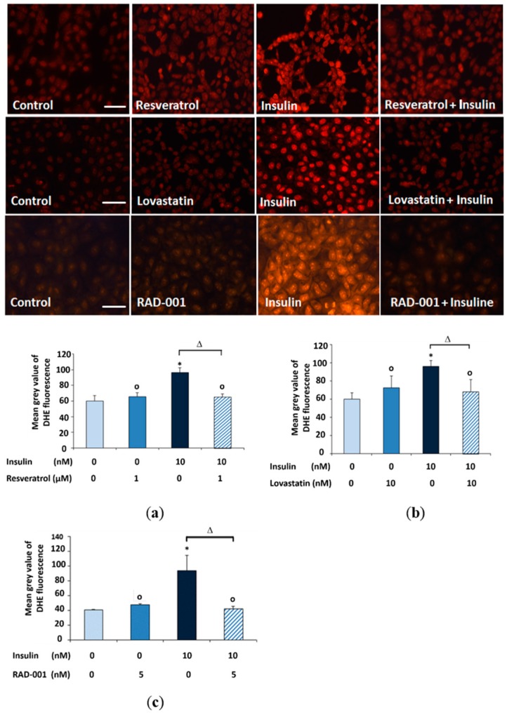
Figure 1.
Microscopic detection of superoxide formation using the dye DHE in NRK cells treated for 15 min with (a) 1 μM resveratrol; (b) 10 nM lovastatin and (c) 5 nM RAD-001 then addition of 10 nM insulin for 30 min in the presence of DHE. Quantification of DHE fluorescence was done by measuring the mean grey value of 200 cells using image j software. (*) Significantly different from control, (Δ) significantly different from insulin and (o) not significantly different from control. Scale bars 50 μm.

