Abstract
BACKGROUND
The nose plays a critical role in determining the external appearance of an individual. We studied the craniofacial anthropometrics by CT scanning since previous studies in the field were conducted in Saudi populations using photometric analysis.
OBJECTIVES
Obtain objective and quantitative data that can help surgeons plan cosmetic procedures for the nose.
DESIGN
A cross-sectional analytical study.
SETTING
Department of Otorhinolaryngology, Head and Neck Surgery, King Abdulaziz University Hospital, King Saud University, Riyadh, Saudi Arabia from February 2015 to December 2015.
PATIENTS AND METHODS
Facial CT scans were performed on native Saudis who underwent CT of the paranasal sinuses.
MAIN OUTCOME MEASURE(S)
Three anthropometric parameters: the nasofrontal angle, the pyramidal angle, and the linear distance between the nasion and the tip of the nasal bone.
RESULTS
In 160 native Saudis (86 males and 74 females) who underwent CT, the mean nasofrontal angle was 125.3° in males and 135.6° in females. The mean linear distance between the nasion and the tip of the nasal bone was 23.0 mm for males and 20.9 mm for females. The mean nasal pyramidal angle was 110.8° in males and 111.9° for females at the level of the nasal root, 105.6° in males and 104.8° in females at the mid-level of the nasal bone, and 116.8° males and 107.9° in females at the level of the tip of the nasal bone.
CONCLUSION
Nasal bone lengths and angles can be obtained accurately from CT scans. These angles differ in different ethnic groups.
LIMITATIONS
The sample represents native Saudis but not a cross section of the Saudi population. The relatively small sample size is a limitation of the study, but we consider these to be initial findings.
The bone of the nose is a crucial structure in forming the shape of the entire nose; hence, it performs specific anatomical and physiological functions. The nose also plays a critical role in determining the external appearance of an individual.1 The dorsum of the nose is considered the key measure for evaluation and aesthetic management of the nose. Effort should be made to individualize the analysis before an intervention to achieve a harmonic appearance for the candidate.
Craniofacial anthropometry has been commonly used in the field of anthropology and medicine. For that reason, it has actively contributed to establishing diagnostic and treatment guidelines in the field of plastic and cosmetic specialties.1 Previous studies in the field of craniofacial anthropometrics were conducted primarily using biometric or photometric analysis. However, there are limitations and difficulties analyzing the nasal bone without soft tissue analysis. Computed tomography of the face can now be used to analyze the nasal bone from different chosen angles.1 To the best of our knowledge, there has been no radiological analysis of nasal bone angles measurements in Saudi Arabia.2 People of Arab origin have characteristic nasal features2 and the popularity of rhinoplasty procedures has increased, so it is important for clinicians to understand not only ethnic differences but also the bony structures of the nose. Analysis of preoperative CT scans has shown a wide variation in lateral nasal wall anatomy and angulation.3 CT scans are used for assessing plans for surgical maneuvers in candidates for rhinoplasty despite the advent of fiberoptic scopes, due to the uncertainties that exist for both scopes and standard x-rays.3 In the era of advancements in CT scan technology, it is worth studying nasal structures, including the bones, to create and establish objective and quantitative data that help surgeons plan cosmetic procedures for the nose.4 The outcome of rhinoplasty procedures depends on the surgical skills as well as analysis of nasal anatomy before surgical intervention.5 We measured three anthropometric parameters: the nasofrontal angle, the nasal pyramid, and the linear distance between the nasion and the tip of the nasal bone. We compared the radiological and photometric characteristics of the native Saudi nose to the characteristics of the Asian nose, as reported, using data from the literature, to find out similarities and ethnic differences.
PATIENTS AND METHODS
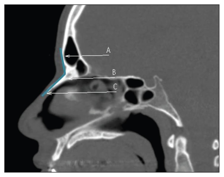
The cross-sectional analytical study was performed from February 2015 to December 2015 in the Department of Otorhinolaryngology, Head and Neck Surgery, King Abdulaziz University Hospital, King Saud University, Riyadh, Saudi Arabia. Subjects in the study included all patients seen during this time period. Approval from the Institutional Review Board (IRB) was obtained. Data was collected from native Saudis who underwent computed tomography of the paranasal sinuses (CT-PNS) for diagnosis of paranasal sinus disease. We collected three anthropometric parameters that are associated with rhinoplasty from these radiological images: the nasofrontal angle, the pyramidal angle, and the linear distance from nasion to the tip of nasal bone. Our exclusion criteria included age younger than 18 years and/or the presence of facial trauma, or a previous nasal surgery. The anthropometric points that we measured in the study included the following: the nasofrontal angle, which is the angle formed between the nasal bone and the frontal bone; the pyramidal angle, which is the angle formed between the two nasal bones; and the linear distance between the nasion and the tip of the nasal bone.
At the midsagittal view of all radiological images, point “A” was set as the most protuberant point of the glabella. “B” at the center of nasofrontal suture, the nasion. “C” was set at the tip of the nasal bone (Figure 1). On the axial cut of the CT scan, the pyramidal angle was measured at three points along the nasal bone, at the nasal root, at the nasal bone tip, and midway between the nasal bone tip and the nasal root. Point “D” was set at these three levels, the level of the nasal root, the middle level and the level at the nasal bone tip. Then point “E” and “F” were placed at the tangent level derived from point “D” to right and left nasal bones, respectively. The angles between D, E and F were calculated (Figure 2). Three-dimensional CT scans were done using a Brilliance iCT machine (Royal Philips Electronics of the Netherlands, Amsterdam). Images were taken in spiral mode with a slice of 1mm×1mm thickness. Measurements were done using the software built in the CT machine.
Figure 1.
Mid-sagittal computed tomography scan of the face. A: Glabella; B: Nasion; C: Tip of nasal bone.
Figure 2.
Axial computed tomography scan of the face. D: Mid nasal bone; E: Right nasal bone; F: Left nasal bone
RESULTS
We included 195 native Saudis who underwent CT scan evaluation for PNS (n=195). Thirty-five persons were excluded from the study based on the exclusion criteria mentioned above. One hundred sixty (86 males and 74 females) were included in the statistical analysis. Ages were distributed as follows: 6 persons (18–20 years of age), 53 (21–29 years of age), 54 (30–39 years of age), 27 (40–49 years of age), and 20 were 50 years of age and older.
The nasofrontal angle and the linear distance between the landmarks
Males had a mean nasofrontal angle of 125.3° (range 102.3°–149.4°), whereas overall mean nasofrontal angle in females was 135.6° (range 111.6°–152.2°). The mean nasofrontal angle in males (18–20 years of age) was 130.3° while it was 132.7° for females. The mean nasofrontal angle in males (21–29 years of age) was 127.9°, and 136.8° in females of the same age group. For the males (30–39 years of age), the mean nasofrontal angle was 123.0°, while for females the mean was 136.4°. For the age group (40–49 years of age), the mean nasofrontal angle for males was 122.4°, and it was 132.9° for females. Those older than 50 had a mean nasofrontal angle of 124.6° for males while 137.1° for females (Table 1). The mean linear distance between the nasion and tip of nasal bone was 23.0° in males, and 20.9° in females (Table 2).
Table 1.
Summary of measurements of the pyrimidal angles for 195 patients.
| Age group | Mean angle | |||||||
|---|---|---|---|---|---|---|---|---|
| Nasal bone tip | Mid nasal bone | Nasal root | NasoFrontal angle | |||||
| Male | Female | Male | Female | Male | Female | Male | Female | |
|
| ||||||||
| 18–20 | 103.2 | 96.5 | 104.2 | 92.5 | 106.6 | 103.7 | 130.3 | 132.7 |
| 21–29 | 116.5 | 107.5 | 106.2 | 108.0 | 107.3 | 112.9 | 127.9 | 136.9 |
| 30–39 | 119.4 | 108.5 | 105.8 | 104.6 | 113.7 | 111.1 | 123.0 | 136.4 |
| 40–49 | 112.7 | 108.4 | 109.3 | 103.8 | 110.7 | 112.6 | 122.4 | 132.9 |
| >50 | 121.3 | 109.1 | 100.8 | 104.1 | 114.7 | 112.6 | 124.6 | 137.1 |
| Range of angles for all patients | 93.1–162.9 | 71.8–136.5 | 73.3–128.6 | 71.3–137.9 | 84.2–132.8 | 91.0–137 | 102.3–149.4 | 111.6–152.2 |
| Mean of angles for all patients | 116.8 | 107.9 | 105.6 | 104.8 | 110.8 | 111.9 | 125.3 | 135.6 |
Values are degrees of angle.
Table 2.
Mean distance between the nasion and nasal bone tip.
| Age group | Mean distance | |
|---|---|---|
| Male | Female | |
|
| ||
| 18–20 | 21.5 | 22.9 |
| 21–29 | 23.2 | 21.3 |
| 30–39 | 24.6 | 20.7 |
| 40–49 | 21.0 | 21.1 |
| >50 | 22.4 | 19.7 |
| Mean distance (n=118) | 23.0 | 20.9 |
Values are millimeters.
Pyramidal angles of nasal bones
The mean pyramidal nasal angle at the level of the nasal root was 110.8° (range, 84.2°–132.8°) for males and 111.9° for females (range, 91°–137°). At the level of the middle nasal bone, the mean pyramidal angle for males was 105.6° (range, 73.3°–128.6°), and the mean was 104.8° for females (range, 71.3°–137.9°). At the tip of the nasal bone, the mean angle was 116.8° (range 93.1°–162.9°) for males and 107.9° (range 71.8°–136.5°) for females.
DISCUSSION
An important factor in performing rhinoplasty in a Middle Eastern population is to understand their physical and social characteristics.6 Although the role of CT scans in evaluating the preoperative plan for rhinoplasty patients is limited, we used our clinical series of CT-PNS data to find unique radiological features of the native Saudi nose and we compared the results of the Saudi population with the Asian community. Until recently, anthropometry was used medically as a tool to help plastic surgeons in the evaluation of the human face, or a part of it, as normal or abnormal, beautiful or aesthetically disadvantaged, and improved or not improved.6 This evaluation is usually performed quantitatively by comparing the individual to an ideal subject.6 However, the 3-dimensional stereoscopic location of facial structures are discrepant between the radiological anthropometry (2 dimension) and biometric (radiographic) anthropometry measurements.1 However, radiological advancement using 3-dimensional CT scans provides a more accurate assessment of the nasal bone and the angles of the nose. Our data fills a gap in the research, providing reference criteria for radiological measurements of the noses of Saudi people.
The mean nasofrontal angle in males was 125.3°, whereas the mean in females was 135.6°. In a study by Al-Qattan et al utilizing photographic analysis, the mean nasofrontal angle in Saudi males was 135.9°, and the mean angle was 145.9° in females.6 They concluded that the Saudi population had a large nasofrontal angle compared to all other races.6 The distance from the nasion to the tip of the nasal bone was reported by Al-Qattan et al to be 19.6 mm in males, and 18.2 mm in females.6 However, our study showed that males had a distance of 23.0 mm, and females had a distance of 20.9 mm. Moon et al measured the nasofrontal angle by CT analysis in 100 healthy young Korean men and women of similar age group who had no history of nasal surgery or deformity. Their study showed the mean radiological nasofrontal angle in men was 131.1°, and 140.7° for females.1 Additionally, their study showed the mean distance between the nasion and the nasal bone tip in men was 21.3 mm, and the mean distance for females was 18.0 mm.1 To the best of our knowledge, there has been no measurement of the pyramidal angle of the nasal bone for the Saudi population. In reference to the pyramidal angle reported by Moon et al, the mean angle at the root of the nasal bone was 112.9° for males and 103.3° for females.1 However, we found the mean pyramidal angle at the root of the nose for males was 110.8°, and the mean angle was 111.9 in females.
We believe that an analysis of the nasal bone alone is crucial but not sufficient for preoperative assessment and surgical planning. In particular, soft tissue/skin envelope thickness should be taken into consideration for corrective rhinoplasty procedures, as poor surgical outcomes have been associated with thick skin.7 Although they have been studied previously, structures adjacent to the nasal bones are important to review in patients who have distorted nasal bone frameworks prior to surgical intervention to avoid unwanted complications.3
In summary, we present measurements and angles at the level of nasal bones in Saudis. These measurements were different from other ethnic groups. Males tend to have longer nasal bones compared with females.
Footnotes
Conflict of interest
The authors have declared no conflicts of interest.
REFERENCES
- 1.Moon KM, Cho G, Sung HM, Jung MS, Tak KS, Jung S-W, et al. Nasal anthropometry on facial computed tomography scans for rhinoplasty in Koreans. Arch Plas Surg. 2013;40:610–615. doi: 10.5999/aps.2013.40.5.610. [DOI] [PMC free article] [PubMed] [Google Scholar]
- 2.Alharethy S, Aldaghri F, Mesallam TA, Farahat M, Bukhari MA. Nasal bone length in Saudi rhinoplasty: a clinical-radiological study. Ann Saudi Med. 2014 Jan-Feb;34(1):65–7. doi: 10.5144/0256-4947.2014.65. [DOI] [PMC free article] [PubMed] [Google Scholar]
- 3.Daniel RK, Ethier R. Rhinoplasty: a CT-scan analysis. Plast Reconstr Surg. 1987 Aug;80(2):175–84. [PubMed] [Google Scholar]
- 4.Lee SH, Yang TY, Han GS, Kim YH, Jang TY. Analysis of the nasal bone and nasal pyramid by three-dimensional computed tomography. Eur Arch Otorhinolaryngol. 2008 Apr;265(4):421–4. doi: 10.1007/s00405-007-0476-9. Epub 2007 Oct 11. [DOI] [PubMed] [Google Scholar]
- 5.Azizzadeh B, Mashkevich G. Middle Eastern rhinoplasty. Facial Plast Surg Clin North Am. 2010 Feb;18(1):201–6. doi: 10.1016/j.fsc.2009.11.013. [DOI] [PubMed] [Google Scholar]
- 6.Al-Qattan MM, Alsaeed AA, Al-Madani OK, Al-Amri NA, Al-Dahian NA. Anthropometry of the Saudi Arabian nose. J Craniofac Surg. 2012 May;23(3):821–4. doi: 10.1097/SCS.0b013e31824dfa69. [DOI] [PubMed] [Google Scholar]
- 7.Cho GS, Kim JH, Yeo NK, Kim SH, Jang YJ. Nasal skin thickness measured using computed tomography and its effect on tip surgery outcomes. Otolaryngol Head Neck Surg. 2011 Apr;144(4):522–7. doi: 10.1177/0194599811398936. Epub 2011 Feb 14. [DOI] [PubMed] [Google Scholar]


