Abstract
BACKGROUND
The paranasal sinuses and mastoid air cells vary considerably in size and shape from person to person. The main structures are pneumatic. In this study, we investigated the relationship between right and left sides and evaluated the volume changes according to age and sex.
METHODS
Of all patients attending the radiology department, 91 cases without paranasal sinuses and mastoid air cells pathology (i.e., inflammation, operation or trauma) were selected for evaluation. Axial computed tomography (CT) scans were obtained for both paranasal sinuses and temporal bones. In all scans, the volumes of each area (maxillary sinus, frontal sinus, sphenoid sinus and mastoid air cell) were calculated and analyzed statistically.
RESULTS
The volumes of paranasal sinuses and mastoid air cells increased with age and women had a lower mean volume. There was a positive correlation between right-left and ipsilateral structures (paranasal sinuses and mastoid air cells).
CONCLUSIONS
These results are helpful in understanding the normal and pathological conditions of the paranasal sinuses and the mastoid air cells.
Paranasal sinus anatomy is complex and rather variable from person to person. Significant differences in structure between the two sides may also exist in the same person. Therefore, a detailed knowledge of the anatomy of the sinuses is critical in performing procedures such as functional endoscopic sinus surgery.1,2
The main characteristics of these structures are pneumatic, and the initiation and process of pneumatization differ from person to person.2–7 Genetic diseases, environmental conditions and past infections may affect this process.4,6,8 There is still no consensus about when pneumatization begins and how it develops in temporal bone.8 Diamant and other investigators first studied the mastoid air cells on a scientific and methodological basis and first used this information in practice.6, 9, 10–14 Several studies on the pneumatization of temporal bone have been published.8,11,15 In their book, Graney et al15 reported several researchers’ results on measurement of paranasal sinuses at and after birth. Until the use of computed tomography (CT) by Haunsfield for diagnosis in 1972, x-ray films were used for the measurement of area, volume and shape of paranasal sinuses and the mastoid air cells.10,12–14 Since then the use of CT has been common in studies.11,12–17–23 CT scans and MR images illustrate the range of normal radiologic findings associated with the developmental process, with emphasis placed on the types of findings that, although normal, create potential interpretation difficulties.23 In this study, we have investigated the volumes obtained from CT images of paranasal sinuses and the mastoid air cells in terms of the following features: (i) differences based on age groups, (ii) the relationship between paranasal sinuses and the mastoid air cells based on age and sex, (iii) differences between the right and left sides in both sexes. The results will be helpful in understanding normal and pathological conditions of the paranasal sinuses and the mastoid air cells.
Methods
This study was based on a retrospective review of the paranasal sinuses and temporal bone CT scans 91 cases (47 men and 44 women) selected from 317 patients with temporal bone and paranasal sinuses CT analysis (General Electric Prospeed Helical CT, Milwaukee, USA) because there was no history of ear disease in any of the subjects and clinical examination revealed no ear, nose or throat abnormality. Persons with congenitally absent sinuses were excluded. Subjects were divided into five-year age groups (total 5 groups) starting at the age of 5 years.10 For symmetry and to prevent rotation, the head was fixed face up in the supine and neutral position for each CT analysis. The scan mode was helical. After lateral scenograms, examinations consisted of 2-mm axial cross sections (120 kv, 160 mA) for the temporal bone and 5-mm axial cross sections (120 kv, 160 mA) for the paranasal sinuses.24 All cross sections were parallel to the infraorbital line. While the cross sectional series for temporal bone were taken upstream of the superior semicircular channel to the basal curl of the cochlea as far as the mastoid air cells, the series for paranasal sinuses were taken from the base of the maxillary sinus to the end of the frontal sinus.
The areas of each tomographic slice were determined by carefully tracing their outlines using the area measurement function of the CT scanner. For consistency and reliability, the same person carried out CT scanning twice for each section, and a mean value was obtained. The volume of CT in each section was calculated by multiplying the area (cm2) by the slice thickness (0.2 cm). By summing all volumes of every slice based on the Cavalieri principle for volume calculation, the total volume of areas was expressed in cubic centimeters as mean ±SD.25
SPSS for Windows 11.0 was used for the statistical assessment. Descriptive statistics are provided as the mean (X) with standard deviation (SD). Analytic assessment was done by the Student t-test and Kruskal-Wallis test and a P value less than 0.05 accepted as statistically significant.
Results
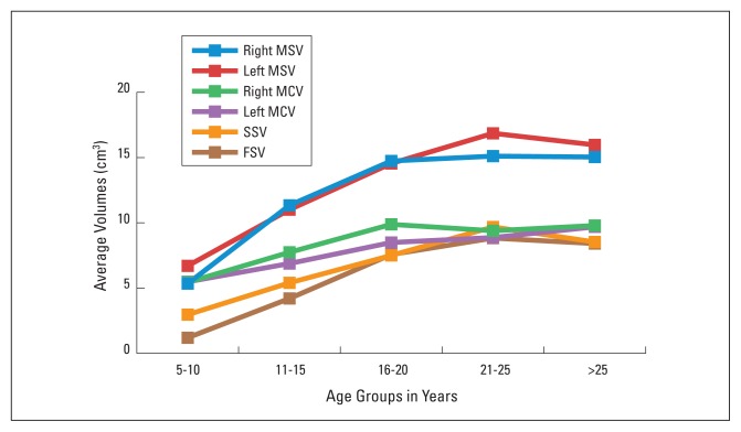
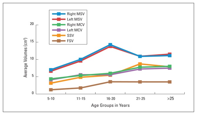
The 91 cases (47 men and 44 women) ranged in age from 5 years to 55 years. Average volumes of the paranasal sinuses and mastoid air cells by age group are shown in Table 1 for men and in Table 2 for women. In men, the highest averages for all paranasal sinuses were mostly in the 21 to 25 year age group. The highest averages for the right and left MCV were in the 16 to 20 and >25 year age groups, respectively. In women, the highest averages for right, left and total MSV were in the 16 to 20 year age group, while the highest averages for right, left and total MCV were in the >25 year age group. Average volumes for the paranasal sinuses and mastoid air cells for all men and women are shown in Table 3. It is evident that the volumes of paranasal sinuses and mastoid air cells increase with age in both sexes.
Table 1.
Average volumes (cm3) of paranasal sinuses and mastoid air cells based on age groups in men.
| 5–10 years old | 11–15 years old | 16–20 years old | 21–25 years old | >25 years old | P value | ||||||
|---|---|---|---|---|---|---|---|---|---|---|---|
| n | Mean±SD | n | Mean±SD | n | Mean±SD | n | Mean±SD | n | Mean±SD | ||
|
| |||||||||||
| Right MSV | 9 | 5.34 ± 0.56 | 10 | 11.34 ± 3.100 | 8 | 14.74 ± 5.79 | 10 | 15.11 ± 4.95 | 10 | 15.04 ± 5.20 | 0.000 |
|
| |||||||||||
| Left MSV | 9 | 6.70 ± 1.10 | 10 | 11.01 ± 2.54 | 8 | 14.55 ± 4.72 | 10 | 16.86 ± 5.77 | 10 | 15.97 ± 6.65 | 0.000 |
|
| |||||||||||
| Right MCV | 9 | 5.44 ± 1.30 | 10 | 7.74 ± 2.11 | 6 | 9.88 ± 5.95 | 10 | 9.39 ± 5.53 | 10 | 9.79 ± 2.82 | 0.021 |
|
| |||||||||||
| Left MCV | 9 | 5.48 ± 1.36 | 10 | 6.88 ± 2.85 | 8 | 8.49 ± 6.47 | 10 | 8.87 ± 5.77 | 10 | 9.69 ± 2.96 | 0.112 |
|
| |||||||||||
| SSV | 2.96 ± 2.53 | 10 | 5.40 ± 1.96 | 8 | 7.50 ± 3.21 | 10 | 9.68 ± 2.62 | 9 | 8.53 ± 4.19 | 0.001 | |
|
| |||||||||||
| FSV | 1.19 ± 0.69 | 9 | 4.20 ± 3.98 | 8 | 7.57 ± 5.72 | 10 | 8.83 ± 4.46 | 10 | 8.41 ± 4.03 | 0.008 | |
|
| |||||||||||
| Total MSV | 9 | 12.04 ± 2.22 | 10 | 22.43 ± 5.50 | 8 | 29.30 ± 10.25 | 10 | 31.97 ± 8.97 | 10 | 30.98 ± 11.41 | 0.000 |
|
| |||||||||||
| Total MCV | 9 | 10.89 ± 2.53 | 10 | 14.62 ± 4.74 | 8 | 15.91 ± 13.24 | 10 | 17.27 ± 9.66 | 10 | 19.48 ± 5.62 | 0.092 |
MSV, maxillar sinus volume; MCV, mastoid air cell volume; SSV, sphenoid sinus volume; FSV, frontal sinus volume.
Table 2.
Average volumes (cm3) of paranasal sinuses and mastoid air cells based on age groups in women.
| 5–10 years old | 11–15 years old | 16–20 years old | 21–25 years old | >25 years old | P value | ||||||
|---|---|---|---|---|---|---|---|---|---|---|---|
| n | Mean±SD | n | Mean±SD | n | Mean±SD | n | Mean±SD | n | Mean±SD | ||
|
| |||||||||||
| Right MSV | 9 | 7.03 ± 2.02 | 9 | 10.03 ± 4.41 | 9 | 14.29 ± 3.42 | 8 | 10.89 ± 4.50 | 9 | 11.13 ± 4.52 | 0.012 |
|
| |||||||||||
| Left MSV | 9 | 6.60 ± 2.25 | 9 | 9.57 ± 4.48 | 9 | 13.78 ± 3.41 | 8 | 10.92 ± 3.63 | 9 | 11.53 ± 5.45 | 0.011 |
|
| |||||||||||
| Right MCV | 8 | 4.38 ± 1.35 | 9 | 5.36 ± 1.83 | 7 | 5.99 ± 2.74 | 8 | 7.76 ± 3.96 | 9 | 8.02 ± 3.84 | 0.106 |
|
| |||||||||||
| Left MCV | 9 | 4.12 ± 1.22 | 9 | 5.57 ± 2.08 | 7 | 5.58 ± 2.80 | 8 | 7.24 ± 3.59 | 9 | 7.49 ± 2.99 | 0.076 |
|
| |||||||||||
| SSV | 8 | 3.14 ± 2.30 | 9 | 4.85 ± 1.09 | 9 | 5.43 ± 2.59 | 8 | 8.71 ± 2.44 | 9 | 7.88 ± 2.99 | 0.001 |
|
| |||||||||||
| FSV | 4 | 1.23 ± 0.29 | 8 | 1.75 ± 1.42 | 9 | 3.54 ± 2.25 | 7 | 3.51 ± 3.11 | 8 | 3.50 ± 2.41 | 0.046 |
|
| |||||||||||
| Total MSV | 9 | 13.63 ± 4.16 | 9 | 19.62 ± 8.74 | 9 | 28.08 ± 6.68 | 8 | 21.81 ± 7.83 | 9 | 22.66 ± 9.75 | 0.010 |
|
| |||||||||||
| Total MCV | 9 | 8.11 ± 2.67 | 9 | 10.9 ± 3.64 | 7 | 11.56 ± 5.17 | 8 | 15.00 ± 6.99 | 9 | 15.52 ± 6.71 | 0.044 |
MSV, maxillar sinus volume; MCV, mastoid air cell volume; SSV, sphenoid sinus volume; FSV, frontal sinus volume.
Table 3.
Mean volumes (cm3) of paranasal sinuses and mastoid air cells.
| All subjects (n=91) | Men (n=47) | Women (n=44) | Men vs. Women | |||||
|---|---|---|---|---|---|---|---|---|
| n | Mean±SD | n | Mean±SD | n | Mean±SD | t | P | |
|
| ||||||||
| Right MSV | 91 | 11.54 ± 5.10 | 47 | 12.36 ± 5.62 | 44 | 10.67 ± 4.39 | 1.592 | 0.115 |
|
| ||||||||
| Left MSV | 91 | 11.82 ± 5.38 | 47 | 13.09 ± 5.85 | 44 | 10.47 ± 5.51 | 2.379 | 0.020 |
|
| ||||||||
| Right MCV | 86 | 7.40 ± 3.73 | 45 | 8.39 ± 3.1 | 41 | 6.33 ± 3.14 | 2.641 | 0.010 |
|
| ||||||||
| Left MCV | 89 | 7.00 ± 3.80 | 47 | 7.91 ± 4.32 | 42 | 5.99 ± 2.81 | 2.442 | 0.017 |
|
| ||||||||
| SSV | 89 | 6.43 ± 3.41 | 46 | 6.83 ± 3.73 | 43 | 6.00 ± 3.02 | 1.143 | 0.256 |
|
| ||||||||
| FSV | 76 | 4.97 ± 4.31 | 40 | 6.86 ± 4.83 | 36 | 2.87 ± 2.29 | 4.516 | 0.000 |
|
| ||||||||
| Total MSV | 91 | 23.37 ± 10.11 | 47 | 15.72 ± 8.06 | 42 | 12.19 ± 5.75 | 2.071 | 0.041 |
|
| ||||||||
| Total MCV | 89 | 14.05 ± 7.24 | 47 | 25.46 ± 10.94 | 44 | 21.14 ± 8.74 | 2.355 | 0.021 |
MSV, maxillar sinus volume; MCV, mastoid air cell volume; SSV, sphenoid sinus volume; FSV, frontal sinus volume.
In the statistical comparison between sexes, there was no significant difference for right MSV and SSV. Significant differences were detected for other parameters. The strongest correlation and most significant differences were observed in the right MSV-left MSV and in the right MCV-left MCV (r=0.822; P<0.001) in both sexes (Table 4).
Table 4.
Correlation constants (r) of paranasal sinus and mastoid air cell parameters in men and women.
| Men | Women | |||
|---|---|---|---|---|
| r | P | r | P | |
|
| ||||
| Right MSV-Left MSV | 0.822 | 0.0001 | 0.922 | 0.0001 |
|
| ||||
| Right MSV-Right MCV | 0.243 | 0.107 | 0.248 | 0.118 |
|
| ||||
| Right MSV-Left MCV | 0.266 | 0.077 | 0.252 | 0.108 |
|
| ||||
| Right MCV-Left MCV | 0.935 | 0.0001 | 0.835 | 0.000 |
|
| ||||
| Right MSV-SSV | 0.407 | 0.005 | 0.372 | 0.014 |
|
| ||||
| Left MSV-SSV | 0.438 | 0.002 | 0.374 | 0.013 |
|
| ||||
| Right MSV-FSV | 0.339 | 0.033 | 0.374 | 0.025 |
|
| ||||
| Left MSV-FSV | 0.386 | 0.014 | 0.330 | 0.049 |
|
| ||||
| Total MCV-Right MSV | 0.138 | 0.355 | 0.272 | 0.081 |
|
| ||||
| Total MCV-Left MSV | 0.227 | 0.125 | 0.314* | 0.043 |
|
| ||||
| Total MCV- SSV | 0.398 | 0.006 | 0.427 | 0.005 |
|
| ||||
| Total MCV- FSV | 0.228 | 0.156 | 0.497 | 0.003 |
|
| ||||
| FSV-SSV | 0.306 | 0.058 | 0.229 | 0.186 |
|
| ||||
| Total MSV- SSV | 0.441 | 0.002 | 0.381 | 0.012 |
|
| ||||
| Total MSV- FSV | 0.388 | 0.013 | 0.361 | 0.030 |
MSV, maxillar sinus volume; MCV, mastoid air cell volume; SSV, sphenoid sinus volume; FSV, frontal sinus volume.
Discussion
The volumes of the paranasal sinuses and mastoid air cells are reported to increase with age.2,5,8 Our results also show that the volumes of paranasal sinuses and mastoid air cells increase regularly with age in both sexes. Diamant reported that development of sinus pneumatization ends at the age of 10 years in women and at the age of 15 years in men.9 Rubensohn agreed with this conclusion, stating that development happens in a regular manner with an average growth of 1.5 cm2 in women and 1 cm2 in men.12 Based on his observation, Rubensohn also suggested that the development of mastoid air cells was different at different ages and sexes during childhood.12 On the other hand, Viraspongse et al, in their study of 100 cases, reported that pneumatization of temporal bone did not change significantly with sex and age.8 Our study was similar to Rubensohn’s study for age and sex differences. The reason for the difference between the Rubensohn and Viraspongse et al’s studies could be the environmental conditions suggested by Wittmaack33 and genetic contributions to pneumatization proposed by Cheatle.34
The mean MCV in our study was 14.05±7.24 cm3 compared with a mean MCV of 10.43±6.66 cm3, 12.22±9.79 cm3, 8.4±3.6 cm3 and 6 cm3 in Park et al,26 Fleshberg et al,11 Colhoun et al,21 and Isono et al’s 19 studies, respectively. In addition, while the mean values for the right and left MCV in our study were 7.40±3.73 cm3 and 7.00±3.80 cm3 (Table 3), compared with 6.08±2.52 cm3 in the right and 6.19±2.93 cm3 in the left in Pata et al.27 Surprisingly, while Isono et al19 and Luntz et al28 observed that there were no differences between right and left MCVs in males and females and while Tos et al6 also reported that MCV remained the same in right and left sides, there were significant differences for right and left MCV between the sexes in our study (P=0.010 for the right and P=0.017 for the left (Table 3). Also, as seen Table 1, while there was a significant difference for right MCV among the different age groups in men, the same difference was not present for left MCV in men (P=0.021 vs. P=0.112). Furthermore, no statistically significant difference was found among the different age groups for right and left MCV in women (P=0.106 vs. P=0.076).
In their examination of the paranasal sinuses, Ariji et al29 and Ikeda et al30 reported that the volumes of the paranasal sinuses increased up to the age of 20 years, but then decreased. Similarly, Schatz et al31 observed that the ethmoid, maxillary and sphenoid sinuses exhibited an increase in volume for a period up to 15 years, afterwards maintaining similar values. In comparison with the volumes of the paranasal sinuses in our and some other investigators’ studies, interesting points were as follows:
For the SSV, Antoniades et al.7 reported that the sphenoid sinus started to develop very quickly after the age of 3 and spread back towards the sella turcica at around the age of 7 years, reaching adult form at around the age of 12.2 Amedee et al2 and Yonetsu et al24 reported a maximum average SSV of 7.5 cm3 and 8.2±0.5 cm3, respectively. Additionally, Yonetsu et al24 observed in the same study that thereafter, the volume decreased gradually, with the average volume in the seventh decade of life being 71% of the maximum level. In our study, SSV increased in both sexes up to 25 years (SSV=8.71±2.44 cm3) and decreased after the age of 25.
For the MSV, Colhoun et al21 and Amedee et al2 reported a mean MSV of 20.05±9.2 cm3 and 14.75 cm3, respectively. In their study, Ariji et al29 observed no significant sex differences and reported a correlation in MSV between the two sides. Similarly, Nowak et al32 reported that the sinus maxillaries of the left side were greater than that of the right side in both male and female patients, and that the sinus maxillaries were greater in female patients than in male patients. Thomas and Raman17 determined that there was a significant relationship among MSV, FSV and MCV as well as a good correlation between FSV and MSV on the same side. Based on their observations, they suggested that paranasal sinuses and mastoid air cells might develop by different processes. In our study, men had a greater difference in MSV than women among the different age groups (Tables 1 and 2).
For the FSV, there is limited information in the literature. Amedee et al2 reported FSVs of 6–7 cm3. In our study, the mean FSV was 4.79±4.31cm3 and the mean values of the sexes were strikingly different (P=0.0001, Table 3).
In our study, there was a direct proportional relationship between age and the volume increase in both sexes as well as a difference in the average volumes between sexes (women’s lower than men’s). In a comparison of men and women, while the highest volume increase for paranasal sinuses (MSV, SSV and FSV) in men appeared in the 21 to 25 year age group, the highest volume for MCV occurred in the >25 year group. On the other hand, while the highest volume increase for MSV and FSV in women was observed in the 16 to 20 year age group, the highest volume was seen for MCV and SSV in the 21 to 25 year age group.
In conclusion, the volumes of the paranasal sinuses and mastoid air cells increase regularly with age in both sexes. The strongest correlation for these volume increases was observed in right-left MSV and in right-left MCV. In the light of these results, it would be useful to have more details for volumes of paranasal sinuses and mastoid air cells by CT scanning. The results will be helpful in understanding normal and pathological conditions of the paranasal sinuses and the mastoid air cells.
Figure 1.
Comparison of the volumes of paranasal sinuses and mastoid air cells based on age groups in men.
Figure 2.
Comparison of the volumes of paranasal sinuses and mastoid air cells based on age groups in women.
Acknowledgement
We thank Dr. Kaya Sarac in Department of Radiology for his assistance in the taking of CT images. Also, we thank to Dr. Yuksel Yildiz for his assistance in the preparation of manuscript.
References
- 1.Miller AJ, Amedee RG. Functional anatomy of the paranasal sinuses. J La State Med Soc. 1997;149(3):85–90. [PubMed] [Google Scholar]
- 2.Amedee R. Sinus Anatomy and Function. In: Bailey BJ, editor. Head and Neck Surgery-Otolaryngology. Vol. 1. Philadelphia: J.B. Lippincott Company; 1993. pp. 342–349. [Google Scholar]
- 3.Moore KL. Clinically Oriented Anatomy. 3rd edition. Baltimore: Williams and Wilkin; 1992. pp. 758–763. [Google Scholar]
- 4.Tos M. Manuel of Middle Ear Surgery. New York: Thieme Medical Publishers; 1995. Mastoid Surgery and Reconstructive Procedures; pp. 54–56. [Google Scholar]
- 5.Williams PL, Warwick R, Bannister LH, Dyson M. Gray’s Anatomy. 36th edition. London: Churchill Livingstone; 1995. pp. 1149–1198. [Google Scholar]
- 6.Tos M, Strangerup SE. The causes of asymmetry of the mastoid air cell system. Acta Otolaryngol. 1985;99(5–6):564–570. doi: 10.3109/00016488509182262. [DOI] [PubMed] [Google Scholar]
- 7.Antoniades K, Vahtsevanos K, Psimopoulou M, Karakasis D. Agenesis of sphenoid sinus: Case report. ORL. J Otorhinolaryngol Relat Spec. 1996;58(6):347–349. doi: 10.1159/000276868. [DOI] [PubMed] [Google Scholar]
- 8.Virapongse C, Sarwar M, Bhimani S, Sasaki C, Shapiro R. Computed tomography of temporal bone pneumatization: Normal pattern and morphology. AJR Am J Roentgenol. 1985;145(3):473–481. doi: 10.2214/ajr.145.3.473. [DOI] [PubMed] [Google Scholar]
- 9.Diamant M. The “Pathological size” of the mastoid air cell system. Acta Otolaryngol. 1965;60:1–10. doi: 10.3109/00016486509126982. [DOI] [PubMed] [Google Scholar]
- 10.Chatterjee D, Ghosh TB, Ghosh BB. Size variation of mastoid air cell system in Indian people at different age groups: a radiographic planimetric study. J Laryngol Otol. 1990;104(8):603–605. doi: 10.1017/s0022215100113349. [DOI] [PubMed] [Google Scholar]
- 11.Flisberg K, Zsigmond M. The size of the mastoid air cell system. Planimetry-direct volume determination. Acta Otolaryngol. 1965;60:23–29. doi: 10.3109/00016486509126985. [DOI] [PubMed] [Google Scholar]
- 12.Rubensohn G. Mastoid pneumatization in children at various ages. Acta Otolaryngol. 1965;60:11–14. doi: 10.3109/00016486509126983. [DOI] [PubMed] [Google Scholar]
- 13.Sade J, Fuchs C, Luntz M. The pars flaccida middle ear pressure and mastoid pneumatization index. Acta Otolaryngol. 1996;116(2):284–287. doi: 10.3109/00016489609137842. [DOI] [PubMed] [Google Scholar]
- 14.Todd NW, Pitts RB, Braun IF, Heinded H. Mastoid size determined with lateral radiographs and computerized tomography. Acta Otolaryngol. 1987;103(3–4):226–231. [PubMed] [Google Scholar]
- 15.Graney DO, Rice DH. Sinus Anatomy, Head and Neck Surgery-Otolaryngology. 2nd edition. Vol. 1. New York: Mosby-Year Book; 1993. pp. 901–906. [Google Scholar]
- 16.Walander A. Considerations on variation of size of frontal sinuses. Acta Otolaryngol. 1965;60:15–22. doi: 10.3109/00016486509126984. [DOI] [PubMed] [Google Scholar]
- 17.Thomas A, Raman R. A comparative study of the pneumatization of the mastoid air cells and the frontal and maxillary sinuses. AJNR Am J Neuroradiol. 1989;10(5 suppl):88–92. [PMC free article] [PubMed] [Google Scholar]
- 18.Azuma H, Isono M, Murata K, Ito A, Kimura H. Morphological characterization and classification of air cells in temporal bone by digital processing of CT images. Nippon Jibiinkoka Gakkai Kaiho. 1997;100(1):13–19. doi: 10.3950/jibiinkoka.100.13. (abstract) [DOI] [PubMed] [Google Scholar]
- 19.Isono M, Murata K, Azuma H, Ishikawa M. Computerized assessment of the mastoid air cell system. Auris Nasus Larynx. 1999;26(2):139–145. doi: 10.1016/s0385-8146(98)00055-8. [DOI] [PubMed] [Google Scholar]
- 20.Som PM. CT of the paranasal sinuses. Neuroradiology. 1985;27(3):189–201. doi: 10.1007/BF00344487. [DOI] [PubMed] [Google Scholar]
- 21.Colhoun EN, O’Neıll G, Francis KR, Hayward C. A comparison between area and volume measurements of mastoid air spaces in normal temporal bones. Clin Otolaryngol. 1988;13(1):59–63. doi: 10.1111/j.1365-2273.1988.tb00282.x. [DOI] [PubMed] [Google Scholar]
- 22.Zinreich SJ. Paranasal sinus imaging. Otolaryngol Head Neck Surg. 1990;103:863–868. doi: 10.1177/01945998901030S505. [DOI] [PubMed] [Google Scholar]
- 23.Scuderi AJ, Harnsberger HR, Boyer RS. Pneumatization of the paranasal sinuses: normal features of importance to the accurate interpretation of CT scans and MR images. ARJ Am J Roentgenol. 1993;160:1101–1104. doi: 10.2214/ajr.160.5.8470585. [DOI] [PubMed] [Google Scholar]
- 24.Yonetsu K, Watanabe M, Nakamura T. Age-Related Expansion and Reduction in Aeration of Sphenoid Sinus: Volume Assessment by Helical CT Scanning. AJNR Am J Neuroradiol. 2000;21(1):179–182. [PMC free article] [PubMed] [Google Scholar]
- 25.Gundersen HJG, Jensen EB. The efficiency of systematic sampling in stereology and its prediction. J Microsc. 1987;147:229–63. doi: 10.1111/j.1365-2818.1987.tb02837.x. [DOI] [PubMed] [Google Scholar]
- 26.Park MS, Yoo SH, Lee DH. Measurement of surface area in human mastoid air cell system. J Laryngol Otol. 2000;114(2):93–96. doi: 10.1258/0022215001904969. [DOI] [PubMed] [Google Scholar]
- 27.Pata YS, Akbas Y, Unal M, Duce MN, Akbas T, Micozkadıoglu D. The relationship between presbycusis and mastoid pneumatization. Yonsei Med J. 2004;45(1):68–72. doi: 10.3349/ymj.2004.45.1.68. [DOI] [PubMed] [Google Scholar]
- 28.Luntz M, Malatskey S, Tan M, Bar-Meir E, Ruimi D. Volume of mastoid pneumatization: three-dimensional reconstruction with ultrahighresolution computed tomography. Ann Otol Rhinol Laryngol. 2001;110:486–490. doi: 10.1177/000348940111000516. [DOI] [PubMed] [Google Scholar]
- 29.Ariji Y, Ariji E, Yoshiura K, Kanda S. Computed tomographic indices for maxillary sinus size in comparison with the sinus volume. Dentomaxillofac Radiol. 1996;25(1):19–24. doi: 10.1259/dmfr.25.1.9084281. [DOI] [PubMed] [Google Scholar]
- 30.Ikeda A. Volumetric measurement of the maxillary sinus by coronal CT scan. Nippon Jibiinkoka Gakkai Kaiho. 1996;99(8):1136–1143. doi: 10.3950/jibiinkoka.99.1136. (abstract) [DOI] [PubMed] [Google Scholar]
- 31.Schatz CJ, Becker TS. Normal CT anatomy of the paranasal sinuses. Radiol Clin North Am. 1984;22(1):107–118. [PubMed] [Google Scholar]
- 32.Nowak R, Mehls G. X-rayfilm analysis of the sinus paranasales from cleft patients (in comparison with a healthy group) Anat Anz. 1977;142(5):451–470.b. (abstract) [PubMed] [Google Scholar]
- 33.Wittmaack K. The significance of middle ear inflammation of infancy (abstracted by Keen JA) J Laryngol Otol. 1931;46:782–784. [Google Scholar]
- 34.Cheatle AH. The etiology and prevention chronic middle ear suppuration. Acta Otolaryngol. 1923;5:283–294. [Google Scholar]


