Abstract
Investigation of the chemical constituents from the fruits of Citrus medica L. var. sarcodactylis Swingle has led to the characterization of a new sesquiterpene 1 along with thirty-two known compounds. The structure of 1 was established on the basis of 2D NMR spectroscopic and mass spectrometric analyses, and the known compounds were identified by comparison of their physical and spectroscopic data with those reported in the literature. In addition, most of the isolated compounds were evaluated for the activity assayed by the in vitro inhibition of superoxide anion generation and elastase release by human neutrophils. The results showed that only 6,7-dimethoxycoumarin (5) exhibited significant inhibition of superoxide anion generation, with IC50 value of 3.8 ± 1.4 μM.
Keywords: Citrus medica L. var. sarcodactylis Swingle, Rutaceae, sesquiterpene, citrumedin-C, superoxide anion formation, elastase release
1. Introduction
Citrus medica L. var. sarcodactylis Swingle belongs to the genus Citrus (Rutaceae), and is commonly distributed in South and Southeast Asia. The plant is cultivated in the tropics and sub-tropics, such as India, Sri Lanka, Thailand, Vietnam, China, Japan, and Taiwan. Various species of Citrus have been used as foods, and their fruits, leaves, and roots have also been used as folk medicine or spice in Taiwan [1]. Citrus is one genus of the economically and medicinally important Rutaceae family, which has shown extensive biological and pharmacological activities, including antitumor [2,3,4,5,6,7], antiallergic [8], antioxidant [9,10,11,12], antiplatelet aggregation [13], anti-microbial [14,15,16,17], and anti-inflammatory activity [18,19,20,21,22]. Moreover, Citrus species have been reported to contain various bioactive coumarins, flavonoids, tetranortriterpenoids, monoterpenoids, and acridone alkaloids [23,24,25,26,27,28,29,30,31,32,33,34]. Previously, several anti-inflammatory compounds have been isolated from the stems and root barks of the titled plant [18]. In order to investigate the bioactive constituents from different parts of C. medica var. sarcodactylis, the fruits of this plant were selected for investigation. As a result, a new sesquiterpene 1 (Figure 1) and thirty-two known compounds have been characterized in the present work. The structural elucidation of 1 is described herein.
Figure 1.
Structures of compounds 1 (relative configuration) and 5.
2. Results and Discussion
2.1. Purification and Characterization
The fresh fruits of C. medica var. sarcodactylis were pulverized into powder and extracted five times with methanol under reflux, and the combined extracts were concentrated to give a deep brown syrup. The crude extract was suspended in water and partitioned with CHCl3 to afford CHCl3 layer and water-soluble layer, respectively. Each layer was subjected to purification by a combination of conventional chromatographic techniques to result in one new compound (1). In addition, thirty-two known compounds were identified to be 5,7-dimethoxycoumarin (2) [3], xanthyletin (3) [18], oxypeucedanin hydrate (4) [35], 6,7-dimethoxycoumarin (5) [18], skimmin (6) [36], haploperoside A (7) [37], leptodactylone (8) [18], 7-methoxycoumarin (9) [18], scopoletin (10) [36], cis-head-to-tail-limittin dimer (11) [38], umbelliferone (12) [36], nordentatin (13) [18], limonin (14) [18], nomilin (15) [18], citrusin (16) [3], obacunone (17) [39], 3-(2-O-β-d-glucopyranosyl-4-methoxyphenyl)-propanoic acid (18) [40], cis-p-coumaric acid (19) [18], methyl vanillate (20) [41], methyl benzoate (21) [42], methyl paraben (22) [43], 4-hydroxy-phenethyl alcohol (23) [44], methyl-4-hydroxycinnamate (24) [43], coniferin (25) [45], syringin (26) [45], (E)-6-hydroxy-2,6-dimethylocta-2,7-dienoic acid (27) [46], a mixture of stigmasterol (28) [3] and β-sitosterol (29) [3], β-sitosteryl glucoside (30) [47], chrysoeriol 8-C-glucoside (31) [48], 1,2,3,4-tetrahydro-β-carboline-3-carboxylic acid (32) [49], and citrylidene malonic acid (33) [50], respectively. The structures of these compounds were identified by comparison of their physical and spectroscopic data with the values reported in the literature.
2.2. Structural Elucidation of Compound 1
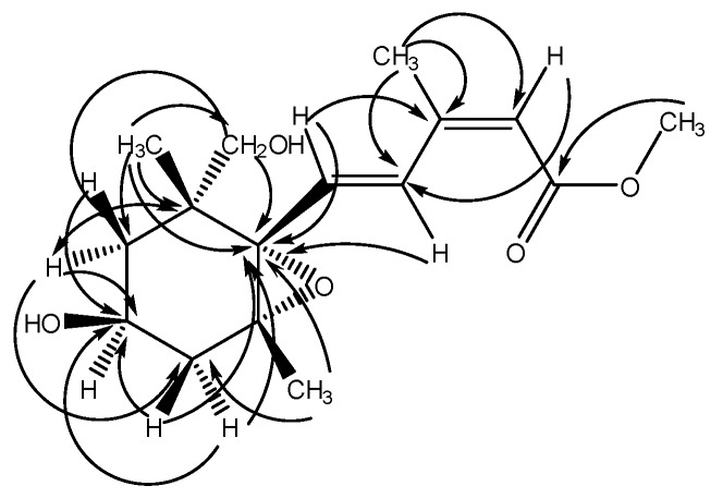
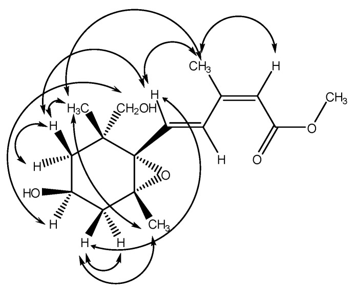
Citrumedin-C (1) was isolated as an optically active colorless powder. The HR-EI-MS analysis of 1 showed a molecular ion peak ([M]+) at m/z 296.1623, which was in agreement with the molecular formula C16H24O5. The UV spectrum appeared to show the maximum absorption at 266 nm. The IR spectrum revealed the presence of hydroxyl (3399 cm−1) and carbonyl groups (1701 cm−1). The 1H-NMR spectrum of 1 exhibited signals for one vinyl proton at δ 5.79 (1H, s), two trans olefinic protons at δ 8.00 (1H, d, J = 15.7 Hz) and 6.55 (1H, d, J = 15.7 Hz), two methyl singlets at δ 1.16 (3H) and 0.94 (3H), one methyl group attached to a double bond at δ 2.10 (3H, s), one methoxy group at δ 3.70 (3H, s), one oxymethine at δ 4.12 (1H, m), two oxymethylene protons at δ 3.82 (1H, d, J = 7.5 Hz) and 3.72 (1H, d, J = 7.5 Hz), one geminally coupled methylene at δ 2.03 (1H, dd, J = 13.7, 7.0 Hz) and 1.73 (1H, dd, J = 13.7, 10.4 Hz), and one methylene group attached with a methine at δ 1.86 (1H, dd, J = 13.5, 6.9 Hz) and 1.68 (1H, dd, J = 13.5, 13.5 Hz), respectively. The 13C-NMR spectrum exhibited a carbonyl signal at δ 168.2 (s); four olefinic carbons at δ 152.0 (s), 135.7 (d), 131.7 (d), and 118.3 (d); and two oxygenated carbons at δ 87.8 (s) and δ 83.3 (s) (Table 1), and these results were confirmed by the HSQC analysis. The COSY correlations of H-4′ (δ 4.12) to H-3′ (δ 2.03 and 1.73) and H-5′ (δ 1.86 and 1.68) suggested the presence of the partial structure (-CH2-CH(-O-)-CH2-). The six-membered C-ring was established by the HMBC correlations from H-5′ (δ 1.86) to C-6′ (δ 49.2) and C-1′ (δ 83.3), and from H-3′ (δ 2.03) to C-2′ (δ 87.8) and C-1′ (δ 83.3) (Table 1, Figure 2). The HMBC spectrum of 1 also showed the conjugated crosspeaks of H-5 (δ 6.55) to C-3 (δ 152.0)/C-1′ (δ 83.3), CH3-3 (δ 2.10) to C-2 (δ 118.3)/C-3 (δ 152.0)/C-4 (δ 131.7), and OCH3 (δ 3.70) to C-1 (δ 168.2), indicating that the partial structure (-CH=CH-C(-CH3)=CH-C(=O)-O-CH3) was substituted at C-1′. The HMBC correlations of H-8′ (δ 0.94) with C-6′ (δ 49.2)/C-5′ (δ 44.6)/C-1′ (δ 83.3)/C-9′ (δ 77.3), and of H-9′ (δ 3.72) with C-5′ (δ 44.6)/C-1′ (δ 83.3) revealed that the quaternary C-6′ was substituted with both methyl and hydroxymethyl groups. In addition, the correlations of the oxygenated quaternary carbon C-2′ (δ 87.8) and C-1′ (δ 83.3) with H-7′ (δ 1.16) supported that the C-2′ was substituted with a methyl group. According to the chemical shifts and the degree of unsaturation, it also suggested the presence of an epoxide ring at C-1′ and C-2′, and a hydroxyl group connected at C-4′, and these were further confirmed with the 2D spectroscopic analytical data. Thus, the structure of 1 was similar to abscisic acid (ABA) and xanthoxin in the phytohormone, which played the key role in biotic and abiotic stress responses [51]. The relative stereochemistry was confirmed by a NOESY experiment, which showed correlations of CH3-8′/CH3-7′, H-4′/H-9′, H-3′ (δ 1.73)/H-5, and H-3′ (δ 1.73)/CH3-7′, respectively (Figure 3). Therefore, the side chain at C-1′, two methyl substituents at C-2′ and C-6′, and the hydroxyl group at C-4′ were all in cis configuration. Conclusively, the structure of citrumedin-C (1) was assigned as shown based on the above-mentioned observations (See Supplementary Materials).
Table 1.
1H- and 13C-NMR spectral data of 1 (CD3OD, 400 MHz).
| Position | δH (mult., J in Hz) | δC |
|---|---|---|
| 1 | 168.2 | |
| 2 | 5.79 (1H, s) | 118.3 |
| 3 | 152.0 | |
| 4 | 8.00 (1H, d, 15.7) | 131.7 |
| 5 | 6.55 (1H, d, 15.7) | 135.7 |
| 1′ | 83.3 | |
| 2′ | 87.8 | |
| 3′ | 2.03 (1H, dd, 13.7, 7.0) 1.73 (1H, dd, 13.7, 10.4) | 46.0 |
| 4′ | 4.12 (1H, m) | 66.0 |
| 5′ | 1.86 (1H, dd, 13.5, 6.9) 1.68 (1H, dd, 13.5, 13.5) | 44.6 |
| 6′ | 49.2 | |
| 7′ | 1.16 (3H, s) | 19.6 |
| 8′ | 0.94 (3H, s) | 16.4 |
| 9′ | 3.82 (1H, d, 7.5) 3.72 (1H, d, 7.5) | 77.3 |
| CH3-3 | 2.10 (3H, s,) | 21.2 |
| OCH3 | 3.70 (3H, s) | 51.6 |
Figure 2.
HMBC Correlations of 1.
Figure 3.
NOESY Correlations of 1.
2.3. The Inhibitory Activity of Superoxide Anion Generation and Elastase Release
Most of the isolated compounds (compounds 2–6, 10, 12, 14–18, and 31) were examined for their inhibition of superoxide anion generation and elastase release by human neutrophils in response to N-formyl-l-methionyl-phenylalanine/cytochalasin B (fMLP/CB) [52,53]. Only compound 5 (Figure 1) displayed inhibition percentages greater than 50% at the test concentration of 10 µM, and in the concentration range used, this compound displayed inhibitory effects in a dose-dependent manner. Compound 5 displayed significant inhibition of superoxide anion generation with IC50 value of 3.8 ± 1.4 μM, compared to the reference compound LY294002 with IC50 values of 0.4 ± 0.02 μM [52] (Table 2).
Table 2.
Inhibitory effects of isolated compounds on superoxide anion generation and elastase release by human neutrophils in response to N-formyl-l-methionyl-phenylalanine/cytochalasin B (fMLP/CB).
| Compound | IC50 (μM) a | |
|---|---|---|
| Superoxide Anion Generation | Elastase Release | |
| 5 | 3.8 ± 1.4 *** | >10 |
| LY294002 b | 0.4 ± 0.02 *** | 1.5 ± 0.3 *** |
a Concentration necessary for 50% inhibition (IC50). Results are presented as means ± SD (n = 3–4). *** p < 0.001 compared with the control (DMSO). b A phosphatidylinositol-3-kinase inhibitor was used as a positive control.
3. Materials and Methods
3.1. General Information
Melting points were determined using an MP-S3 apparatus (Yanaco, Tokyo, Japan). Optical rotations were measured using a P-2000 digital polarimeter (JASCO, Tokyo, Japan). UV spectra were recorded at room temperature on a U-0080-D spectrophotometer (Hitachi, Tokyo, Japan). IR spectra were obtained with an FT-IR Spectrum RX I spectrophotometer (PerkinElmer, Waltham, MA, USA). The EI-MS and HR-EI-MS were obtained on a VG-70-250S mass spectrometer. The 1H- and 13C-NMR, DEPT, COSY, HMQC, NOESY, and HMBC experiments were recorded on a Bruker AMX-400 spectrometer. Standard pulse sequences and parameters were used for the NMR experiments, and all chemical shifts were reported in parts per million (ppm, δ). Column chromatography (CC) was performed on silica gel (70–230 mesh and 230–400 mesh, Merck, Darmstadt, Germany), Diaion HP-20 (Mitsubishi, Tokyo, Japan), and C 18 (Sigma-Aldrich, St. Louis, MO, USA) gels, respectively, and preparative TLC (thin-layer chromatography) was conducted on Merck precoated silica gel 60 F254 plates, using UV light to visualize the spots. All solvents of extraction and isolation were purchased from Merck (Darmstadt, Germany).
3.2. Materials
The fruits of Citrus medica L. var. sarcodactylis Swingle was collected from the markets of Hualien County, Taiwan in September 2002, and identified by Prof. Chang-Sheng Kuoh. A voucher specimen (TSWu 20020923) has been deposited in the Herbarium of National Cheng Kung University, Tainan, Taiwan.
3.3. Extraction and Isolation
The fresh whole fruits of Citrus medica L. var. sarcodactylis Swingle (7.97 kg) were pulverized into small pieces and extracted with methanol under reflux (40 L × 5 h × 5). The resulting solution was then filtered and concentrated in vacuo to yield a crude extract. The MeOH extract (696 g) was suspended in distilled water and successively partitioned with chloroform yielding a chloroform layer (26 g) and water-soluble layer (670 g). The chloroform layer (26 g) was chromatographed directly on silica gel and eluted with a gradient of n-hexane and ethyl acetate to afford eight fractions. Fraction 3 was rechromatographed on silica gel and eluted with solvent of n-hexane–acetone (25:1) to give a mixture of β-sitosterol (28) and stigmasterol (29, 227 mg, Rf = 0.6), and citrylidene malonic acid (33, 1 mg, Rf = 0.4). Purification of fraction 4 by column chromatography with silica gel was eluted by gradient solvent mixture of chloroform and ethyl acetate (20:1 to 1:1) to afford 7-methoxycoumarin (9, 5 mg, Rf = 0.8), nordentatin (13, 1 mg, Rf = 0.7), nomilin (15, 19 mg, Rf = 0.6), methyl vanillate (20, 1 mg, Rf = 0.5), 4-hydroxy-phenethyl alcohol (23, 1 mg, Rf = 0.3), and methyl-4-hydroxycinnamate (24, 2 mg, Rf = 0.2), respectively. Separation of fraction 5 by column chromatography with silica gel eluted by chloroform and methanol (20:1) solvent mixture yielded xanthyletin (3, 13 mg, Rf = 0.7), leptodactylone (8, 1 mg, Rf = 0.5), umbelliferone (12, 11 mg, Rf = 0.4), and methyl paraben (22, 1 mg, Rf = 0.3). Fraction 6 was purified by silica gel CC eluted by a gradient solvent mixture of chloroform and methanol (20:1 to 1:1) to afford 5,7-dimethoxycoumarin (2, 198 mg, Rf = 0.6), 6,7-dimethoxycoumarin (5, 24 mg, Rf = 0.6), scopoletin (10, 21 mg, Rf = 0.5), cis-head-to-tail-limittin dimer (11, 1 mg, Rf = 0.3), and limonin (14, 59 mg, Rf = 0.2). Fraction 7 underwent a series of chromatographic separations on silica gel using chloroform/ methanol (15:1) as eluent to afford oxypeucedanin hydrate (4, 12 mg, Rf = 0.8), citrusin (16, 15 mg, Rf = 0.6), obacunone (17, 16 mg, Rf = 0.5), and (E)-6-hydroxy-2,6-dimethylocta-2,7-dienoic acid (27, 1 mg, Rf = 0.3). Recrystallization of fraction 8 produced the solid β-sitosteryl glucoside (30, 23 mg).
The water layer was subjected directly to Diaion HP-20 column chromatography, eluted by water and gradient with methanol to give six fractions. Fraction 4 was chromatographed over Sephadex LH-20 eluted with gradient solvent mixture of water and methanol to give skimmin (6, 17 mg, Rf = 0.7), haploperoside A (7, 1 mg, Rf = 0.6), methyl benzoate (21, 1 mg, Rf = 0.5), coniferin (25, 27 mg, Rf = 0.3), and syringin (26, 7 mg, Rf = 0.2). Fraction 5 was chromatographed on silica gel and eluted with solvent mixture of chloroform and methanol (5:1) to afford 3-(2-O-β-d-glucopyranosyl-4-methoxyphenyl)propanoic acid (18, 11 mg, Rf = 0.5) and 1,2,3,4-tetrahydro-β-carboline-3- carboxylic acid (32, 3 mg, Rf = 0.3). Fraction 6 was rechromatographed on silica gel and eluted with mixture of chloroform and methanol (9:1) to give citrumedin-C (1, 1 mg, Rf = 0.7), cis-p-coumaric acid (19, 1 mg, Rf = 0.6), and chrysoeriol 8-C-glucoside (31, 8 mg, Rf = 0.2).
Citrumedin-C (1): colorless powder, mp 138–139 °C; [α]d +50.5 (CHCl3, c = 0.07); UV λmax (MeOH) nm (log ε) 266 (4.38); IR(KBr) νmax cm−1 3399, 2920, 2874, 1701, 1601, 1447, 1373, 1234, 1165; 1H- and 13C-NMR data, see Table 1. EI-MS m/z 296 ([M+], 5), 278 (14), 264 (7), 246 (13), 220 (12), 188 (22), 169 (13), 154 (37), 135 (29), 122 (100); HR-EI-MS m/z [M]+ (Calcd. for C16H24O5 296.1624, Found 296.1623).
3.4. Bioactivity Examination
3.4.1. Preparation of Human Neutrophils
A study involving human neutrophils was approved by the Institutional Review Board at Chang Gung Memorial Hospital, Taoyuan, Taiwan, and was conducted according to the Declaration of Helsinki (2013). The written informed consent was obtained from each healthy donor before blood was drawn. Blood was drawn from healthy human donors (20–30 years old) by venipuncture into heparin-coated vacutainer tubes, using a protocol approved by the Institutional Review Board at Chang Gung Memorial Hospital. Blood samples were mixed gently with an equal volume of 3% dextran solution. Neutrophils were isolated with a standard method of dextran sedimentation prior to centrifugation in a Ficoll Hypaque gradient and hypotonic lysis of erythrocytes. The leukocyte-rich plasma was collected after sedimentation of the red cells for 30 min at room temperature, and was transferred to 20 mL Ficoll solution (1.08 g/mL) and spun down at 400 g for 40 min at 20 °C. The granulocyte/erythrocyte pellets were resuspended in ice-cold 0.2% NaCl to lyse erythrocytes [52]. After 30 s, the same volume of 1.6% NaCl solution was added to reconstitute the isotonic condition. Purified neutrophils were pelleted and then resuspended in a calcium (Ca2+)-free Hank’s balanced salt solution (HBSS) buffer at pH 7.4, and were maintained at 4 °C before use.
3.4.2. Inhibition of Superoxide Anion Generation
The assay of the generation of superoxide anion was based on the superoxide dismutase (SOD)-inhibitable reduction of ferricytochrome c [52,54]. In brief, after supplementation with 0.6 mg/mL ferricytochrome c and 1 mM Ca2+, neutrophils (6 × 10⁵ cells/mL) were equilibrated at 37 °C for 2 min and incubated with drugs or an equal volume of vehicle (0.1% DMSO, negative control) for 5 min. Cells were activated with 100 nM fMLP during the preincubation of 1 µg/mL cytochalasin B (fMLP/CB) for 3 min. Changes in the absorbance with a reduction in ferricytochrome c at 550 nm were continuously monitored in a double-beam, six-cell positioner spectrophotometer with constant stirring (Hitachi U-3010). Calculations were based on differences in the reactions with and without SOD (100 U/mL) divided by the extinction coefficient for the reduction of ferricytochrome c (ε = 21.1/mM/10 mm at the concentration of 1 mM in cuvette with 1-cm optical path length).
3.4.3. Inhibition of Elastase Release
Elastase release was measured by degranulation of azurophilic granules as described previously [52,54]. Experiments were performed using MeO-Suc-Ala-Ala-Pro-Val-p-nitroanilide as the elastase substrate. Briefly, after supplementation with MeO-Suc-Ala-Ala-Pro-Val-p-nitroanilide (100 µM), neutrophils (6 × 10⁵ cells/mL) were equilibrated at 37 °C for 2 min and incubated with test compounds or an equal volume of vehicle (0.1% DMSO, negative control) for 5 min. Cells were activated by 100 nM fMLP and 0.5 µg/mL cytochalasin B, and changes in absorbance at 405 nm were continuously monitored to assay elastase release. The results were expressed as the percent of elastase release in the fMLP/CB-activated, drug-free control system.
3.4.4. Statistical Analysis
Normal distribution with Shapiro–Wilk was performed. The results are expressed as the mean ± SD and analyzed by analysis of variance (ANOVA) with post-hoc Bonferroni multiple comparisons tests. Calculations of 50% inhibitory concentrations (IC50) were computer-assisted (PHARM/PCS v.4.2). Statistical comparisons were made between groups using the Student’s t test. Values of p less than 0.05 were considered to be statistically significant.
Acknowledgments
This study is sponsored by the Ministry of Science and Technology, Taiwan, granted to T.-S.W. and Y.-Y.C. Authors are also thankful to Chang Gung Memorial Hospital (CMRPD1B0281~3, CMRPF1D0442~3, CMRPF 1F0011~3, CMRPF1F0061~3 and BMRP450 granted to T.-L.H.) for the partial financial support for the present research.
Supplementary Materials
1H-, 13C-NMR and 2D spectroscopic data of 1 are available online.
Author Contributions
Yu-Yi Chan and Ping-Chung Kuo performed the research and recorded the spectra. Tsong-Long Hwang had done the inhibition experiments on human neutrophils. Tian-Shung Wu designed the research and provided the research outlines. Yu-Yi Chan, Ping-Chung Kuo and Hsin-Yi Hung completed the manuscript draft. All authors read and approved the final manuscript.
Conflicts of Interest
The authors declare no conflict of interest.
Footnotes
Sample Availability: Samples of the purified compounds are available from the authors.
References
- 1.Chiu N.Y., Chang K.H. The Illustrated Medicinal Plants of Taiwan. 1st ed. Volume III. SMC Publishing; Taipei, Taiwan: 1987. p. 121. [Google Scholar]
- 2.Wang Y., Qian J., Cao J., Wang D., Liu C., Yang R., Li X., Sun C. Antioxidant capacity, anticancer ability and flavonoids composition of 35 Citrus (Citrus reticulata Blanco) varieties. Molecules. 2017;22:1114. doi: 10.3390/molecules22071114. [DOI] [PMC free article] [PubMed] [Google Scholar]
- 3.Chan Y.Y., Wu T.S., Kuo Y.H. Chemical constituents and cytotoxicity from the stem bark of Citrus medica. Heterocycles. 2009;78:1309–1316. [Google Scholar]
- 4.Roohbakhsh A., Parhiz H., Soltani F., Rezaee R., Iranshahi M. Molecular mechanisms behind the biological effects of hesperidin and hesperetin for the prevention of cancer and cardiovascular diseases. Life Sci. 2015;124:64–74. doi: 10.1016/j.lfs.2014.12.030. [DOI] [PubMed] [Google Scholar]
- 5.Murthy K.N., Jayaprakasha G.K., Patil B.S. Cytotoxicity of obacunone and obacunone glucoside in human prostate cancer cells involves Akt-mediated programmed cell death. Toxicology. 2015;329:88–97. doi: 10.1016/j.tox.2015.01.008. [DOI] [PubMed] [Google Scholar]
- 6.Leclere L., Fransolet M., Cote F., Cambier P., Arnould T., Van Cutsem P., Michiels C. Heat-modified Citrus pectin induces apoptosis-like cell death and autophagy in HepG2 and A549 cancer cells. PLoS ONE. 2015;10:e0115831. doi: 10.1371/journal.pone.0115831. [DOI] [PMC free article] [PubMed] [Google Scholar]
- 7.Zeng Z.Y., Chen Y., Zou L., Zhang Y., Hu F., Feng J., Shen J., Wei B. Naringin inhibits growth and induces apoptosis by a mechanism dependent on reduced activation of NF-κB/COX-2 caspase-1 pathway in HeLa cervical cancer cells. Int. J. Oncol. 2014;45:1929–1936. doi: 10.3892/ijo.2014.2617. [DOI] [PubMed] [Google Scholar]
- 8.Huber R., Stintzing F.C., Briemle D., Beckmann C., Meyer U., Gründemann C. In vitro antiallergic effects of aqueous fermented preparations from Citrus and Cydonia fruits. Planta Med. 2012;78:334–340. doi: 10.1055/s-0031-1280455. [DOI] [PubMed] [Google Scholar]
- 9.Mokbel M.S., Hashinaga F. Evaluation of the antioxidant activity of extracts from buntan (Citrus grandis Osbeck) fruit tissues. Food Chem. 2006;94:529–534. doi: 10.1016/j.foodchem.2004.11.042. [DOI] [Google Scholar]
- 10.Tsai H.L., Chang S.K., Chang S.J. Antioxidant content and free radical scavenging ability of fresh red pummelo [Citrus grandis (L.) Osbeck] juice and freeze-dried products. J. Agric. Food Chem. 2007;55:2867–2872. doi: 10.1021/jf0633847. [DOI] [PubMed] [Google Scholar]
- 11.Kim G.N., Shin J.G., Jang H.D. Antioxidant and antidiabetic activity of Dangyuja (Citrus grandis Osbeck) extract treated with Aspergillus saitoi. Food Chem. 2009;117:35–41. doi: 10.1016/j.foodchem.2009.03.072. [DOI] [Google Scholar]
- 12.Parhiz H., Roohbakhsh A., Soltani F., Rezaee R., Iranshahi M. Antioxidant and anti-inflammatory properties of the Citrus flavonoids hesperidin and hesperetin: An updated review of their molecular mechanisms and experimental models. Phytother. Res. 2015;29:323–331. doi: 10.1002/ptr.5256. [DOI] [PubMed] [Google Scholar]
- 13.Yu H.Y., Park S.W., Chung I.M., Jung Y.S. Anti-platelet effects of yuzu extract and its component. Food Chem. Toxicol. 2011;49:3018–3024. doi: 10.1016/j.fct.2011.09.038. [DOI] [PubMed] [Google Scholar]
- 14.Shende S., Ingle A.P., Gade A., Rai M. Green synthesis of copper nanoparticles by Citrus medica Linn. (Idilimbu) juice and its antimicrobial activity. World J. Microbiol. Biotechnol. 2015;31:865–873. doi: 10.1007/s11274-015-1840-3. [DOI] [PubMed] [Google Scholar]
- 15.Chubukov V., Mingardon F., Schackwitz W., Baidoo E.E., Alonso-Gutierrez J., Hu Q., Lee T.S., Keasling J.D., Mukhopadhyay A. Acute limonene toxicity in Escherichia coli is caused by limonene hydroperoxide and alleviated by a point mutation in alkyl hydroperoxidase AhpC. Appl. Environ. Microbiol. 2015;81:4690–4696. doi: 10.1128/AEM.01102-15. [DOI] [PMC free article] [PubMed] [Google Scholar]
- 16.Wu M.C., Li H.C., Wu P.H., Huang P.H., Wang Y.T. Assessment of oligogalacturonide from Citrus pectin as a potential antibacterial agent against foodborne pathogens. J. Food Sci. 2014;79:1541–1544. doi: 10.1111/1750-3841.12526. [DOI] [PubMed] [Google Scholar]
- 17.Vollmerhausen T.L., Ramos N.L., Dzung D.T., Brauner A. Decoctions from Citrus reticulata Blanco seeds protect the uroepithelium against Escherichia coli invasion. J. Ethnopharmacol. 2013;150:770–774. doi: 10.1016/j.jep.2013.09.050. [DOI] [PubMed] [Google Scholar]
- 18.Chan Y.Y., Li C.H., Shen Y.C., Wu T.S. Anti-inflammatory principles from the stem and root barks of Citrus medica. Chem. Pharm. Bull. 2010;58:61–65. doi: 10.1248/cpb.58.61. [DOI] [PubMed] [Google Scholar]
- 19.La V.D., Zhao L., Epifano F., Genovese S., Grenier D. Anti-inflammatory and wound healing potential of Citrus auraptene. J. Med. Food. 2013;16:961–964. doi: 10.1089/jmf.2013.0029. [DOI] [PubMed] [Google Scholar]
- 20.Impellizzeri D., Bruschetta G., Di Paola R., Ahmad A., Campolo M., Cuzzocrea S., Esposito E., Navarra M. The anti-inflammatory and antioxidant effects of bergamot juice extract (BJe) in an experimental model of inflammatory bowel disease. Clin. Nutr. 2014;33:749–753. doi: 10.1016/j.clnu.2014.11.012. [DOI] [PubMed] [Google Scholar]
- 21.Mitoshi M., Kuriyama I., Nakayama H., Miyazato H., Sugimoto K., Kobayashi Y., Jippo T., Kuramochi K., Yoshida H., Mizushina Y. Suppression of allergic and inflammatory responses by essential oils derived from herbal plants and Citrus fruits. Int. J. Mol. Med. 2014;33:1643–1651. doi: 10.3892/ijmm.2014.1720. [DOI] [PubMed] [Google Scholar]
- 22.Noh H.J., Hwang D., Lee E.S., Hyun J.W., Yi P.H., Kim G.S., Lee S.E., Pang C., Park Y.J., Chung K.H., et al. Anti-inflammatory activity of a new cyclic peptide, citrusin XI, isolated from the fruits of Citrus unshiu. J. Ethnopharmacol. 2015;163:106–112. doi: 10.1016/j.jep.2015.01.024. [DOI] [PubMed] [Google Scholar]
- 23.Hattori S., Shimokoriyama M., Kanao M. Studies on flavanone glycosides. IV. The glycosides of ripe fruit peel and flower petals of Citrus aurantium L. J. Am. Chem. Soc. 1952;74:3614–3615. doi: 10.1021/ja01134a043. [DOI] [Google Scholar]
- 24.Wu T.S., Kuoh C.S., Furukawa H. Acridone alkaloids and a coumarin from Citrus grandis. Phytochemistry. 1983;22:1493–1497. [Google Scholar]
- 25.McPhail A.T., Ju-ichi M., Fujitani Y., Inoue M., Wu T.S., Furukawa H. Isolation and structures of citropone-A and -B from Citrus plants, first examples of naturally-occurring homoacridone alkaloids containing a tropone ring system. Tetrahedron Lett. 1985;26:3271–3272. doi: 10.1016/S0040-4039(00)98169-3. [DOI] [Google Scholar]
- 26.Wu T.S., Huang S.C., Jong T.T., Lai J.S., Furukawa H. Honyumine, a new linear pyranoacridone alkaloids from Citrus grandis Osbeck. Heterocycles. 1986;24:41–43. doi: 10.3987/R-1986-01-0041. [DOI] [Google Scholar]
- 27.Wu T.S. Baiyumine-A and -B, two acridone alkaloids from Citrus grandis. Phytochemistry. 1987;26:871–872. doi: 10.1016/S0031-9422(00)84813-X. [DOI] [Google Scholar]
- 28.Wu T.S., Huang S.C., Jong T.T., Lai J.S., Kuoh C.S. Coumarins, acridone alkaloids and a flavone from Citrus grandis. Phytochemistry. 1988;27:585–587. doi: 10.1016/0031-9422(88)83146-7. [DOI] [Google Scholar]
- 29.Wu T.S. Alkaloids and coumarins of Citrus grandis. Phytochemistry. 1988;27:3717–3718. doi: 10.1016/0031-9422(88)80815-X. [DOI] [Google Scholar]
- 30.Huang S.C., Chen M.T., Wu T.S. Alkaloids and coumarins from stem bark of Citrus grandis. Phytochemistry. 1989;28:3574–3576. doi: 10.1016/0031-9422(89)80402-9. [DOI] [Google Scholar]
- 31.Wu T.S., Huang S.C., Lai J.S. Stem bark coumarins of Citrus grandis. Phytochemistry. 1994;36:217–219. [Google Scholar]
- 32.Takemura Y., Ju-ichi M., Ito C., Furukawa H., Tokuda H. Studies on the inhibitory effects of some acridone alkaloids on Epstein-Barr virus activation. Planta Med. 1995;61:366–368. doi: 10.1055/s-2006-958104. [DOI] [PubMed] [Google Scholar]
- 33.Marıín F.R., Martinez M., Uribesalgo T., Castillo S., Frutos M.J. Changes in nutraceutical composition of lemon juices according to different industrial extraction systems. Food Chem. 2002;78:319–324. doi: 10.1016/S0308-8146(02)00102-4. [DOI] [Google Scholar]
- 34.Giuffrè A.M., Zappia C., Capocasale M. Physico-chemical stability of blood orange juice during frozen storage. Int. J. Food Prop. 2017 in press. [Google Scholar]
- 35.Harkar S., Razdan T.K., Waight E.S. Steroids, chromone and coumarins from Angelica officinalis. Phytochemistry. 1984;23:419–426. doi: 10.1016/S0031-9422(00)80344-1. [DOI] [Google Scholar]
- 36.Wu T.S., Chang F.C., Wu P.L., Kuoh C.S., Chen I.S. Constituents of Leaves of Tetradium Glabrifolium. J. Chin. Chem. Soc. 1995;42:929–934. doi: 10.1002/jccs.199500128. [DOI] [Google Scholar]
- 37.Sendker J., Böker I., Lengers I., Brandt S., Jose J., Stark T., Hofmann T., Fink C., Abdel-Aziz H., Hensel A. Phytochemical characterization of low molecular weight constituents from marshmallow roots (Althaea officinalis) and inhibiting effects of the aqueous extract on human hyaluronidase-1. J. Nat. Prod. 2017;80:290–297. doi: 10.1021/acs.jnatprod.6b00670. [DOI] [PubMed] [Google Scholar]
- 38.He H.Y., Ling L., Zhou M. Isolation and structure elucidation of two dimeric limettins from fingered citron. Youji Huaxue. 1987;3:193–196. [Google Scholar]
- 39.Sugimoto T., Miyase T., Kuroyanagi M., Ueno A. Limonoids and quinolone alkaloids from Evodia rutaecarpa bentham. Chem. Pharm. Bull. 1988;36:4453–4461. doi: 10.1248/cpb.36.4453. [DOI] [Google Scholar]
- 40.Aquino R., Cáceres A., Morelli S., Rastrelli L. An extract of Tagetes lucida and its phenolic constituents as antioxidants. J. Nat. Prod. 2002;65:1773–1776. doi: 10.1021/np020018i. [DOI] [PubMed] [Google Scholar]
- 41.Chan Y.Y., Juang S.H., Huang G.J., Liao Y.R., Chen Y.F., Wu C.C., Chang H.T., Wu T.S. The Constituents of Michelia compressa var. formosana and their bioactivities. Int. J. Mol. Sci. 2014;15:10926–10935. doi: 10.3390/ijms150610926. [DOI] [PMC free article] [PubMed] [Google Scholar]
- 42.Boyd D.R., Sharma N.D., Harrison J.S., Malone J.F., McRoberts W.C., Hamilton J.T.G., Harper D.B. Enzyme-catalysed synthesis and reactions of benzene oxide/oxepine derivatives of methyl benzoates. Org. Biomol. Chem. 2008;6:1251–1259. doi: 10.1039/b718375e. [DOI] [PubMed] [Google Scholar]
- 43.Huang C.H., Chan Y.Y., Kuo P.C., Chen Y.F., Chang R.J., Chen I.S., Wu S.J., Wu T.S. The constituents of roots and stems of Illigera luzonensis and their anti-platelet aggregation effects. Int. J. Mol. Sci. 2014;15:13424–13436. doi: 10.3390/ijms150813424. [DOI] [PMC free article] [PubMed] [Google Scholar]
- 44.Claydon N., Frederick J., Pople G.M. Elm bark beetle boring and feeding deterrents from Phomopsis oblonga. Phytochemistry. 1985;24:937–943. doi: 10.1016/S0031-9422(00)83157-X. [DOI] [Google Scholar]
- 45.Steevesa V., Försterb H., Pommerb U., Savidgea R. Coniferyl alcohol metabolism in conifers—I. Glucosidic turnover of cinnamyl aldehydes by UDPG: Coniferyl alcohol glucosyltransferase from pine cambium. Phytochemistry. 2001;57:1085–1093. doi: 10.1016/S0031-9422(01)00107-8. [DOI] [PubMed] [Google Scholar]
- 46.Marco J.A., Sanz-Cervera J.F., Sancenon F., Jakupovic J., Rustaiyant A., Mohamadit F. Oplopanone derivatives monoterpene glycosides from Artemisia sieberi. Phytochemistry. 1993;34:1061–1067. doi: 10.1016/S0031-9422(00)90714-3. [DOI] [Google Scholar]
- 47.Chan Y.Y., Leu Y.L., Wu T.S. The constituents of the leaves of Aristolochia heterophylla Hemsl. Chem. Pharm. Bull. 1999;47:887–889. doi: 10.1248/cpb.47.887. [DOI] [PubMed] [Google Scholar]
- 48.Gattuso G., Barreca D., Caristi C., Gargiulli C., Leuzzi U. Distribution of flavonoids and furocoumarins in juices from cultivars of Citrus bergamia Risso. J. Agric. Food Chem. 2007;55:9921–9927. doi: 10.1021/jf072065s. [DOI] [PubMed] [Google Scholar]
- 49.Yuan Y., Win Aung K.K., Ran X.K., Wang X.T., Dou D.Q., Dong F. A new sesquiterpene lactone from yacon leaves. Nat. Prod. Res. 2017;31:43–49. doi: 10.1080/14786419.2016.1212028. [DOI] [PubMed] [Google Scholar]
- 50.Yang S., Wang L., Guo X.J., Lou H.X., Ren D.G. A new flavonoid glycoside and other constituents from Dracocephalum moldavica. Nat. Prod. Res. 2013;27:201–207. doi: 10.1080/14786419.2012.666746. [DOI] [PubMed] [Google Scholar]
- 51.Sakai K., Takahashi K., Nukano T. Convenient synthesis of optically active abscisic acid and xanthoxin. Tetrahedron. 1992;48:8229–8238. doi: 10.1016/S0040-4020(01)80491-9. [DOI] [Google Scholar]
- 52.Yang S.C., Chung P.J., Ho C.M., Kuo C.Y., Hung M.F., Huang Y.T., Chang W.Y., Chang Y.W., Chan K.H., Hwang T.L. Propofol inhibits superoxide production, elastase release, and chemotaxis in formyl peptide-activated human neutrophils by blocking formyl peptide receptor 1. J. Immunol. 2013;190:6511–6519. doi: 10.4049/jimmunol.1202215. [DOI] [PubMed] [Google Scholar]
- 53.Yu H.P., Hsieh P.W., Chang Y.J., Chung P.J., Kuo L.M., Hwang T.L. 2-(2-Fluorobenzamido) benzoate ethyl ester (EFB-1) inhibits superoxide production by human neutrophils and attenuates hemorrhagic shock-induced organ dysfunction in rats. Free Radic. Biol. Med. 2011;50:1737–1748. doi: 10.1016/j.freeradbiomed.2011.03.026. [DOI] [PubMed] [Google Scholar]
- 54.Strehmel N., Böttcher C., Schmidt S., Scheel D. Profiling of secondary metabolites in root exudates of Arabidopsis thaliana. Phytochemistry. 2014;108:35–46. doi: 10.1016/j.phytochem.2014.10.003. [DOI] [PubMed] [Google Scholar]
Associated Data
This section collects any data citations, data availability statements, or supplementary materials included in this article.


