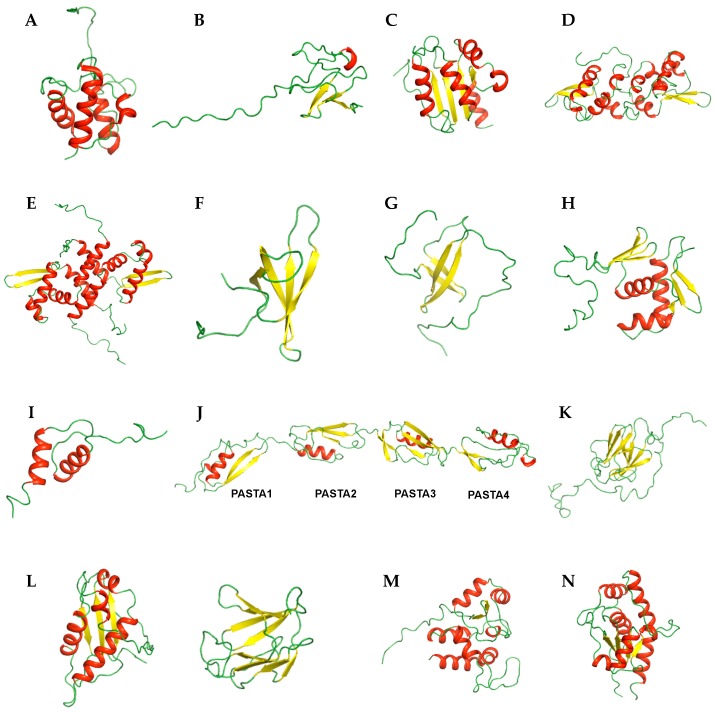
Figure 1.
Ribbon representation of NMR structures of M. tuberculosis proteins. Transport-related proteins (A) Rv2244 (PDB ID 1KLP); (B) Rv3250c (PDB ID 2KN9); (C) Rv1739c (PDB ID 2KLN). Transcription-related proteins (D) Rv1994c (PDB ID 2JSC); (E) MT3852 (PDB ID 2LKP); (F) Rv0639 (PDB ID 2MI6); (G) Rv2050 (PDB ID 2M4V). Nucleotide-binding proteins (H) J113_05350 (PDB ID 2RV8); (I) Rv3597c (PDB ID 2KNG); Ser/Thr Protein kinase-related proteins (J) Rv0014c (PDB ID 2KUI); (K) Rv1827 (PDB ID 2KFU); (L) Rv0020c (PDB ID 2LC0 (Left) and 2LC1 (Right)); (M) Rv2175c (PDB ID 2KFS); (N) Rv2234 (PDB ID 2LUO). Secondary structural elements, α-helix, β-sheet, and loop are colored in red, yellow, and green, respectively.

