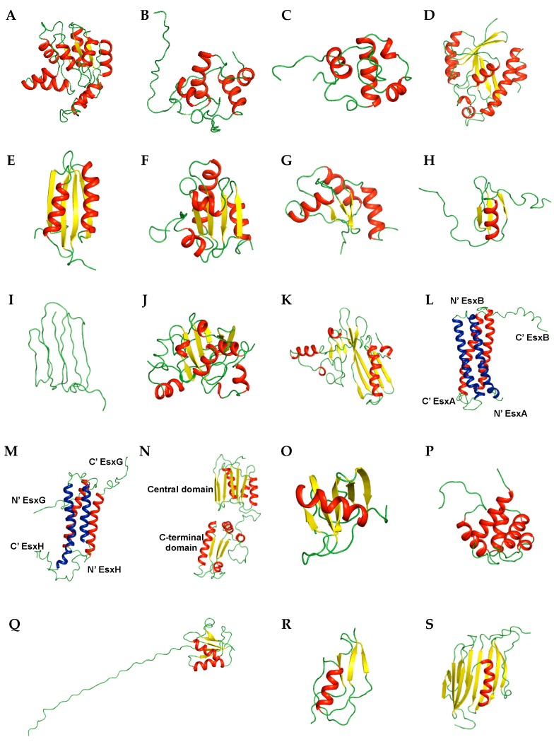
Figure 2.
Ribbon representation of NMR structures of M. tuberculosis proteins. Enzymes and related proteins (A) Rv0733 (PDB ID 1P4S); (B) Rv1009 (PDB ID 1XSF); (C) Rv1884c (PDB ID 2N5Z); (D) Rv1014c (PDB ID 2JRC); (E) MT1859 (PDB ID 2LQJ); (F) Rv3914 (PDB ID 2L59); (G) Rv3198.1 (PDB ID 2LQQ). Siderophore-related proteins (H) Rv2377c (PDB ID 2KHR); (I) Rv0451c (PDB ID 2LW3). Secreted proteins (J) Rv2875 (PDB ID 1NYO); (K) Rv1980c (PDB ID 2HHI); (L) Rv3875/Mb3904 (PDB ID 1WA8); (M) Rv0287/Rv0288 (PDB ID 2KG7). Membrane proteins (N) Rv0899 (PDB ID 2L26). Uncharacterized proteins (O) Rv2302 (PDB ID 2A7Y); (P) Rv0543c (PDB ID 2KVC). Other proteins (Q) Rv0431 (PDB ID 2M5Y); (R) Rv3682 (PDB ID 2MGV); (S) Rv2171 (PDB ID 2NC8). The same colors as used in Figure 1 are employed. Two helix-turn-helix hairpins of (L) and (M), originated from different proteins were colored in blue (EsxA (L) and EsxH (M) and red (EsxB (L) and EsxG (M)), respectively.

