Figure 3.
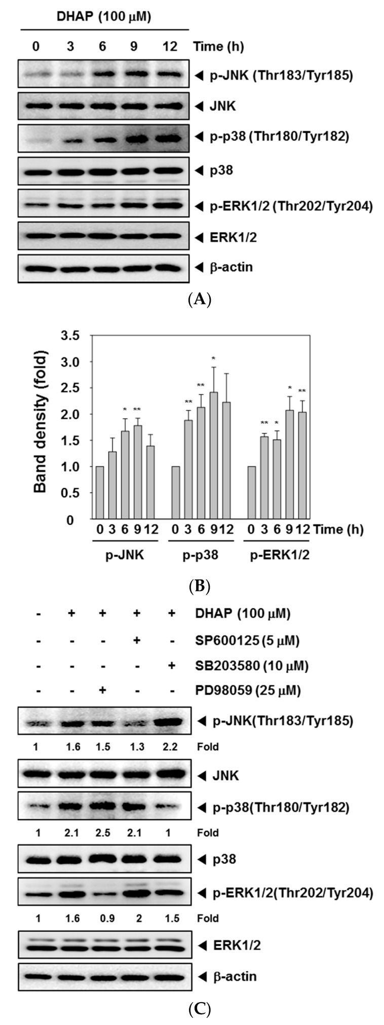
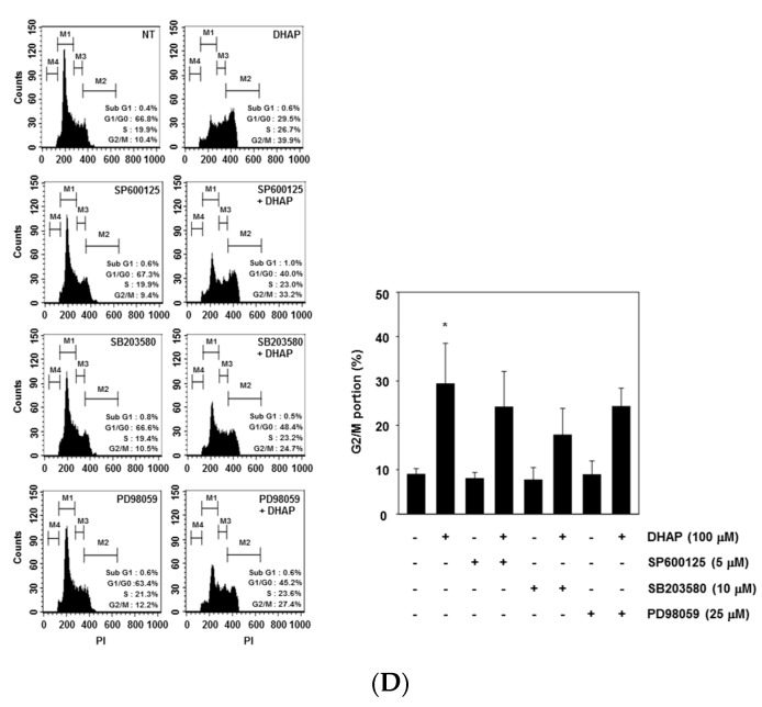
Effect of DHAP on MAPK activation in U266 cells. (A) The cells were treated with 100 μM of DHAP for the time periods stated; whole-cell extracts were then prepared and analyzed via Western blot analysis for p-p38 (Thr180/Tyr182), p-JNK (Thr183/Tyr185), and p-ERK1/2 (Thr202/Tyr204) by Western blot analysis. To confirm equal protein loading, the immunoblot was stripped and reprobed for JNK, p38, and ERK1/2. The results shown are representative of the three independent experiments; (B) The ratios of phosphorylated proteins to non-phosphorylated proteins were measured and the band density values were expressed as mean ± SE; (C) U266 cells were pretreated with SP600125 (5 μM), SB203580 (10 μM), or PD98059 (25 μM) for 30 min, then incubated with DHAP (100 μM) for 12 h. The expression of p-p38 (Thr180/Tyr182), p-JNK (Thr183/Tyr185), and p-ERK1/2 (Thr202/Tyr204) was ascertained via Western blot analysis. To confirm equal protein loading, the immunoblot was stripped and reprobed for total JNK, p38, and ERK1/2 proteins. Densitometric quantitation in fold change of each band has been indicated below the gel; (D) U266 cells were pretreated with SP600125 (5 μM), SB203580 (10 μM), or PD98059 (25 μM) for 30 min, then incubated with DHAP (100 μM) for 48 h. Cellular DNA staining with PI and flow cytometric analysis was performed to ascertain the cell cycle distribution. * p < 0.05, ** p < 0.01, vs. control.


