Abstract
Bacterial and human voltage-gated sodium channels (Navs) exhibit similar cation selectivity, despite their distinct EEEE and DEKA selectivity filter signature sequences. Recent high-resolution structures for bacterial Navs have allowed us to learn about ion conduction mechanisms in these simpler homo-tetrameric channels, but our understanding of the function of their mammalian counterparts remains limited. To probe these conduction mechanisms, a model of the human Nav1.2 channel has been constructed by grafting residues of its selectivity filter and external vestibular region onto the bacterial NavRh channel with atomic-resolution structure. Multi-μs fully atomistic simulations capture long time-scale ion and protein movements associated with the permeation of Na+ and K+ ions, and their differences. We observe a Na+ ion knock-on conduction mechanism facilitated by low energy multi-carboxylate/multi-Na+ complexes, akin to the bacterial channels. These complexes involve both the DEKA and vestibular EEDD rings, acting to draw multiple Na+ into the selectivity filter and promote permeation. When the DEKA ring lysine is protonated, we observe that its ammonium group is actively participating in Na+ permeation, presuming the role of another ion. It participates in the formation of a stable complex involving carboxylates that collectively bind both Na+ and the Lys ammonium group in a high-field strength site, permitting pass-by translocation of Na+. In contrast, multiple K+ ion complexes with the DEKA and EEDD rings are disfavored by up to 8.3 kcal/mol, with the K+-lysine-carboxylate complex non-existent. As a result, lysine acts as an electrostatic plug that partially blocks the flow of K+ ions, which must instead wait for isomerization of lysine downward to clear the path for K+ passage. These distinct mechanisms give us insight into the nature of ion conduction and selectivity in human Nav channels, while uncovering high field strength carboxylate binding complexes that define the more general phenomenon of Na+-selective ion transport in nature.
Author summary
Ion channels can rapidly and selectively conduct an ionic species, essential for the firing of neurons, where sodium and potassium channels respond to changes in membrane potential to release stores of sodium and potassium ions in succession. The ability of a protein pore to discriminate between these two nearly identical ions has remained an intriguing problem for decades. In particular, the origins of sodium selectivity have been obscured by diverse protein chemistries that exhibit sodium-selective conduction in prokaryotes and eukaryotes. Here we use multi-microsecond atomistic simulations to observe and contrast the permeation mechanisms of sodium and potassium ions in model human and bacterial sodium channels. These channels exhibit shared features of conduction, centered on the involvement of charged protein groups that form complexes with the smaller sodium ion. In the human channel model, we observe a special role for the signature lysine that allows sodium to pass, but partially blocks potassium. As sodium channels are vital to heartbeat, sensation, muscle contraction and brain activity, these insights could assist developments in improved therapeutics for disorders such as epilepsy and chronic pain, with mechanisms relevant to a range of ion transport processes in biology and materials.
Introduction
Voltage-gated sodium (Nav) channels are widely distributed in the central and peripheral nervous systems where they participate in essential functions, including heartbeat, muscle contraction and brain activity [1, 2]. Dysfunctional Navs are associated with several physiological disorders, including epileptic seizures and chronic pain [3, 4], and they are therefore a major target for new drugs [5]. However, our understanding of the fundamental mechanisms governing these channels remains incomplete, largely due to the lack of high-resolution structural data on mammalian Navs. There have, however, been recent breakthroughs in the structural determination for several Nav channels that enable investigations into molecular mechanisms.
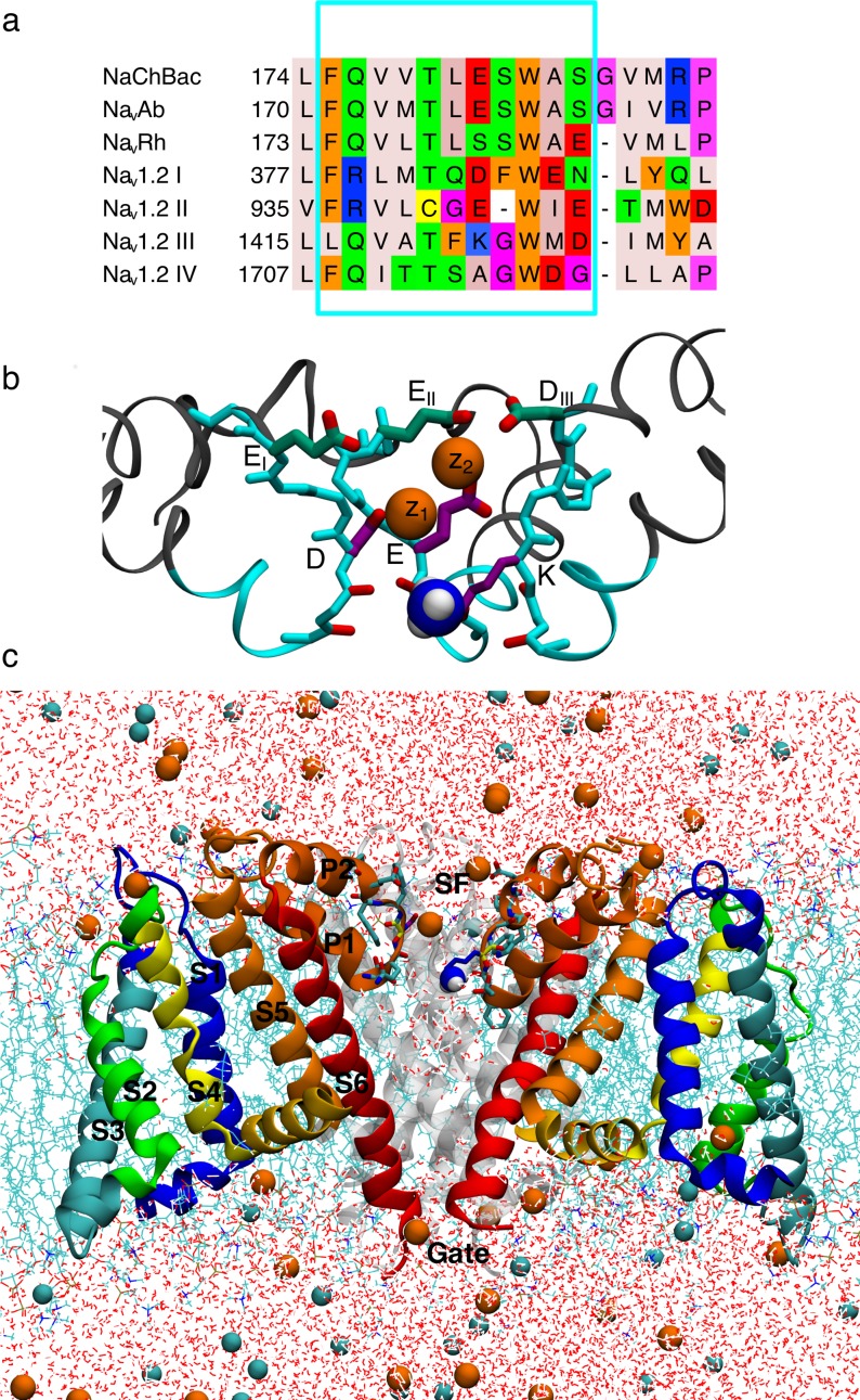
The first high-resolution structure of a Nav channel was the X-ray structure of the bacterial channel NavAb [6], which was followed by numerous other high resolution structures, including for NavRh [7], NavAep1 [8] and NavMs [9, 10]. Although no mammalian Nav structure is yet available, two eukaryotic Nav structures have recently been solved using cryo EM; NavPaS from cockroach [11] and a newly resolved EeNav1.4 from electric eel [12], released after the simulations in this study were completed. NavPaS has a 36–46% sequence identity to human Nav channels, but is 300–500 residues shorter. NavPaS has several residues in and around the SF unresolved, has reduced charge in the vestibule of the SF compared to human Navs and a resolution of 3.8 Å [11]. EeNav1.4 has a 65% sequence identity to the human Nav1.4 and a resolution of 4 Å [12]. The lower resolution of these structures is significant given the small size of the Nav SF (diameter of ~3.6 Å at its narrowest point [12]), and thus while these new structures help guide and validate studies, there remains much to be learned from the existing high-resolution bacterial structures. Bacterial and human Nav channels share several features including Na+-selective conduction, voltage-dependent activation, pore-based inactivation and drug modulation [13–16]. While there is only a 25–30% sequence identity between bacterial and human Navs [10], there exists evidence for overall shared structure [13]. Human Navs consist of four domains, DI-DIV, linked together to form one long polypeptide chain, whereas the simpler bacterial channels are made up of four identical subunits [13] (Fig 1A). Each of these domains/subunits consists of 6 helical trans-membrane spanning segments, S1-S6, where S1-S4 make up a voltage sensor domain (VSD) and S5-S6 the pore domain (PD). Between these two latter segments is a P-loop that includes a narrow ion selectivity filter (SF) [13] (Fig 1B and 1C), which establishes an ion preference “fingerprint” (permeability Li+~Na+ > K+~Cs+~Rb+) that is the same for both bacterial and human Navs [1, 17, 18]. Both eukaryotic and bacterial Navs are Na+ over K+ selective with eukaryotic Navs selecting for Na+ with PNa+/PK+ ~10–30 [1, 19] and bacterial Navs with PNa+/PK+ ~5–170 [17, 18, 20]. Due to the structural and functional similarities, the bacterial Nav channels offer an excellent template, or scaffold, to support investigation into the core functional activities of mammalian Navs.
Fig 1. Human Nav1.2 model ion channel system.
a) Jalview [81] sequence alignment of bacterial NaChBac, NavAb and NavRh, with the four domains of human Nav1.2 (labeled DI-DIV). Amino acids are colored according to their properties using the Zappo coloring scheme [81]. Cyan box selection marks the grafted Nav1.2 residues. Red box selection marks the EEEE/DEKA signature sequences responsible for selectivity. b) SF and vestibule of the NavRh/Nav1.2 model with grafted residues in cyan. The residues in the inner DEKA and outer EEDD rings are indicated in purple and green, respectively, and the charged ammonium group of the Lys is shown as blue and white balls. c) NavRh/Nav1.2 channel embedded in a hydrated DPPC bilayer (cyan sticks), surrounded by water (red and white sticks) with Na+ and Cl- ions (orange and cyan balls). Three out of four monomers are shown (front subunit removed for clarity; rear subunit in gray), with VSD in green/blue/yellow and the PD in orange/red ribbons.
Despite overall conservation of structure and function, the channels present distinct SF sequences (Fig 1A), with bacterial Nav channels making use of a ring of four Glu side chains (EEEE), to create a high field strength binding site (SHFS) thought to favor Na+ [13], whereas all mammalian Nav utilize a ring of Glu, Asp, Lys and Ala (DEKA; Fig 1A) [13, 19, 21], containing not only acidic residues, but also a basic lysine side chain that is known to be crucial to selectivity in mammalian channels [19, 21]. So how do these different channels achieve similar ion selectivity, and does the existence of disparate sequences mean that bacterial and mammalian channels impose their ion preference via different molecular strategies [18]? We seek to reveal and compare the fundamental rules of selective ion permeation for the whole family of sodium channels, by exploring ion conduction mechanisms in a human Nav channel to compare to previous simulations in bacterial channels.
There are 9 different Navs comprising Nav1.1 to Nav1.9 [22], with highly conserved sequences throughout these different subtypes [23]. Nav1.2 is abundant in the human nervous system and has been investigated extensively using methods of mutagenesis and electrophysiology (e.g. [21, 24, 25]). Here we take advantage of the conserved structural and functional features between mammalian and bacterial channels to generate a model of Nav1.2, which incorporates the key SF and vestibular regions, into a high-resolution bacterial structure. Such grafting has previously been successful experimentally for imparting Ca2+ selectivity on a bacterial Nav channel [26]. The bacterial NavRh structure was chosen as the scaffold due to its higher sequence similarity in and around the SF (Fig 1A; only shown for NavAb, NavRh and Nav1.2, however, it can be seen that the same is true also for NavMs, NavAe and NavCt [16]). In particular, other bacterial Navs have one residue extra just above the SF (Gly182; NavAb numbering) compared to Nav1.2 (Fig 1A). Furthermore, those channels have an arginine (Arg185; NavAb numbering) close to the SF that was found to interfere with the side chains of the Nav1.2 SF in our separate models using NavAb as a scaffold (not shown). We also note that proposed open structures of a bacterial Nav have been presented [9, 16, 27], but our simulations (not reported here) have suggested that the proposed open NavMs structure does not stay open without strong constraints, as discovered previously [28]. Furthermore, previous studies of ion selectivity in bacterial Navs using a closed lower gate have shown good sampling of ion translocation throughout the SF [18, 29–31], and are thus capable of shedding light on selectivity mechanisms by revealing the underlying equilibrium free energy surfaces governing multi-ion movements. There is minimal thermodynamic perturbation for ions in the SF and cavity due to the closed lower gate, with the free energy of an ion inside open and closed NavAb pores having been shown to be similar [27].
Previous simulations of bacterial channels have taught us much about the potential behavior of the human sodium channel. The bacterial SF contains a highly conserved ring of four Glu side chains forming a high field strength site (SHFS) [6]. Early simulation studies revealed ion binding sites and indications of a multi-ion conduction mechanism [28, 31–36]. These studies suggested favoring of a partly-hydrated Na+ due to the SF geometry in the NavAb crystal structure and the strength of interaction of ions with glutamate side chains [28, 31, 36]. They also demonstrated the ability of the SF to bind 2 ions concurrently within the SHFS site, with higher affinity for Na+ potentially creating a reduced permeation barrier [36]. Simulations have shown that the SF is flexible and wide enough to house multiple Na+ ions [37], and is occupied by an average of two to three ions [29, 30, 38]; although simulations under significant membrane potentials have indicated reduced occupancy [28]. However, microsecond-length MD simulations have shown that the symmetric arrangement of Glu side chains in the crystal structure is broken on long time scales, affecting ion occupancy and having significant implications for the permeation mechanism [29, 30, 36, 39]. These studies demonstrated that there is coupling between ion translocation and SF conformation, with isomerization of glutamate side chains catalyzing Na+ conduction. These long unbiased simulations have described a stable 2-ion state, where the ions are trapped [30]. While some studies have focused only on this 2-ion state [31, 36], it has been shown that it is when a third ion enters the SF that efficient knock-on of the bottom ion into the central cavity occurs [18, 29, 30]. The top ion can either knock-on or pass-by the middle ion in SHFS, with both permeation pathways experiencing similar energy barriers (S2 Fig; [30]). Regardless, binding of Na+ to the SHFS Glu side chains is central to understanding Na+ over K+ selectivity [18, 30, 31, 36].
Conduction is reliant on the flexibility of these side chains [29, 30, 36], where Na+ ions form favorable 2 ion-2 carboxylate clusters (see S2 Fig, states C2 and C3) that are not stable for K+ [30]. Human Nav channels do not possess a symmetric E ring, but contain a set of carboxylates that might mediate similar complex formation. In particular, the DEKA signature sequence (Fig 1, right inset) contains two carboxylates that may be sufficient. However, human channels also possess a well-conserved [24] charged ring in the outer vestibule, immediately adjacent to the SF, consisting of Glu, Glu, Asp and Asp residues (EIEIIDIIIDIV; with the exception of Nav1.7 that contains EIEIIIIIIDIV) [40]. The residues in these two rings have been shown to be asymmetrical in position [41] and highly flexible [42], and thus may facilitate multi-ion conduction, akin to the bacterial channels.
Substitution experiments have shown that the DEKA-ring is crucial for the selection of Na+ ions in mammalian Nav channels [19, 21, 25], with at least one of the D and E necessary to preserve selectivity [19]. The most important carboxylate in the DEKA ring is the E, which together with the K (residue 1422 in human Nav1.2), maintains wildtype (WT) selectivity, despite mutation of the D [19]. However, if the positions of these two residues (E and K) are swapped, into DKEA, selectivity is reduced nearly 4-fold [25], showing the importance of their precise locations, potentially due to interactions with surrounding amino acids, or implying a specific coordinating complex for Na+ during conduction. Mutagenesis has also shown that both the charge and length of the K1422 are necessary for maintaining Na+ selectivity; substitution of Lys with neutral side chains causing a complete loss of Na+ selectivity, and substitution with negatively charged side chains even reversing selectivity [19]. Previous computational studies examining the role of Lys in conduction have also been based on models using bacterial structures, and concluded on a passive blocking role for Lys. Xia et al. replaced the ring of four Ser in the bacterial channel NavRh with DEKA and performed MD simulations to find that only when the Lys was constrained to EIV was the SF permeable to Na+ and K+ ions [43, 44]. Mahdavi et al. threaded the amino acids from rat Nav1.4 onto the backbone of a pore only model of NavAb, also suggesting that ion translocation requires displacement of Lys out of the permeation pathway, by hydrogen bonding to the neighboring DIV Ser [45]. We will show in this study, using extended length simulations, that Lys in fact plays an active role in conduction (not just based on pore occlusion) that is quite different for Na+ and K+ ions.
The vestibular EEDD-ring has also been shown to be important for ion conduction, possibly because it increases the electrostatic attraction of extracellular cations [24, 25, 46, 47]. However, all four residues are not equally important, with cysteine mutations showing the greatest decrease in conduction occurs when domain II Glu is mutated (effect on conduction: EII>EI> DIV~DIII) [25, 41]. Furthermore, mutagenesis experiments have shown that replacement of EEDD residues can be detrimental to Na+ selectivity. In particular PNa/PK has been shown to decrease when DIV is replaced with Cys [40, 41, 48], while other experiments have shown little or no effect [25, 49]. Functionally, therefore, the exact roles of the outer ring carboxylates in selectivity are not well defined. Previous models based on bacterial structures have implied a greater thermodynamic preference for Na+ in the lower SF, and an additional outer binding site at the EEDD-ring [43, 45]. However, we will show that on long timescales, SF rearrangements allow for stable complexes with carboxylates from both the SHFS and outer ring that are important to Na+ permeation.
Experiments have suggested that Navs possess multiple binding sites inside the SF [50–53]. However, the evidence for their ability to bind multiple ions at the same time is inconclusive [50–55]. Investigations of flux coupling suggest a flux ratio exponent close to unity for mammalian Nav channels, which is generally thought to imply a 1-ion conduction mechanism [51, 54, 55]. Furthermore, anomalous mole fraction effects, considered evidence of ion cooperation during conduction, have only been observed in bacterial [18] and not mammalian Navs [53]. Such effects are normally expected in single-file channels that impose ion correlation [56], but may also arise from preferential and localized ion binding [57–59]. Single-file conduction is unlikely in Nav channels given their size [60] and flexibility [42]. However, absence of an anomalous mole fraction effect does not rule out ion cooperativity. The fact that anomalous mole fraction effects are seen in bacterial Nav channels in the absence of bona fide single-file permeation, as suggested by several simulation studies, as well as the decoupling of ion and water fluxes [28], demonstrates limitations in the ability of this measure to identify a multi-ion mechanism. Likewise, there exists contradictory evidence from single channel conductance measurements for mammalian Nav channels, which have shown a saturating dependence on concentration in some experiments, indicative of a single ion mechanism [50–52], whereas others, where a wider range of concentrations have been examined, witness a complex relationship that may be explained by a multi-ion conduction mechanism [53]. Thus the experimental evidence is inconclusive when it comes to the ion occupancy and potential multi-ion conduction mechanism in mammalian Navs. Molecular dynamics (MD) simulations can therefore help us elucidate the mechanisms involved in ion selectivity in Navs.
Bacterial and human Nav channels therefore both possess several carboxylate side chains in (or adjacent to) the SF, allowing for multiple high field strength sites that may bind one or more ions, possibly leading to increased Na+ selectivity [61]. In a multi-ion/multi-ligand complex there will be competition between favorable ion-ligand and unfavorable ligand-ligand and ion-ion energies [62], needing long time scale simulations to capture the ion and protein configurations [29, 30]. The mechanism is further complicated by the presence of the Lys, which may increase the sampling challenges due to side chain isomerizations. We thus turn to long MD simulations to capture those ion and slowly interconverting protein movements using the DE Shaw Anton supercomputer. We have performed multi-μs simulations where Na+ and K+ are permitted to freely diffuse into and out of the SF of the NavRh/Nav1.2 model, observing the involvement of DEKA and outer EEDD rings, as well as participation of the signature Lys in multi-ion binding complexes, revealing distinct permeation mechanisms for Na+ and K+ ions.
Methods
We have created a model of human Nav1.2 that includes the most important residues for ion permeation and selectivity, using a structurally well-defined bacterial channel as a scaffold. Although other parts of the channel may be important for attracting ions [11], the main functional region involves the SF and outer vestibular regions [24]. The sequence alignment in Fig 1 was used to create a model of human Nav1.2 based on the bacterial NavRh structure (PDB:4DXW [7]), due to their higher sequence homology in and around the SF region, as discussed above. The mutated region (framed in cyan in Fig 1A), spanning residues 174–184 (NavRh numbering used herein), was chosen to introduce the SF and vestibular sequence with minimal perturbation. This includes the human DEKA and EEDD rings (residues 180 and 183/184) in the upper SF. Furthermore, residues 174–177 are located behind the SF and may interact with the side chains of the SF. In particular, within that range is NavRh Q175, which is a positively charged Arg in Nav1.2 DI and DII, and may influence both structure and ion conduction. To minimize perturbation, side chains of the chosen amino acids were removed from NavRh and the corresponding Nav1.2 side chains rebuilt manually using the CHARMM program [63]. Herein the residues from the DEKA ring will be referred to by their 1-letter abbreviations. To avoid confusion with the symbol for Lys (K), potassium will always be referred to in ionic form, K+. The Asp and Glu residues from the vestibular EEDD ring will be referred to according to their domain numbers (EI, EII, DIII and DIV).
Between the DEKA ring and the EEDD ring the Nav1.2 sequence is one residue shorter in DII than in the bacterial channels. This residue (number 181) was removed, and neighboring residues (180–182) joined with constrained MD simulation (see below). We note that the original NavRh SF structure was closed at the lower gate as well as having a slightly collapsed SF. The collapsed SF has been proposed to be associated with the orientation of the Ser181 side chains, blocking the SF [64], however, after removing the Ser side chains as well as patching Nav1.2 residues and equilibration (described below), the filter forms an open SF conformation that allows us to study ion permeation (Fig 1B and 1C). Other changes to the model, including the rebuilding of missing intracellular loops connecting S2 and S3 helices in the VSD using Rosetta [65], and the maintenance of a key hydrogen bond between Thr178 and Trp182 that has been postulated to be important for keeping the shape of the SF in bacterial Nav [6, 7], are described in the Supplementary Information.
It is difficult to predict or measure the pKa shift of Lys in a non-aqueous microenvironment with the coming and going of conducting ions, such as in the SF of a Nav channel [66]. Large pKa shifts have been recorded in channel environments, and protonation states depend on the exact environment and ion occupancy, which will fluctuate over time [67]. The dependence of Nav conduction on the protonation states of key residues in the SF has therefore been investigated in several studies of bacterial Navs [30, 36, 68]. Furthermore, quantum mechanical calculations using simplified models of the DEKA ring alone have demonstrated that a thermodynamic preference for Na+ may be achieved with either protonation state of the Lys [61]. However, the signature Lys has been shown to be crucial to Na+ selectivity [19], intuitively suggesting that its charge may be important. To cover both possibilities, we have examined Na+ ion movements with both charged and neutral lysine, which will help us isolate the role of that charge in ion conduction.
The proteins were embedded in lipid bilayers of dipalmitoyl-phosphatidylcholine (DPPC), being the best characterized lipid for MD simulations [69], with explicit TIP3P water molecules [70] and 150 mM of NaCl or KCl solution. Systems were built and pre-equilibrated with CHARMM [71] and further equilibrated using NAMD [72] prior to unbiased production simulations carried out on the purpose-built supercomputer Anton [73, 74] for 4 μs for NavRh/Nav1.2 with charged Lys both for NaCl and KCl, and 2 μs for neutral Lys (less time required due to better sampling of ion movements), totaling 10 μs. Simulations all used the CHARMM36 [69] lipid and CHARMM22 protein and ion parameters [72] with CMAP corrections [75], chosen to provide direct comparison to our past simulations of the bacterial NavAb channel [30]. However, attention to ion-carboxylate parameters was given to ensure accurate interactions for Na+ and K+ inside the SF, with standard parameters for the ion-carboxylate interaction shown to lead to reasonable agreement with both ab initio MD free energies of binding and osmotic pressure data [76]. Descriptions of both ion-carboxylate and corrected ion-carbonyl interactions are discussed in the Supplementary Information.
The free energy map, or potential of mean force (PMF), for ion movement was calculated for each system using trajectory data with specific pore ion occupancies, from unbiased simulations as W({zi}) = −kBT ln ρ({zi}) + C, where ρ is the unbiased probability distribution as a function of reaction coordinate(s) {zi}, being the vertical positions of one or more ions (z1, z2, or z3; defined with z1 being the lower-most ion), or their centroids (e.g. z12 = (z1 + z2)/2), each relative to the center of mass of the protein, zref, and where C is a constant. See Supplementary Information for more details, including error calculations.
The most important states involved in the permeation of Na+ and K+ ions across the SF were identified from the PMFs and cluster analysis. The frames were broken down according to ion occupancy as well as ion-ion and ion-carboxylate distances (S1 Table). Different states were identified according to how many carboxylates were interacting with the ion/ions using a cutoff defined from the radial distribution functions in S3 Fig, with further details of the clustering method described in the Supplementary Information. These states include complexes involving one or more carboxylate groups, Na+ or K+ ions and/or the signature Lys ammonium group. To evaluate the relative binding affinities of these states, Free Energy Perturbation (FEP) [77] calculations were performed to obtain the relative binding free energies of Na+ and K+ to each particular complex. Details of these, and other calculations, including structural comparisons of available X-ray and Cryo-Em structures, and continuum electrostatic calculations to examine ion-binding propensities, are provided in the Supplementary Information.
Results and discussion
A stable human Nav model with asymmetric and flexible selectivity filter
After patching of Nav1.2 SF and vestibular residues into the bacterial channel, followed by equilibration, the overall structures of the PD and SF were stable and settled down to RMSD 2–3 Å (S4 Fig) after initial changes during the first μs of simulation, with the level dependent on the choice of Lys protonation state (see below). Late changes are seen, especially for simulations in KCl, due to the onset of permeation following conformational changes within the SF, described below. The first 1 μs of the Nav1.2 model with charged Lys with NaCl and KCl and the first 0.25 μs of the shorter simulation with uncharged Lys have been discarded as equilibration (S5 Fig). We observe that the Nav1.2 SF is relaxing into an asymmetrical shape where the DEKA and EEDD-ring span ~10–15 Å, allowing interactions with residues other than their immediate neighbors (S5 Fig), explaining the increased RMSD. Previous experiments involving cysteine mutations have shown that the residues in the filter of an Nav channel are indeed asymmetrical in height and that the inner and outer rings span up to ~15 Å [41]. This asymmetry destabilizes the Thr/Cys178-Trp182 bond, important for keeping the shape of the SF in bacterial channels [6, 7], which are seen to break and form several times during the simulations.
Flexibility has been shown to be a critical property of the bacterial Nav channel SF to facilitate ion conduction [29, 30]. Furthermore, pairwise cysteine mutations have suggested significant flexibility (up to 7 Å) of the residues in the SF of a mammalian Nav [48] [42]. We observe high flexibility in the NavRh/Nav1.2 SF (S6 Fig, with mean SF backbone fluctuations being approximately twice as large as those for NavAb (RMSF~1.5 Å and RMSF~0.8 Å for NavRh/Nav1.2 and NavAb, respectively) (S6A and S6B Fig). These RMSF values (S6A Fig) are mostly due to asymmetric subunit movements, with the RMSF values halved when computed based on subunit-by-subunit orienting (S6B Fig).
The recent eukaryotic NavPaS [11] and EeNav1.4 [12] cryo EM structures offer related structural data to validate our model, although at limited resolution where several SF side chains were not resolved, preventing immediate conclusions about specific interactions. Structural alignment between the NavRh/Nav1.2 model and the NavPaS or EeNav1.4 structures (S1 Fig) shows structural similarities between the proteins, with key residues at the same positions. Due to the highly dynamic nature of the SF and its side chains, structural comparisons are limited and will depend on the choice of structure from the model simulation. Overall, however, structures are consistent, with the RMSD of the SF and vestibular region (residue 178 to 184) backbone being 2.0 Å and 2.3 Å when aligning to all four subunits (S1A Fig), and 1.8 Å and 2.1 Å when aligning subunit by subunit (S1A Fig), for NavPaS and EeNav1.4, respectively. The residues of the DEKA and EEQD/EEDD rings are asymmetrical in position in all three structures, with these residues spanning 10–15 Å in the model, ~10 Å in NavPaS and ~14 Å in EeNav1.4 (S1 Fig). The spread of these side chains in both our model and the NavPaS and EeNav1.4 structures, combined with the high flexibility and lack of experimental resolution in this region, indicates that SF dynamics may play an important role in conduction.
Selectivity filter with neutral lysine exhibits bacterial-like multi-ion complex formation and conduction
The DEKA Lys has been shown to be crucial to Na+ selectivity in mammalian Nav channels [19]. However, the protonation states of amino acids in a dynamic microenvironment, like that seen here within the SF), are hard to pinpoint experimentally [66] and may have large fluctuations [67]. We have therefore investigated ion permeation in Nav1.2 not only with a protonated (Lys+) but first also with a deprotonated Lys (Lys0). Importantly, this allows us to isolate the role that the positive charge may play in the SF of Nav1.2 for Na+ permeation, and offers a comparison to existing bacterial Nav simulations.
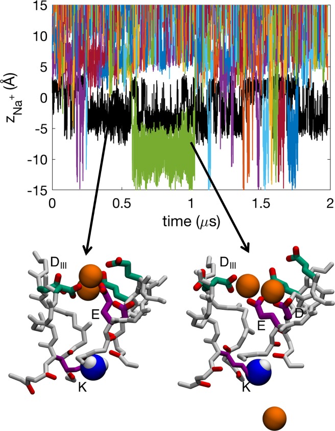
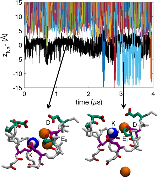
Permeation of Na+ in the SF of the Nav1.2 model when its DEKA Lys is neutral involves both 2-ion and 3-ion conduction mechanisms with an average number of 2.1±0.2 Na+ ions in the SF, similar to (albeit slightly less than, though within the errors) that seen in the bacterial channel NavAb (2.3±0.5 ions; [30]). The ions are partly hydrated as they permeate the SF (S7 Fig). In the time series for ion movements shown in Fig 2, several ions (colored lines) are observed to enter and exit the central cavity (below −5 Å), often coexisting within the SF (S1 Movie). We see 61 complete permeation events through the SF (inward or outward moving involving 16 distinct ions) and the ions appear to move independently of the neutral Lys side chain (black line). Snapshots in Fig 2 show representative configurations of a 2-ion and 3-ion state, where we see multi Na+-ion/multi-carboxylate complexes similar to those in for the bacterial Nav ([30]; see also Fig 3E and 3F, to be discussed below). However, in the Nav1.2 channel it most commonly not only involves carboxylates only from the inner DEKA-ring, but also from the outer EEDD-ring, whose carboxylates reach down to help coordinate the ions, as opposed to only using two carboxylates from the inner ring, as was the case of the bacterial NavAb channel [30].
Fig 2. Time series showing the z positions of deprotonated Lys0 (K; black) and Na+ ions (colors) in the Nav1.2 SF.
Insets show snapshots of Na+ ions (orange balls) in representative configurations with active residues from the DEKA (purple) and EEDD (green) rings labeled. The neutral amine group of the Lys shown is shown as blue and white balls. See also sample trajectory in S1 Movie.
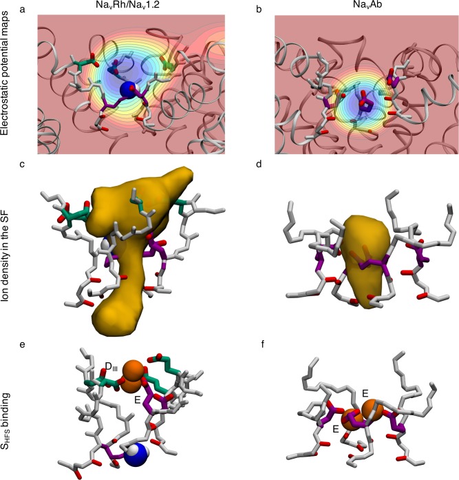
Fig 3. Comparison of electrostatic potential maps.
Electrostatic maps of a) the Nav1.2 SF with deprotonated Lys0, compared to b) bacterial NavAb. Each contour represents 10 kcal/mol/Å/e. Density maps of Na+ occupancy in c) the Nav1.2 SF with Lys0 and d) NavAb. Multi ion-multi carboxylate binding sites in e) Nav1.2 and f) NavAb. The residues in the inner DEKA and outer EEDD rings are indicated in purple and green, respectively, and the uncharged amine group of the Lys is shown as blue and white balls.
This cooperation between the DEKA and EEDD-rings creates a longer SF binding region with a broad span of negative electrostatic potential (Fig 3A and 3B), leading to less distinct binding sites. While NavAb has all of its charged side chains in one symmetrical ring, Nav1.2 has its charges spread out in two asymmetrical and highly flexible rings creating a broad SHFS (ion density map shown in Fig 3C), compared to NavAb (Fig 3D). Commonly a binding site is created with two Na+ and one or two carboxylates from the DEKA and EEDD rings (S1 Table; column 2 and S8 Fig). The specific residues involved vary and we see several distinct carboxylate-ion complexes forming during the simulation (S1 Table; column 2 and S8 Fig).
Double ion occupancy dominates in the SF, but 3-ion occupancy is also common (S8A Fig). When the SF is occupied by a single ion it is most commonly unbound, however, it is also likely to be bound to one or more carboxylate side chains (S8B Fig). When there are two ions in the filter they most likely form single ion complexes with multiple carboxylates (S8C Fig). They are also often singly bound or bound together in a tight multi-ion/multi-carboxylate complex (defined by the radial distribution function in S3 Fig; with ions within 4.7 Å of each other and 2 or more carboxylate groups (based on central C atom) from the DEKA and EEDD-rings within 3.8 Å of the ions). When there are three ions in the SF, two of these are most likely to be bound together in a tight multi-carboxylate complex (S8D Fig). We have previously shown that multi-ion/multi-carboxylate complexes play a role in Na+ selectivity in acid sensing ion channels [76]. When these complexes are present, the ions are mostly bound by E and/or D from the DEKA ring, together with the outer ring DIII (S1 Table; column 2). This cooperativity of inner and outer charge rings is common and while all carboxylate side chains from either of the two rings ring are not necessary, at least one is needed for Na+ binding in the SHFS. In particular, the DEKA ring is involved ~80% of the time, the EEDD ring ~80% of the time, and both rings cooperatively involved ~60% of the time. This need for only one of the carboxylates in the DEKA ring to maintain cation permeability has previously been implicated by mutagenesis experiments [19].
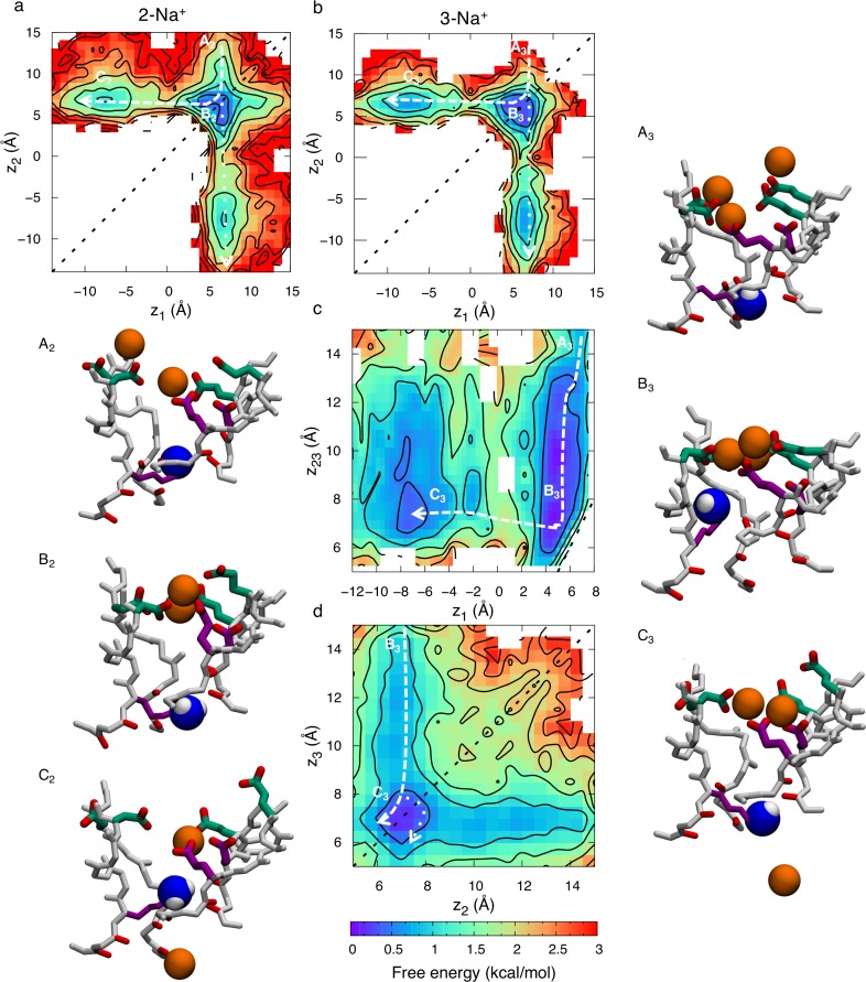
Despite the longer SF binding region in Nav1.2 with neutral Lys, we see a conduction mechanism resembling that of the bacterial Nav, relying on multi ion-multi carboxylate clusters for an efficient knock-on conduction. When there are 2 ions within the SF, at z1 (lower) and z2 (upper), the free energy map (as function of z1 and z2) in Fig 4A exhibits three states; A2, B2 and C2. In state A2, the upper ion is in the vestibular region of the SF (z2~14Å) and the lower ion is collectively bound by the inner (purple side chains) and outer rings (green side chains) (z1~7Å). In state B2 the top ion has joined the bottom ion and they are both bound by the inner and outer rings (z1~z2~7Å) forming cooperative tight multi-ion/multi-carboxylate clusters, commonly made up by D, E and DIII, and reminiscent of those seen in the bacterial Nav (compare Fig 3E and 3F). In state C2 the top ion has either pushed the bottom ion into the cavity by Coulomb repulsion (dashed line) or it has passed by the bottom ion and entered the cavity itself (dotted line crossing the diagonal, y = x, represented by black dashed line), in both cases translocating from z2~7 Å to z2~-10 Å, while the other ion remains collectively bound to the inner and outer rings. These distinct permeation pathways are equally likely, with the largest barrier encountered being 1.9±0.8 kcal/mol.
Fig 4. Permeation of Na+ in the Nav1.2 SF with deprotonated Lys0.
2D free energy projections showing a) 2-ion conduction mechanism, and b, c & d) 3-ion conduction mechanism (graphed for the two bottom ions in b, all three ions in c, and the two top ions in d); where z1, z2, and z3 correspond to the z positions of the bottom, middle and top ions, respectively, and where z23 is the z position of the COM of the top two ions. Each contour represents 0.5 kcal/mol. Snapshots, with the inner DEKA ring in purple, the outer EEDD-ring in green, and the uncharged amine group of the Lys shown as blue and white balls, indicate the corresponding Na+ ion (orange ball) movements. State labels (A-D) include a subscript 2 or 3, representing the 2-ion or 3-ion mechanisms, respectively.
Fig 4B–4D show free energy projections for the 3-ion occupancy state, with ion positions specified as z1 (lower ion), z2 (middle ion), z3 (upper ion) or z23 (COM of the two upper ions). Here state A3 has one ion in the middle of the two rings (z1~7 Å) and one ion in the vestibule (z2~14 Å), and an additional ion entering from the bulk above (z3~15 Å). In Fig 4B and 4C we see how the upper ion pushes the middle ion downward to bind together with the lower ion at the inner ring (z1~z2~6 Å), represented by state B3, before eventually entering this site itself and pushing the bottom ion into the cavity, state C3, completing the conduction event. The broad free energy surface in panel c is due to the range of binding sites offered by the inner and outer carboxylate rings, as well as the fact that the centroid of 2 ions may span a wide range. Fig 4D shows another projection involving only the top two ions (z2 and z3). The dashed line in Fig 4D shows how the top ion moves from state B3 to state C3 by entering the SHFS and knocking the bottom ion downward. The dotted line shows an alternative path where the top ion instead passes by the middle ion before knocking the bottom ion into the cavity. Regardless of the permeation pathway, the largest barrier experienced during permeation through the SF in the 3-ion state is 1.4±0.6 kcal/mol, similar to that experienced for Na+ in the bacterial channel (S2 Fig). These results demonstrate that low barrier conduction may occur via knock-on or pass-by mechanisms in either 2- or 3-ion occupancy states for Nav1.2, but where 3-ion conduction is energetically more favorable, with reduced activation barrier. However, considering a 2-ion occupancy is more common (S8 Fig), the 2-ion mechanism is likely also contributing significantly to the overall ion flux.
We therefore have observed that the mammalian SF conducts Na+ ions well with a neutral/deprotonated Lys in the DEKA ring, providing a useful comparison to the bacterial channel, with similar multi-ion/multi-carboxylate complexes forming during permeation (although involving both SF DEKA and vestibular EEDD carboxylates). However, because Lys is so important to permeation and selectivity experimentally [19, 21, 25], but when neutral apparently plays only a passive role that would not implicate it, this suggests that we must turn our attention to the protonated Lys case.
Charged lysine forms a high field-strength complex to facilitate Na+ conduction
When Lys is charged, there is an average of 1.5±0.4 Na+ ions in the SF and permeation involves either 1 or 2-ion conduction (S2 Movie). This reduction in ion occupancy (compared to the neutral Lys case above and the bacterial channel) is presumed to be due to the reduced negative charge inside the SF. If the ammonium group of the Lys was considered as another ion, we would have, in total, a similar ion occupancy of 2–3 ions in the SF. (n.b. Lys+ may be considered another ion, as Nav channels conduct ammonium ions nearly as well as Na+, with relative permeability PNH4+/PNa+ = 0.16 [60]). The time series in Fig 5 reveals that several ions (colored lines) enter and leave the SF, but it is not until the second half of the 4 μs simulation that ions begin to cross the charged Lys and enter the cavity beneath, after which we observe 28 complete permeation events (defined as any ion crossing downward or upward past the Lys, and involving 6 distinct ions that experience repeat crossings).
Fig 5. Time series of z positions for protonated Lys+ (K; black) and Na+ ions (colors) in the Nav1.2 SF.
Insets show snapshots of Na+ ions (orange balls) in representative configurations with active residues from the inner DEKA (purple) and outer EEDD (green) rings labeled. The charged ammonium group of the Lys is shown as blue and white balls. See also sample trajectory in S2 Movie.
Snapshots in Fig 5 show representative configurations of 1- and 2-ion carboxylate complexes. As with the neutral Lys case, we see multi-ion/multi-carboxylate complex formation where the residues from the EEDD ring (green) bend down into the SF region, leading to the binding of ions together with the carboxylates from the DEKA ring (purple; see also S1 Table; column 3). The DEKA ring is involved in binding ~50% of the time, the EEDD-ring ~90% of the time, and both rings cooperatively involved ~40% of the time. These complexes are most commonly (69±11%) involving D from DEKA and EII from EEDD. The ammonium group of the Lys from DEKA is also involved, binding 61±10% of the time to the E or D from the DEKA-ring. The salt bridge between Lys and one or both of these residues has previously been suggested by mutagenesis experiments as being important for Na+ selectivity [19], and may be responsible for the importance of the precise sequence position of the Lys in the DEKA ring [25]. We also observe that when ions enter the SF, the ammonium group of Lys (black line in Fig 5) is displaced downward; being knocked-on, like any other ion. Thus, the charged Lys is intimately involved in the multi-ion mechanism. Importantly, it also participates in a Lys+Na+/carboxylate complex, similar to the 2-ion/multi-carboxylate clusters, commonly coordinated by D and EII and sometimes additionally by E (S1 Table; column 5). The ability to form these complexes is likely dependent on the location of the Glu and Lys in the DEKA ring, and swapping their positions might affect this complex formation thus decreasing selectivity, as indicated by mutagenesis experiments [25]. We will see below how this tight cluster plays an important role in the conduction mechanism.
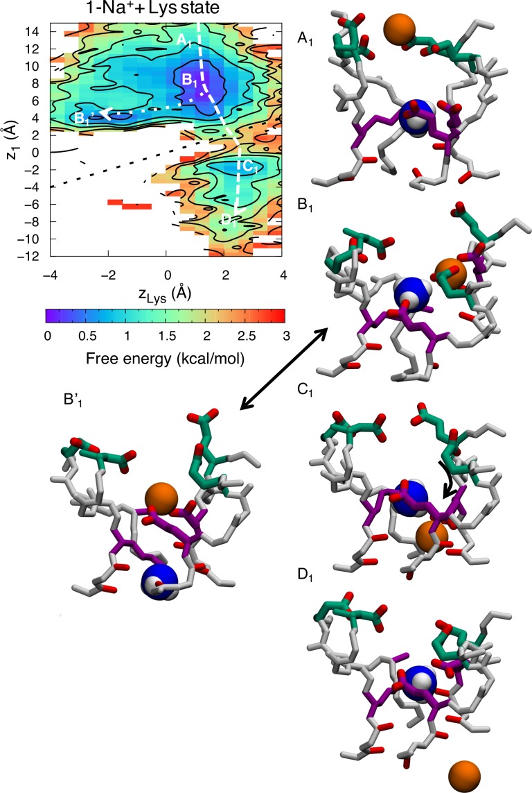
Free energy projections reveal the relationship between the ions and the position of the ammonium group of Lys, zLys, demonstrating conduction with 1-ion and 2-ion occupancies in the SF. When there is a lone ion in the SF, at z1, the free energy map (as function of z1 and zLys) in Fig 6 exhibits four states; A1, B1, C1 and D1. In state A1, the ion is in the outer vestibular region of the SF (z1~14Å) and the Lys is bound to one of the inner ring carboxylates (zLys~1 Å). In state B1, the ion has entered deeper into the SF and is bound to the inner and outer rings (z1~6 Å). The dotted line shows an alternative state, B1’, in which the ion entering the filter replaces the ammonium group of the Lys, which is displaced downward (from z1~6 Å & zLys~1 Å to z1~4 Å & zLys~-2 Å). It is interesting to note that there is no conduction occurring when this happens. The ion is bound tightly to the DEKA ring and the ammonium group of the Lys is too repulsive for it to pass by in the lower part of the filter, where the electrostatic potential is not as negative. However, the dashed line shows how the ion can cross the ammonium group of the Lys in a region of higher field strength created by multiple carboxylates (z~2 Å), leading to state C1. In state C1 the ammonium group of the Lys remains bound to the inner ring (zLys~2 Å) and the ion is bound to the backbone carbonyl of the two residues underneath the DEKA ring (z1~-2 Å). In state D1 the ion has left the SF and entered the cavity (z1~-8 Å), completing the permeation event. The dashed arrow shows the minimum free energy path for conduction, where the largest barrier encountered is 1.7±0.3 kcal/mol.
Fig 6. Single ion permeation for Na+ in the Nav1.2 SF with protonated Lys+.
2D free energy projections showing 1-ion conduction mechanism where z1 corresponds to the z position of the ion and zLys the z position of the Lys+ ammonium group. Snapshots, with the inner DEKA ring in purple, the EEDD ring in green and the charged ammonium group of the Lys shown as blue and white balls, indicate the corresponding Na+ ion (orange balls) movements.
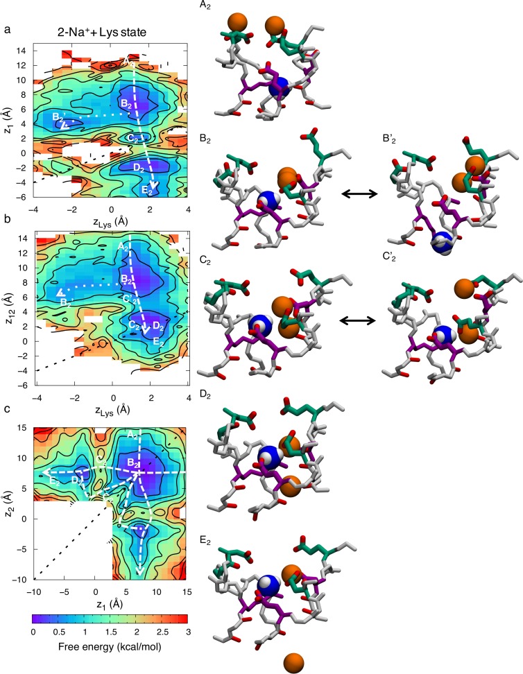
The 2-ion conduction mechanism is summarized in Fig 7. The free energy projection in Fig 7A shows the position of the bottom ion (z1) as a function of the Lys (zLys), Fig 7B shows the COM of the two ions (z12) as a function of the Lys, while Fig 7C shows bottom versus top ions. In this 2-ion case, we see five distinct states; state A2 is like the corresponding 1-ion state A1, but with an additional ion entering from the bulk above (z12~14 Å). In Fig 7B we can see how the two ions enter deeper into the SF and bind collectively to the inner and outer rings, just above the Lys in state B2 (z1~z2~7 Å & zLys~2 Å). There is an additional state B2’, related to B2, where the ammonium group of the Lys is displaced downward (from z1~6 Å & zLys~2 Å to z1~4 Å & zLys~-2 Å; dotted line). However, just as for the 1-ion state, this downward Lys displacement is not sufficient for conduction, instead requiring that the ammonium group of Lys rise up and join one of the ions in a new intermediate stable state C2 as shown in Fig 7A (z1~zLys~2 Å). This state only appears when there are 2 Na+ ions in the filter and seems to be vital for efficient permeation, because it allows the ion to pass the Lys with reduced energetic barrier (dashed line). The ion then binds below the Lys to the backbone carbonyls of the lower SF represented by state D2 (z1~-2 Å & zLys~2 Å). In state E2 the top ion pushes the bottom ion into the cavity and binds to the inner ring (z1~-6 Å, z2~6 Å & zLys~2 Å). Fig 7C shows only the two ions, at z1 (lower) and z2 (upper), and the corresponding states. The top ion can either enter and knock on or pass by the bottom ion. The dashed arrows show the lowest free energy path for conduction. The greatest barrier to overcome in the 2-ion state is 1.6±0.7 kcal/mol. Importantly, while conduction was seen to be possible with a singly-occupied SF (seen in Fig 6; dashed line), the entrance of a second Na+ ion enables a stable state (Fig 7A; state C2) where the ammonium group of the Lys and the bottom ion are collectively bound by the carboxylates, this allows the ion to pass by the Lys and in to the cavity with a lower barrier. As we shall see below, this efficient conduction mechanism does not exist for K+ ions.
Fig 7. Two-ion permeation for Na+ in the Nav1.2 SF with protonated Lys+.
2D free energy projections showing the 2-ion conduction mechanism for: a) the bottom ion and Lys; b) the two ions and Lys; and c) the two ions; where z1 and z2 correspond to the z positions of the bottom and top ions, z12 the z position of the COM of the two ions, and zLys the z position of the Lys+ ammonium group. Snapshots, with the DEKA ring in purple, EEDD ring in green, and the charged ammonium group of the Lys shown as blue and white balls, indicate the Na+ ion (orange balls) movements.
K+ exhibits less multi-ion complex formation, and is partially blocked by lysine
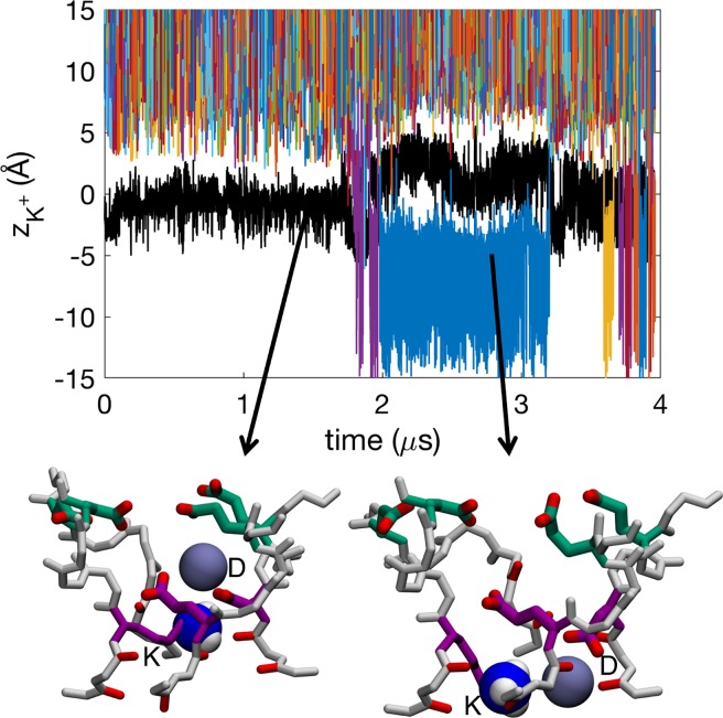
The Nav1.2 SF with charged Lys, in the presence of KCl solution, has an average occupancy of 1.3±0.1 K+ ions, being slightly lower than for Na+ ions, but comparable within errors. Fig 8 shows several ions (colored lines) entering and exiting the SF but not interacting extensively with the Lys (black line). We see 38 complete K+ ion permeation events (involving 13 distinct ions with repeat crossings, predominantly in the latter half of the 4 μs simulation after Lys rotamer change; see below and S3 Movie), being similar but somewhat less than the Na+ case above.
Fig 8. Time series of z positions for protonated Lys+ (K; black) and K+ ions (colors) in the SF of Nav1.2.
Insets show snapshots of K+ ions (purple balls) in representative configurations with active residues from the DEKA (purple) and EEDD (green) rings labeled. The label for the Lys of DEKA, “K”, should not be confused with the potassium ion K+. The charged ammonium group of the Lys shown is shown as blue and white balls. See also sample trajectory in S3 Movie.
Most of the time there exists a single K+ ion in the SF (S9 Fig). When there are two K+ ions in the SF they are generally further from each other, with a mean ion-ion distance of 15.1±0.8 Å, compared to 11.4±1.2 Å for Na+. Snapshots in Fig 8 show representative configurations. The K+ ions most commonly bind to single carboxylates or stay unbound (S9 Fig). The lower occupancy and the larger ion-ion distances mean that we do not see the same tight multi-ion/multi-carboxylate complexes as frequently as we did for Na+ (S9 Fig), and when they do occur they are almost always singly coordinated by outer ring carboxylates (S1 Table; column 4). Instead, a single K+ ion typically enters the SF and binds solely to the D from the DEKA ring (S1 Table; column 6). The DEKA ring is involved in binding ~40% of the time, the EEDD ring ~70% of the time, and both rings cooperatively involved only ~10% of the time. This reduced binding and absence of complex formation deep in the SF slows K+ permeation. We instead observe K+ ions held bound to the D of DEKA, electrostatically repelled by the Lys ammonium group, until the Lys side chain changes rotamer downward to allow ion movement, in stark contrast to the Na+ case above.
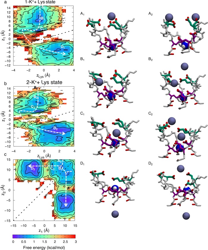
We again see conduction with either 1 or 2 K+ ion occupancy. However, unlike for Na+ ions, we see similar permeation mechanisms when 1 or 2 K+ ions are in the SF. This, together with the large K+- K+ distance of 15.1±0.8 Å, compared to 11.4±1.2 Å for Na+, suggest that the second ion does not participate in conduction but rather is loosely associated around the vestibule region. Fig 9A shows the 1-ion conduction mechanism, with the ion at z1, and the ammonium group of Lys, at zLys. We observe four states in the 2D PMF in Fig 9; A1, B1, C1 and D1. In state A1 the ion is in the vestibular region of the SF (z1~14 Å) and the Lys is bound to one of the inner ring carboxylates (z1~0 Å). The ion then moves down and binds to the outer ring (z1~8 Å) represented by state B1. In state C1, the ion has entered deeper into the SF and is now bound only to the carboxylate of the D1 from the DEKA ring (z1~4 Å). The Lys is still bound to the inner ring in both these two states (zK~0 Å). The ions do not have considerable effect on the position of the ammonium group of the Lys. However, occasionally (~10% of the time) the Lys changes rotamer downward (zLys<-2 Å), allowing leakage of K+ ions and thus permeation, represented by dashed line, leading to state D1. After the conduction event, the ammonium group of the Lys again bends upward to bind to the inner ring carboxylates (zLys~2 Å). For this to happen the K+ ion has to pass by the ammonium group of the Lys in the lower part of the SF (z1~zLys~-2 Å) where the electrostatic potential is less negative, leading to a larger energy barrier of 2.8±0.3 kcal/mol, as seen in Fig 8A along the pathway represented with a dashed arrow. This increased barrier (by 1.1±0.4 kcal/mol) would suggest a relative permeability for the channel of the order of 10, which is consistent with the experimental value for mammalian Nav channels (e.g. PNa+/PK+ ~10 in Nav1.2 from rat [25]). This estimate, however, assumes the rate-limiting step for conduction does not involve Lys rotameric change, which was observed here on the multi-μs timeframe; apparently exceeding the sub-μs scale of permeation, but which may not have been reliably quantified based on 4μs simulation.
Fig 9. Permeation of K+ in the Nav1.2 SF with protonated Lys+.
2D free energy projections showing: a) 1- ion conduction mechanism; b & c) the 2-ion conduction mechanisms (graphed for the bottom ion and Lys in b, and the two ions and Lys in c); where z1 corresponds to the z position of the bottom ion, z2 the z position of the top ion, and zLys the z position of the Lys/K ammonium group. Snapshots for 1-ion (left) and 2-ion (right) mechanisms, with the DEKA ring in purple, EEDD ring in green, and the charged ammonium group of the Lys shown as blue and white balls, indicate K+ ion (purple balls) movements. State labels include a subscript 1 or 2, representing the 1-ion or 2-ion mechanisms, respectively.
The 2-ion conduction mechanism in Fig 9B looks very similar, with the large K+- K+ distance of 15.1±0.8 Å showing that it is the same as the 1-ion conduction mechanism, only with an additional ion in the vicinity. Fig 9B shows the 2D PMF with the bottom ion, at z1, and the ammonium group of Lys, at zLys, being almost identical to the 1-ion case in Fig 9A; supporting the lack of a concerted 2-ion conduction mechanism. The lower ion enters the channel, binds collectively to the DEKA and EEDD rings in state B2, before binding solely to the DEKA ring in state C2. To enter the cavity, the ion has to wait for the Lys to change rotamer, it can then pass the Lys ammonium group in the lower SF, and then move further down into the cavity (state D2). In the meanwhile, the upper ion generally sits in the vicinity of the SF. In Fig 9C we see how the two ions can cross each other in the top part of the channel. The top ion can be anywhere between z2~7 Å and z2~15 Å during conduction, however, with a slight preference around z2~12 Å. The 2-ion energy barrier is similar to the 1-ion case at 2.7±0.3 kcal/mol. The Na+ and K+ barriers differ by 1.1 kcal/mol, however, the slowest coordinate in the K+ translocation pathway appears to involve the structural isomerization of the Lys side chain downward to permit conduction, leading to a permeation event that is more costly for K+.
Key to the low barrier for Na+ permeation is its ability to form tight multi-ion/multi-carboxylate clusters as well as a complex with Lys and 2 carboxylates, allowing pass-by conduction in the SHFS. We do not see the same tight K+-K+ clusters nor the simultaneous binding of K+ and Lys near the DEKA ring. Instead we see a lone K+ ion that is singly bound and forced to pass by the Lys lower in the SF (S9 Fig), where the electrostatic field is less favorable (Fig 3A), making the whole process far less likely.
We now explore the thermodynamic stabilities of the complexes observed during permeation events to understand the causes of the different Na+ and K+ mechanisms that may underlie selectivity.
Multi-ion complex stability underpins selective conduction
Multi-ion complex formation with carboxylates is required for efficient permeation as it helps draw in and stabilize the ions in the SF. This acts as a precursor for the ion+Lys/multi-carboxylate complex that aids in the crossing of the SF. The analysis in S9 Fig suggests that such multi-ion/multi-carboxylate complexes appear more favorable for Na+ over K+. When the SF is occupied by a single ion, Na+ is most commonly bound to one or more carboxylate side chains (45% and 26%), whereas K+ is more likely to be unbound (61%) (S9B Fig), and Na+ is several times more likely than K+ to form multi-carboxylate complexes (26% vs. 6%). Of the multi-ion occupancies observed, Na+ ions are most likely to be either singly bound or in loose multi-carboxylate complexes (42% and 40%), whereas K+ is most likely to be either singly bound or unbound (52% and 36%). Na+ is several times more likely to form tight multi-ion/multi-carboxylate clusters than K+ (12% vs. 3%) (S9C Fig). For Na+ ions, these clusters are predominantly made up by outer ring carboxylates together with either the E or D from the DEKA ring, most commonly EII together with D (S1 Table; column 3). For K+ such complexes are almost always made up only by the outer ring carboxylates (S1 Table; column 4).
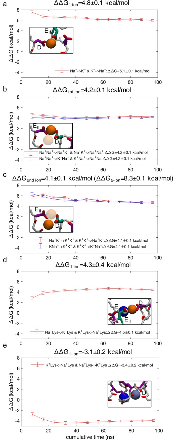
Thus, tight 2-ion/multi-carboxylate complexes, similar to the complex that is formed before the low free energy pass-by conduction events for Na+ (Fig 7; state B2), are more common for Na+ than K+, and may be very important for selectivity (S9 Fig). FEP calculations were used to investigate the relative stabilities of Na+ and K+ in such a complex. The most representative multi-ion/multi-carboxylate complex was identified to be bound collectively by D and EII (Fig 10B and 10C; inset) (69%) (S1 Table; column 3). Results from FEP calculations show preferential binding by Na+ with 4.8±0.1 kcal/mol for single ion occupancy (Fig 10A), showing a strong inherent preference by carboxylates to bind Na+, in agreement with Eisenman high field strength theory on ion selectivity [78]. Quantum mechanical calculations using a model DEKA ring have demonstrated a similar preference for Na+ over K+ (4.8 kcal/mol [79]), revealing consistency between models. The same complex containing two ions shows a large preference of 8.3±0.1 kcal/mol for Na+ over K+ ions (Fig 10B and 10C); 4.2 kcal/mol from the transformation of the 1st ion (Fig 10B) and 4.1 kcal/mol from the transformation the 2nd ion (Fig 10C). This shows that an additional ion creates extra stability for Na+ relative to K+. When another ion is added to the complex, the cumulative ion-carboxylate attraction increases faster than the carboxylate-carboxylate and ion-ion repulsion, more so for Na+ than K+, giving additional stability to Na+ complexes. These complexes create deeper binding in the SF that aids crossing the ammonium group of the Lys. We note that this large cumulative free energy difference between Na+ and K+ ions to form a 2-ion complex with multiple carboxylates is not the key energy controlling the different permeation mechanisms in Figs 7 and 9, but represents the relative stability of a complex that is particular to Na+, and largely unseen for the K+ ion because of its low stability, helping explain the distinct mechanisms.
Fig 10. Free energy perturbation for ion binding selectivity.
a) Single ion Na+→ K+ and K+→ Na+ in an ion-DEII complex; b) 1st ion transformations Na+Na+→K+Na+, Na+Na+→ Na+K+, K+K+→K+Na+ and K+K+→ Na+K+ in the 2-ion-DEII complex (with 2nd ion grayed out) and c) 2nd ion transformations K+Na+→K+K+, Na+K+→ K+K+, K+Na+→Na+Na+ and Na+K+→ Na+Na+ (bottom) in the 2-ion-DEII complex (with 1st ion grayed out). d) single ion transformations Na+→K+ and K+→Na+ for the ion+Lys-DEEII complex; e) single ion transformations K+→ Na+ and Na+→ K+ in an ion+Lys position in the lower SF (note that K++Lys is the starting configuration in this case, leading to a negative free energy corresponding to the transformation K+→ Na+). The value for the bulk reference for Na+→K+ in aqueous solution was found to be 17.7 ± 0.3 kcal/mol. Insets show starting configurations and indicate amino acids involved. Free energy corrections to account for constraints used to maintain complexes (see text) were found to be: a) 0.30±0.02 kcal/mol; b) −0.017±0.042 kcal/mol; c) −0.023±0.075 kcal/mol; d) 0.15±0.42 kcal/mol; and e) 0.34±0.12 kcal/mol. Uncorrected ΔΔG values are displayed within each panel after the specified transformations, with corrected ΔΔG values given at the top of each panel, corresponding to 1-ion, or 1st or 2nd ion of a 2-ion complex (with the cumulative 2-ion value indicated in parenthesis in panel c). Snapshots, with the DEKA ring in purple, EEDD ring in green, and the charged ammonium group of the Lys shown as blue and white balls, indicate starting configurations with Na+ (orange balls) and K+ (purple balls).
During the ion crossing of the charged Lys, the ion creates a stable transition state together with the Lys and multiple carboxylates (Fig 10D; inset). This ion-Lys state is bound collectively by the D and E from the DEKA ring, as well as EII from the outer ring, and shows preferential binding of a single Na+ by 4.3±0.4 kcal/mol (Fig 10D). This ability to create a joint ion-Lys complex to facilitate crossing of the Lys in the SHFS is the salient difference in Na+ and K+ permeation mechanisms.
The K+ ion instead usually resides alone in the SF and crosses the ammonium group of the Lys further down, (Fig 10E; inset), where the electrostatic fields arising from carboxylates in the upper SF is less attractive. If we examine the relative free energies of Na+ and K+ ions in this region of the SF near the Lys, where such crossing occurs for K+, we calculate a similar but reduced value of 3.1±0.2 kcal/mol, still favoring Na+ (Fig 10E). This value remains negative because the ion remains in contact with D and E side chains from the DEKA ring as it passes the Lys side chain. Theoretically, both K+ and Na+ should be able to translocate in this fashion when the Lys moves downward, but this was not observed for Na+. We may understand this from the relative destabilization of Na+ in this region relative to the selective multi-carboxylate complexes that form above it, making downward movement of Na+ less likely.
We therefore hypothesize that selective permeation in Nav1.2 arises from the distinct mechanisms (Lys-complex pass-by Na+ permeation versus electrostatically Lys plugged K+ permeation), which originates from the difference in stabilities of multi-ion and ion-Lys complexes in the high field strength region of the SF. Multi-ion/multi-carboxylate complexes have also been seen to be crucial for Na+ permeation in the bacterial NavAb channel, where a preference for single Na+ ions of -2.2±0.6 kcal/mol has been estimated [30]. In the bacterial channel, we also suggest that optimal conduction requires the greater occupancy of the 3-ion state which is achieved when the crystallographic E177-S178 H-bond is lost on 2 opposite monomers, allowing the SHFS carboxylate groups to reach toward the center of the SF and coordinate 2 Na+ ions concurrently. This two-Na+/two-carboxylate complex offers additional stability, allowing those 2 ions to coexist in SHFS, and we calculate the relative stability of Na+ vs. K+ in SHFS to become -3.8±0.2 kcal/mol in NavAb. This observation suggests that Na+ ions adopt a specific 3-ion arrangement with 2 ions at the level of SHFS, that does not occur in simulations of K+, which closely parallels our observations in the Nav1.2 channel.
Conclusions
The ability to select for native ions while discriminating against other similar ions, is one of the key features of voltage gated ion channels. Ion conduction is a dynamic process that involves conformational isomerizations of the residues of the SF, as well as cooperation between multiple ions, requiring long timescale simulations to extract the underlying energetic landscape. In the absence of a high-resolution structure of a mammalian channel to study selectivity and conduction at the molecular level, we designed a model of the human Nav1.2 by patching the essential sequence of residues in and around the SF, including the key DEKA and EEDD rings, into the NavRh bacterial channel. This NavRh/Nav1.2 channel relaxes into an asymmetrical configuration, where the DEKA and EEDD rings are highly flexible and cooperate to ensure efficient conduction and selectivity for Na+ over K+, in line with previous studies of bacterial Nav channels where similar flexibility has been shown to be critical for ion conduction [29, 30].
The presence of a positively charged Lys in Nav channels has long been a source of intrigue; being essential for selectivity, but its specific role unexplained. Previous computational studies of eukaryotic Nav models point toward a passive, hindering role, where the Lys side chain needs to bind away from the middle of the pore lumen in order to allow conduction of Na+ ions. We reveal a more intimate role for this residue, depending on its protonation state. When Lys is deprotonated (neutral), we observe a 2 to 3-ion occupancy in the SF where both 2- and 3-ion knock on conductions are common, facilitated by tight multi-ion/multi-carboxylate complexes that are made possible by the flexible side chains of the SF. When Lys is instead protonated (charged), we see reduction to 1 to 2-ion occupancy, due to the extra charge introduced by the ammonium group of the Lys. In fact, we see the Lys actively participating in the conduction mechanism with a role similar to that of a permeating ion. In the presence of K+ ions, the conduction mechanism instead relies on single ion permeation, where multi-ion/multi-carboxylate complexes are less likely.
These multi-Na+/multi-carboxylate complexes help attract Na+ in to the SF of Nav1.2, where binding deeper in the SF stabilizes the ions. Interestingly, the carboxylates involved in these key multi-Na+/multi-carboxylate complexes generally include only one carboxylate from DEKA and one or two from the EEDD ring in the vestibular region, rather than both D and E from the DEKA ring. This observation is consistent with mutagenesis experiments showing the need for only one of the D and E from the DEKA ring, supporting the idea of involvement of the outer ring [19]. These Na+ favorable complexes are more commonly bound collectively by D and EII. Cysteine mutations of the residues in the EEDD ring show the greatest decrease in conduction when this domain II Glu (EII in our model) is mutated [25, 41], consistent with our observation that this residue from the outer ring is particularly important.
In the case of charged Lys we see conduction that is possible with a singly occupied SF, however, the binding of a second Na+ ion reduces the conduction barrier by creating an intermediate state that enables efficient multi-ion conduction. The second Na+ ion helps to push the ion already in the SF and stabilize a state where the ammonium group of the Lys and the bottom ion are collectively bound by the carboxylates, allowing the ion to pass by the Lys into the cavity. The facilitating of inward conduction by the presence of an additional external Na+ ion may explain the robust inward rectification of current in mammalian Nav channels seen experimentally [1]. Even though more space for conduction can be created in the SF by the steric unplugging of the ammonium group of the Lys, no conduction occurs in that state. This behavior is consistent and comparable with previous results in NavAb, where pass by of ion by another would only occur at the level of SHFS. When the charged ammonium group of Lys is in the SHFS, where the electrostatic potential is most negative, it creates a smooth electrostatic environment leading into the cavity, whereas when it is in the down state it creates a zone of high electrostatic potential that cuts the cavity off from the SF. The Na+ ion can benefit from the smooth energetic surface by binding at SHFS in a multi-ion complex with Lys, whereas K+ cannot; instead becoming partially electrostatically blocked by the Lys in the lower SF. This illustrates the fundamental role of the Lys as an active participant in the selective conduction mechanism. K+ is far less likely to form these multi-ion/multi-carboxylate complexes; in particular, we see preferential binding of 8.3±0.1 kcal/mol for Na+-Na+ over K+-K+ in these complexes. Furthermore, K+ does not form the stable multi-carboxylate/Lys state. In fact, the ion+Lys/multi-carboxylate cluster favors Na+ over K+ by 4.3±0.4 kcal/mol. The consequence of this difference in affinity is a completely different conduction mechanism for K+, having to pass the ammonium group of Lys in the lower part of the SF, where the electrostatic environment is not as favorable. Interestingly, the apparently independent conduction pathways for Na+ and K+ ions could potentially explain absence of anomalous mole fraction effects in mammalian Nav channels [53]. In the future, simulations of a model human Nav channel in an open conformation, based on constrained-open NavAb or NavMs structures [9, 16, 27], could be used to explore the competition of Na+ and K+ ions under the driving force of a membrane potential.
It has been proposed that mammalian Navs select for Na+ via a different mechanism to bacterial Navs, owing to their divergent SHFS site forming residues (EEEE Vs DEKA) [18, 80]. While this appears to be the case based on our observations, we do see evidence for common features that may be central to Na+ selectivity. Both bacterial and human channels make use of an efficient multi-ion process enabled by flexible carboxylates binding two Na+ ions during ion knock on or pass by conduction events. The efficiency of this multi-ion mechanism is reliant on the formation of 2-Na+/2-carboxylate complexes, whose thermodynamic stabilities are increased for Na+ compared to K+. Even though bacterial channels possess 4 carboxylates at the SHFS site, only two are needed for high affinity binding [30]. Thus, the bacterial channel uses two of its four EEEE Glus, whereas the human channel uses carboxylates both from the DEKA and EEDD rings, leading to a Na+ permeation process that closely resembles our observations in the model human channel. However, the Lys residue in the human DEKA signature participates in the multi-ion knock-on mechanism in ways that impose a unique ion discrimination process, perhaps explaining the higher level of ion selectivity in mammalian Navs [1]. We propose that this additional selectivity is achieved by establishing distinct permeation mechanisms for Na+ and K+ ions, where the Lys expedites Na+ translocation through high field strength complex formation, while partially blocking the K+ ion.
Supporting information
(PDF)
The first column defines which carboxylate(s) the ion(s) are bound to. Columns 2,3 and 4 show the tight 2-ion/multi-carboxylate clusters for the Nav1.2 SF with deprotonated Lys0 in NaCl, protonated Lys+ in NaCl, and protonated Lys+ in KCl solutions, respectively. Columns 5 and 6 show the tight ion+Lys/multi-carboxylate clusters with protonated Lys+ in NaCl and KCl solutions, respectively. Clusters that occur less than 4% of the time are not shown. Most commonly, Na+ is bound to D and/or E together with EII, whereas K+ is bound to the outer EEDD ring or singly bound to only the DEKA D side chain.
(TIFF)
Alignments of model human Nav1.2 (pink) cockroach NavPaS (purple) and eel Nav1.4 (blue), showing DI and DIII on top and DII and DIV at the bottom. Panel a) shows the alignment according to the backbone of the SF and outer vestibule based on all four subunits, while b) shows alignment according to the SF and vestibule subunit by subunit, revealing the similarity of the Nav1.2 model to NavPaS and EeNav1.4 cryoEM structures. Side chains of Glu, Asp and Lys are indicated with sticks.
(TIF)
2D free energy projections showing: a) 2-ion occupancy; and b & c) 3-ion occupancy (graphed for all three ions in b, and the top two ions in c); where z1, z2, and z3 correspond to the z positions of the bottom, middle and top ions, respectively, and where z23 is the z position of the COM of the top two ions. When there are 2 ions in the SF, the ions are trapped and it is not until a third ion enters from above that we can observe either a knock on or pass by conduction. Snapshots, with the EEEE ring in purple, indicate the corresponding Na+ ion (orange balls) movements. State labels include a subscript 2 or 3, representing the 2-ion or 3-ion mechanisms, respectively. Here the state label A has no subscript because A2 and A3 represent the same state, with same 2-ion configuration in the SF, but with approaching 3rd ion from bulk.
(TIF)
Distributions are shown for Na+-Na+ (a), K+-K+ (b), Na+-carboxylate (c) and K+-carboxylate (d). Multi-ion clusters defined by r < 4.7 Å for Na+ and r < 5.5 Å for K+, and carboxylate binding by r < 3.8 Å for Na+ and r < 4.2 Å for K+.
(TIF)
a) Na+ with deprotonated Lys0; b) Na+ with protonated Lys+; and c) K+ with protonated Lys+. Red dashed lines indicate the level of RMSD after the equilibration period. Late changes due to the onset of conduction, particularly for K+, are discussed in the text.
(TIF)
a) Na+ with deprotonated Lys0; b) Na+ with protonated Lys+; and c) K+ with protonated Lys+.
(TIF)
Fluctuations are shown for residues 178 to 184 in NavRh/Nav1.2 (yellow) and NavAb (blue) for: a) the mean of the four subunits (where orientation was according to all 4 subunits); and b) each individual subunit (where orientation was according to the subunit of interest).
(TIF)
Mean ion coordination numbers relative to bulk water in NavRh/Nav1.2 for: a) Na+ with deprotonated Lys0; b) Na+ with protonated Lys+; and c) K+ with protonated Lys+. Bulk hydration numbers are 5.67±0.04 for Na+, and 6.94±0.07 for K+.
(TIF)
a) Distribution of Na+ occupancy and dominant clusters representing complexes with: b) 1-ion occupancies; c) 2 ion occupancies; and d) 3 ion occupancies. In panels c and d, ‘single’ clusters include 1-ion/1-carboxylate complexes only, 'multi' clusters include 1-ion/multi-carboxylate complexes with a loosely associated second ion, and ‘tight multi’ clusters represent 2-ion/multi-carboxylate clusters where both ions are within an ion-ion cut off, defined from the radial distribution function g(r) for ion-ion distance (see text). The channel range is defined by the region -15<z<15 Å.
(TIF)
a) Distribution of ion occupancies. Panels b and c show dominant cluster conformations for: 1-ion (b) and 2 ion (c) occupancies, for Na+ (orange) and K+ (blue). See S8 Fig for definitions.
(TIF)
Panel a shows the convergence of 1D free energy for the ion across the channel, comparing time ranges specified in the legend. The free energy profile converges quickly in the outer vestibule and SF, but more slowly in the central cavity because it takes longer for ions to enter this part of the channel. Panels b-e show convergence of 2D free energy maps corresponding to Fig 4, for the last 2 μs of each simulation (left and right show time ranges 0–1 μs and 0–2 μs, respectively).
(TIF)
Panel a shows the convergence of 1D free energy for the ion across the channel, comparing time ranges specified in the legend. Panels b-e, show convergence of 2D maps corresponding to Figs 6 & 7 (left and right show time ranges 0–3 μs and 0–4 μs, respectively).
(TIF)
Panel a shows the convergence of 1D free energy for the ion across the channel, comparing time ranges specified in the legend. Panels b-d show convergence of 2D maps, corresponding to Fig 9 (left and right show time ranges 0–3 μs and 0–4 μs, respectively).
(TIF)
Sample trajectory revealing interactions of Na+ ions with the Nav1.2 SF, showing multi-ion complexes involving carboxylates from inner DEKA and outer EEDD rings, and knock-on and pass-by conduction events. Na+ ions are shown as orange balls, lysine N atom as a blue ball, and side chains of DEKA and EEDD rings as purple and green sticks, respectively.
(MP4)
Sample trajectory revealing interactions of Na+ ions with the Nav1.2 SF, showing multi-ion complexes involving inner DEKA and outer EEDD rings, and complex formation with the lysine ammonium group, enabling pass-by conduction events. Representations as in S1 Movie.
(MP4)
Sample trajectory revealing interactions of K+ ions with the Nav1.2 SF, showing ion interactions with DEKA and EEDD rings, lack of multi-ion or lysine complex formation, and conduction events requiring unblocking of the SF by downward movement of the lysine ammonium group. Representations as in S1 Movie, with K+ ions shown as purple balls.
(MP4)
Acknowledgments
The authors would like to thank Phuong T. Nguyen (UC Davis) for his assistance in using Rosetta to rebuild missing loops in our Nav1.2 model.
Data Availability
All relevant data are within the paper and its Supporting Information files.
Funding Statement
We acknowledge financial support from the National Institutes of Health (Grant number U01- HL126273-01/02), https://www.nih.gov; the National Health and Medical Research Council of Australia (Grant numbers APP1104259 and APP1141974), https://www.nhmrc.gov.au; the Australian Research Council (Grant number DP170101732), http://www.arc.gov.au; the Medical Advances Without Animals Trust (Australia), http://www.mawa-trust.org.au; and supercomputing support from the Pittsburgh Supercomputing Center, DE Shaw Anton (Grant number PSCA17045P; NRBSC, through NIH RC2GM093307), https://www.psc.edu; and the National Computational Infrastructure (Australia; Grant number dd7), http://nci.org.au. The funders had no role in study design, data collection and analysis, decision to publish, or preparation of the manuscript.
References
- 1.Hille B. Ion channels of excitable membranes: Sinauer Sunderland, MA; 2001. [Google Scholar]
- 2.Catterall AW. Structure and function of voltage-gated ion channels. Annual review of biochemistry. 1995;64(1):493–531. [DOI] [PubMed] [Google Scholar]
- 3.Lossin C, Wang DW, Rhodes TH, Vanoye CG, George AL. Molecular basis of an inherited epilepsy. Neuron. 2002;34(6):877–84. [DOI] [PubMed] [Google Scholar]
- 4.Akopian AN, Souslova V, England S, Okuse K, Ogata N, Ure J, et al. The tetrodotoxin-resistant sodium channel SNS has a specialized function in pain pathways. Nature neuroscience. 1999;2(6):541–8. 10.1038/9195 [DOI] [PubMed] [Google Scholar]
- 5.England S, de Groot MJ. Subtype‐selective targeting of voltage‐gated sodium channels. British journal of pharmacology. 2009;158(6):1413–25. 10.1111/j.1476-5381.2009.00437.x [DOI] [PMC free article] [PubMed] [Google Scholar]
- 6.Payandeh J, Scheuer T, Zheng N, Catterall WA. The crystal structure of a voltage-gated sodium channel. Nature. 2011;475(7356):353–8. 10.1038/nature10238 [DOI] [PMC free article] [PubMed] [Google Scholar]
- 7.Zhang X, Ren W, DeCaen P, Yan C, Tao X, Tang L, et al. Crystal structure of an orthologue of the NaChBac voltage-gated sodium channel. Nature. 2012;486(7401):130–4. 10.1038/nature11054 [DOI] [PMC free article] [PubMed] [Google Scholar]
- 8.Shaya D, Findeisen F, Abderemane-Ali F, Arrigoni C, Wong S, Nurva SR, et al. Structure of a prokaryotic sodium channel pore reveals essential gating elements and an outer ion binding site common to eukaryotic channels. Journal of molecular biology. 2014;426(2):467–83. 10.1016/j.jmb.2013.10.010 [DOI] [PMC free article] [PubMed] [Google Scholar]
- 9.McCusker EC, Bagnéris C, Naylor CE, Cole AR, D'Avanzo N, Nichols CG, et al. Structure of a bacterial voltage-gated sodium channel pore reveals mechanisms of opening and closing. Nature communications. 2012;3:1102–10. 10.1038/ncomms2077 [DOI] [PMC free article] [PubMed] [Google Scholar]
- 10.Sula A, Booker J, Ng LC, Naylor CE, DeCaen PG, Wallace B. The complete structure of an activated open sodium channel. Nature Communications. 2017;8. [DOI] [PMC free article] [PubMed] [Google Scholar]
- 11.Shen H, Zhou Q, Pan X, Li Z, Wu J, Yan N. Structure of a eukaryotic voltage-gated sodium channel at near-atomic resolution. Science. 2017;355(6328):eaal4326 10.1126/science.aal4326 [DOI] [PubMed] [Google Scholar]
- 12.Yan Z, Zhou Q, Wang L, Wu J, Zhao Y, Huang G, et al. Structure of the Nav1. 4-β1 Complex from Electric Eel. Cell. 2017;170(3):470–82. e11. 10.1016/j.cell.2017.06.039 [DOI] [PubMed] [Google Scholar]
- 13.Charalambous K, Wallace BA. NaChBac: the long lost sodium channel ancestor. Biochemistry. 2011;50(32):6742–52. 10.1021/bi200942y [DOI] [PMC free article] [PubMed] [Google Scholar]
- 14.Lee S, Goodchild SJ, Ahern CA. Local anesthetic inhibition of a bacterial sodium channel. The Journal of general physiology. 2012;139(6):507–16. 10.1085/jgp.201210779 [DOI] [PMC free article] [PubMed] [Google Scholar]
- 15.Pavlov E, Bladen C, Winkfein R, Diao C, Dhaliwal P, French RJ. The pore, not cytoplasmic domains, underlies inactivation in a prokaryotic sodium channel. Biophysical journal. 2005;89(1):232–42. 10.1529/biophysj.104.056994 [DOI] [PMC free article] [PubMed] [Google Scholar]
- 16.Bagnéris C, DeCaen PG, Naylor CE, Pryde DC, Nobeli I, Clapham DE, et al. Prokaryotic NavMs channel as a structural and functional model for eukaryotic sodium channel antagonism. Proceedings of the National Academy of Sciences. 2014;111(23):8428–33. [DOI] [PMC free article] [PubMed] [Google Scholar]
- 17.Ren D, Navarro B, Xu H, Yue L, Shi Q, Clapham DE. A prokaryotic voltage-gated sodium channel. Science. 2001;294(5550):2372–5. 10.1126/science.1065635 [DOI] [PubMed] [Google Scholar]
- 18.Finol-Urdaneta RK, Wang Y, Al-Sabi A, Zhao C, Noskov SY, French RJ. Sodium channel selectivity and conduction: Prokaryotes have devised their own molecular strategy. The Journal of general physiology. 2014;143(2):157–71. 10.1085/jgp.201311037 [DOI] [PMC free article] [PubMed] [Google Scholar]
- 19.Favre I, Moczydlowski E, Schild L. On the structural basis for ionic selectivity among Na+, K+, and Ca2+ in the voltage-gated sodium channel. Biophysical journal. 1996;71(6):3110–25. 10.1016/S0006-3495(96)79505-X [DOI] [PMC free article] [PubMed] [Google Scholar]
- 20.Yue L, Navarro B, Ren D, Ramos A, Clapham DE. The cation selectivity filter of the bacterial sodium channel, NaChBac. The Journal of general physiology. 2002;120(6):845–53. 10.1085/jgp.20028699 [DOI] [PMC free article] [PubMed] [Google Scholar]
- 21.Heinemann SH, Terlau H. Calcium channel characteristics conferred on the sodium channel by single mutations. Nature. 1992;356(6368):441–3. 10.1038/356441a0 [DOI] [PubMed] [Google Scholar]
- 22.Frank HY, Catterall WA. Overview of the voltage-gated sodium channel family. Genome biology. 2003;4(3):207–14. [DOI] [PMC free article] [PubMed] [Google Scholar]
- 23.Catterall WA, Perez-Reyes E, Snutch TP, Striessnig J. International Union of Pharmacology. XLVIII. Nomenclature and structure-function relationships of voltage-gated calcium channels. Pharmacological reviews. 2005;57(4):411–25. 10.1124/pr.57.4.5 [DOI] [PubMed] [Google Scholar]
- 24.Terlau H, Heinemann SH, Stühmer W, Pusch M, Conti F, Imoto K, et al. Mapping the site of block by tetrodotoxin and saxitoxin of sodium channel II. FEBS letters. 1991;293(1–2):93–6. [DOI] [PubMed] [Google Scholar]
- 25.Schlief T, Schönherr R, Imoto K, Heinemann SH. Pore properties of rat brain II sodium channels mutated in the selectivity filter domain. European biophysics journal. 1996;25(2):75–91. [DOI] [PubMed] [Google Scholar]
- 26.Tang L, El-Din TMG, Payandeh J, Martinez GQ, Heard TM, Scheuer T, et al. Structural basis for Ca2+ selectivity of a voltage-gated calcium channel. Nature. 2014;505(7481):56–61. 10.1038/nature12775 [DOI] [PMC free article] [PubMed] [Google Scholar]
- 27.Lenaeus MJ, El-Din TMG, Ing C, Ramanadane K, Pomès R, Zheng N, et al. Structures of closed and open states of a voltage-gated sodium channel. Proceedings of the National Academy of Sciences. 2017;114:3051–60. [DOI] [PMC free article] [PubMed] [Google Scholar]
- 28.Ulmschneider MB, Bagnéris C, McCusker EC, DeCaen PG, Delling M, Clapham DE, et al. Molecular dynamics of ion transport through the open conformation of a bacterial voltage-gated sodium channel. Proceedings of the National Academy of Sciences. 2013;110(16):6364–9. [DOI] [PMC free article] [PubMed] [Google Scholar]
- 29.Chakrabarti N, Ing C, Payandeh J, Zheng N, Catterall WA, Pomès R. Catalysis of Na+ permeation in the bacterial sodium channel NaVAb. Proceedings of the National Academy of Sciences. 2013;110(28):11331–6. [DOI] [PMC free article] [PubMed] [Google Scholar]
- 30.Boiteux C, Vorobyov I, Allen TW. Ion conduction and conformational flexibility of a bacterial voltage-gated sodium channel. Proceedings of the National Academy of Sciences. 2014;111(9):3454–9. [DOI] [PMC free article] [PubMed] [Google Scholar]
- 31.Corry B, Thomas M. Mechanism of ion permeation and selectivity in a voltage gated sodium channel. Journal of the American Chemical Society. 2012;134(3):1840–6. 10.1021/ja210020h [DOI] [PubMed] [Google Scholar]
- 32.Carnevale V, Treptow W, Klein ML. Sodium Ion Binding Sites and Hydration in the Lumen of a Bacterial Ion Channel from Molecular Dynamics Simulations. Journal of Physical Chemistry Letters. 2011;2(19):2504–8. [Google Scholar]
- 33.Stock L, Delemotte L, Carnevale V, Treptow W, Klein ML. Conduction in a biological sodium selective channel. J Phys Chem B. 2013;117(14):3782–9. 10.1021/jp401403b [DOI] [PubMed] [Google Scholar]
- 34.Zhang X, Xia M, Li Y, Liu H, Jiang X, Ren W, et al. Analysis of the selectivity filter of the voltage-gated sodium channel NavRh. Cell Res. 2013;23(3):409–22. 10.1038/cr.2012.173 [DOI] [PMC free article] [PubMed] [Google Scholar]
- 35.Qiu H, Shen R, Guo W. Ion solvation and structural stability in a sodium channel investigated by molecular dynamics calculations. Biochim Biophys Acta. 2012;1818(11):2529–35. 10.1016/j.bbamem.2012.06.003 [DOI] [PubMed] [Google Scholar]
- 36.Furini S, Domene C. On conduction in a bacterial sodium channel. PLoS computational biology. 2012;8(4):e1002476 10.1371/journal.pcbi.1002476 [DOI] [PMC free article] [PubMed] [Google Scholar]
- 37.Payandeh J, Minor DL Jr. Bacterial Voltage-Gated Sodium Channels (BacNaVs) from the Soil, Sea, and Salt Lakes Enlighten Molecular Mechanisms of Electrical Signaling and Pharmacology in the Brain and Heart. Journal of Molecular Biology. 2015;427(1):3–30. 10.1016/j.jmb.2014.08.010 [DOI] [PMC free article] [PubMed] [Google Scholar]
- 38.Naylor CE, Bagnéris C, DeCaen PG, Sula A, Scaglione A, Clapham DE, et al. Molecular basis of ion permeability in a voltage‐gated sodium channel. The EMBO journal. 2016:e201593285. [DOI] [PMC free article] [PubMed] [Google Scholar]
- 39.Ke S, Timin EN, Stary-Weinzinger A. Different inward and outward conduction mechanisms in NaVMs suggested by molecular dynamics simulations. PLoS Comput Biol. 2014;10(7):e1003746 10.1371/journal.pcbi.1003746 [DOI] [PMC free article] [PubMed] [Google Scholar]
- 40.Chiamvimonvat N, Perez-Garcia MT, Tomaselli GF, Marban E. Control of ion flux and selectivity by negatively charged residues in the outer mouth of rat sodium channels. The Journal of physiology. 1996;491(1):51–9. [DOI] [PMC free article] [PubMed] [Google Scholar]
- 41.Chiamvimonvat N, Pé MT, Ranjan R, Marban E, Tomaselli GF. Depth asymmetries of the pore-lining segments of the Na+ channel revealed by cysteine mutagenesis. Neuron. 1996;16(5):1037–47. [DOI] [PubMed] [Google Scholar]
- 42.Tsushima RG, Li RA, Backx PH. P-loop flexibility in Na+ channel pores revealed by single-and double-cysteine replacements. The Journal of general physiology. 1997;110(1):59–72. [DOI] [PMC free article] [PubMed] [Google Scholar]
- 43.Xia M, Liu H, Li Y, Yan N, Gong H. The mechanism of Na+/K+ selectivity in mammalian voltage-gated sodium channels based on molecular dynamics simulation. Biophysical journal. 2013;104(11):2401–9. 10.1016/j.bpj.2013.04.035 [DOI] [PMC free article] [PubMed] [Google Scholar]
- 44.Li Y, Liu H, Xia M, Gong H. Lysine and the Na+/K+ Selectivity in Mammalian Voltage-Gated Sodium Channels. PloS one. 2016;11(9):e0162413 10.1371/journal.pone.0162413 [DOI] [PMC free article] [PubMed] [Google Scholar]
- 45.Mahdavi S, Kuyucak S. Mechanism of ion permeation in mammalian voltage-gated sodium channels. PloS one. 2015;10(8):e0133000 10.1371/journal.pone.0133000 [DOI] [PMC free article] [PubMed] [Google Scholar]
- 46.Noda M, Suzuki H, Numa S, Stühmer W. A single point mutation confers tetrodotoxin and saxitoxin insensitivity on the sodium channel II. FEBS letters. 1989;259(1):213–6. [DOI] [PubMed] [Google Scholar]
- 47.Khan A, Romantseva L, Lam A, Lipkind G, Fozzard H. Role of outer ring carboxylates of the rat skeletal muscle sodium channel pore in proton block. The Journal of physiology. 2002;543(1):71–84. [DOI] [PMC free article] [PubMed] [Google Scholar]
- 48.Tsushima RG, Li RA, Backx PH. Altered ionic selectivity of the sodium channel revealed by cysteine mutations within the pore. The Journal of general physiology. 1997;109(4):463–75. [DOI] [PMC free article] [PubMed] [Google Scholar]
- 49.Penzotti JL, Fozzard HA, Lipkind GM, Dudley SC. Differences in saxitoxin and tetrodotoxin binding revealed by mutagenesis of the Na+ channel outer vestibule. Biophysical journal. 1998;75(6):2647–57. 10.1016/S0006-3495(98)77710-0 [DOI] [PMC free article] [PubMed] [Google Scholar]
- 50.Begenisich T, Cahalan M. Sodium channel permeation in squid axons. I: Reversal potential experiments. The Journal of physiology. 1980;307(1):217–42. [DOI] [PMC free article] [PubMed] [Google Scholar]
- 51.Begenisich T, Cahalan M. Sodium channel permeation in squid axons. II: Non‐independence and current‐voltage relations. The Journal of physiology. 1980;307(1):243–57. [DOI] [PMC free article] [PubMed] [Google Scholar]
- 52.French R, Worley J, Wonderlin W, Kularatna AS, Krueger B. Ion permeation, divalent ion block, and chemical modification of single sodium channels. Description by single-and double-occupancy rate-theory models. The Journal of general physiology. 1994;103(3):447–70. [DOI] [PMC free article] [PubMed] [Google Scholar]
- 53.Ravindran A, Kwiecinski H, Alvarez O, Eisenman G, Moczydlowski E. Modeling ion permeation through batrachotoxin-modified Na+ channels from rat skeletal muscle with a multi-ion pore. Biophysical journal. 1992;61(2):494–508. 10.1016/S0006-3495(92)81854-4 [DOI] [PMC free article] [PubMed] [Google Scholar]
- 54.Rakowski RF, Gadsby DC, De Weer P. Single ion occupancy and steady-state gating of Na channels in squid giant axon. The Journal of general physiology. 2002;119(3):235–50. 10.1085/jgp.20028500 [DOI] [PMC free article] [PubMed] [Google Scholar]
- 55.Begenisich T, Busath D. Sodium flux ratio in voltage-clamped squid giant axons. The Journal of general physiology. 1981;77(5):489–502. [DOI] [PMC free article] [PubMed] [Google Scholar]
- 56.Eisenman G, Latorre R, Miller C. Multi-ion conduction and selectivity in the high-conductance Ca2+-activated K+ channel from skeletal muscle. Biophys J. 1986;50(6):1025–34. 10.1016/S0006-3495(86)83546-9 [DOI] [PMC free article] [PubMed] [Google Scholar]
- 57.Gillespie D, Giri J, Fill M. Reinterpreting the anomalous mole fraction effect: the ryanodine receptor case study. Biophysical journal. 2009;97(8):2212–21. 10.1016/j.bpj.2009.08.009 [DOI] [PMC free article] [PubMed] [Google Scholar]
- 58.Gillespie D, Boda D. The anomalous mole fraction effect in calcium channels: a measure of preferential selectivity. Biophysical Journal. 2008;95(6):2658–72. 10.1529/biophysj.107.127977 [DOI] [PMC free article] [PubMed] [Google Scholar]
- 59.Nonner W, Chen DP, Eisenberg B. Anomalous mole fraction effect, electrostatics, and binding in ionic channels. Biophysical Journal. 1998;74(5):2327–34. 10.1016/S0006-3495(98)77942-1 [DOI] [PMC free article] [PubMed] [Google Scholar]
- 60.Hille B. The permeability of the sodium channel to organic cations in myelinated nerve. The Journal of general physiology. 1971;58(6):599–619. [DOI] [PMC free article] [PubMed] [Google Scholar]
- 61.Dudev T, Lim C. Factors governing the Na+ vs K+ selectivity in sodium ion channels. Journal of the American Chemical Society. 2010;132(7):2321–32. 10.1021/ja909280g [DOI] [PubMed] [Google Scholar]
- 62.Roux B. Exploring the ion selectivity properties of a large number of simplified binding site models. Biophysical journal. 2010;98(12):2877–85. 10.1016/j.bpj.2010.03.038 [DOI] [PMC free article] [PubMed] [Google Scholar]
- 63.Brooks BR, Bruccoleri RE, Olafson BD, States DJ, Swaminathan S, Karplus M. CHARMM: A program for macromolecular energy, minimization, and dynamics calculations. Journal of computational chemistry. 1983;4(2):187–217. [Google Scholar]
- 64.Kanai R, Ogawa H, Vilsen B, Cornelius F, Toyoshima C. Crystal structure of a Na+-bound Na+, K+-ATPase preceding the E1P state. Nature. 2013;502(7470):201 10.1038/nature12578 [DOI] [PubMed] [Google Scholar]
- 65.Combs SA, DeLuca SL, DeLuca SH, Lemmon GH, Nannemann DP, Nguyen ED, et al. Small-molecule ligand docking into comparative models with Rosetta. Nature protocols. 2013;8(7):1277–98. 10.1038/nprot.2013.074 [DOI] [PMC free article] [PubMed] [Google Scholar]
- 66.Isom DG, Castañeda CA, Cannon BR. Large shifts in pKa values of lysine residues buried inside a protein. Proceedings of the National Academy of Sciences. 2011;108(13):5260–5. [DOI] [PMC free article] [PubMed] [Google Scholar]
- 67.Cymes GD, Ni Y, Grosman C. Probing the structure and electrostatics of ion-channel pores one proton at a time. Nature. 2005;438(7070):975–89. 10.1038/nature04293 [DOI] [PMC free article] [PubMed] [Google Scholar]
- 68.Domene C, Furini S. Effects of the Protonation States of the EEEE Motif of a Bacterial Na+-Channel on Conduction and Pore Structure. Biophysical Journal. 2014;106(2):130a. [DOI] [PMC free article] [PubMed] [Google Scholar]
- 69.Klauda JB, Venable RM, Freites JA, O’Connor JW, Tobias DJ, Mondragon-Ramirez C, et al. Update of the CHARMM all-atom additive force field for lipids: validation on six lipid types. The journal of physical chemistry B. 2010;114(23):7830–43. [DOI] [PMC free article] [PubMed] [Google Scholar]
- 70.Jorgensen WL, Chandrasekhar J, Madura JD, Impey RW, Klein ML. Comparison of simple potential functions for simulating liquid water. The Journal of chemical physics. 1983;79(2):926–35. [Google Scholar]
- 71.Brooks BR, Brooks CL, MacKerell AD, Nilsson L, Petrella RJ, Roux B, et al. CHARMM: the biomolecular simulation program. Journal of computational chemistry. 2009;30(10):1545–614. 10.1002/jcc.21287 [DOI] [PMC free article] [PubMed] [Google Scholar]
- 72.MacKerell AD Jr, Bashford D, Bellott M, Dunbrack RL Jr, Evanseck JD, Field MJ, et al. All-atom empirical potential for molecular modeling and dynamics studies of proteins†. The journal of physical chemistry B. 1998;102(18):3586–616. 10.1021/jp973084f [DOI] [PubMed] [Google Scholar]
- 73.Shaw DE, Dror RO, Salmon JK, Grossman J, Mackenzie KM, Bank JA, et al., editors. Millisecond-scale molecular dynamics simulations on Anton. High performance computing networking, storage and analysis, proceedings of the conference on; 2009: IEEE.
- 74.Shaw DE, Grossman J, Bank JA, Batson B, Butts JA, Chao JC, et al., editors. Anton 2: raising the bar for performance and programmability in a special-purpose molecular dynamics supercomputer. Proceedings of the international conference for high performance computing, networking, storage and analysis; 2014: IEEE Press.
- 75.MacKerell AD, Feig M, Brooks CL. Extending the treatment of backbone energetics in protein force fields: Limitations of gas‐phase quantum mechanics in reproducing protein conformational distributions in molecular dynamics simulations. Journal of computational chemistry. 2004;25(11):1400–15. 10.1002/jcc.20065 [DOI] [PubMed] [Google Scholar]
- 76.Lynagh T, Flood E, Boiteux C, Wulf M, Komnatnyy VV, Colding JM, et al. A selectivity filter at the intracellular end of the acid-sensing ion channel pore. eLife. 2017;6:e24630 10.7554/eLife.24630 [DOI] [PMC free article] [PubMed] [Google Scholar]
- 77.Kollman P. Free energy calculations: applications to chemical and biochemical phenomena. Chemical reviews. 1993;93(7):2395–417. [Google Scholar]
- 78.Eisenman G, Krasne S, editors. Further considerations on the ion selectivity of carrier molecules and membranes IV International Biophysics Congress Symposium on Membrane Structure and Function, Moscow; 1973. [Google Scholar]
- 79.Dudev T, Lim C. Ion selectivity strategies of sodium channel selectivity filters. Accounts of chemical research. 2014;47(12):3580–7. 10.1021/ar5002878 [DOI] [PubMed] [Google Scholar]
- 80.Ahern CA, Payandeh J, Bosmans F, Chanda B. The hitchhiker’s guide to the voltage-gated sodium channel galaxy. The Journal of general physiology. 2016;147(1):1–24. 10.1085/jgp.201511492 [DOI] [PMC free article] [PubMed] [Google Scholar]
- 81.Waterhouse AM, Procter JB, Martin DM, Clamp M, Barton GJ. Jalview Version 2—a multiple sequence alignment editor and analysis workbench. Bioinformatics. 2009;25(9):1189–91. 10.1093/bioinformatics/btp033 [DOI] [PMC free article] [PubMed] [Google Scholar]
Associated Data
This section collects any data citations, data availability statements, or supplementary materials included in this article.
Supplementary Materials
(PDF)
The first column defines which carboxylate(s) the ion(s) are bound to. Columns 2,3 and 4 show the tight 2-ion/multi-carboxylate clusters for the Nav1.2 SF with deprotonated Lys0 in NaCl, protonated Lys+ in NaCl, and protonated Lys+ in KCl solutions, respectively. Columns 5 and 6 show the tight ion+Lys/multi-carboxylate clusters with protonated Lys+ in NaCl and KCl solutions, respectively. Clusters that occur less than 4% of the time are not shown. Most commonly, Na+ is bound to D and/or E together with EII, whereas K+ is bound to the outer EEDD ring or singly bound to only the DEKA D side chain.
(TIFF)
Alignments of model human Nav1.2 (pink) cockroach NavPaS (purple) and eel Nav1.4 (blue), showing DI and DIII on top and DII and DIV at the bottom. Panel a) shows the alignment according to the backbone of the SF and outer vestibule based on all four subunits, while b) shows alignment according to the SF and vestibule subunit by subunit, revealing the similarity of the Nav1.2 model to NavPaS and EeNav1.4 cryoEM structures. Side chains of Glu, Asp and Lys are indicated with sticks.
(TIF)
2D free energy projections showing: a) 2-ion occupancy; and b & c) 3-ion occupancy (graphed for all three ions in b, and the top two ions in c); where z1, z2, and z3 correspond to the z positions of the bottom, middle and top ions, respectively, and where z23 is the z position of the COM of the top two ions. When there are 2 ions in the SF, the ions are trapped and it is not until a third ion enters from above that we can observe either a knock on or pass by conduction. Snapshots, with the EEEE ring in purple, indicate the corresponding Na+ ion (orange balls) movements. State labels include a subscript 2 or 3, representing the 2-ion or 3-ion mechanisms, respectively. Here the state label A has no subscript because A2 and A3 represent the same state, with same 2-ion configuration in the SF, but with approaching 3rd ion from bulk.
(TIF)
Distributions are shown for Na+-Na+ (a), K+-K+ (b), Na+-carboxylate (c) and K+-carboxylate (d). Multi-ion clusters defined by r < 4.7 Å for Na+ and r < 5.5 Å for K+, and carboxylate binding by r < 3.8 Å for Na+ and r < 4.2 Å for K+.
(TIF)
a) Na+ with deprotonated Lys0; b) Na+ with protonated Lys+; and c) K+ with protonated Lys+. Red dashed lines indicate the level of RMSD after the equilibration period. Late changes due to the onset of conduction, particularly for K+, are discussed in the text.
(TIF)
a) Na+ with deprotonated Lys0; b) Na+ with protonated Lys+; and c) K+ with protonated Lys+.
(TIF)
Fluctuations are shown for residues 178 to 184 in NavRh/Nav1.2 (yellow) and NavAb (blue) for: a) the mean of the four subunits (where orientation was according to all 4 subunits); and b) each individual subunit (where orientation was according to the subunit of interest).
(TIF)
Mean ion coordination numbers relative to bulk water in NavRh/Nav1.2 for: a) Na+ with deprotonated Lys0; b) Na+ with protonated Lys+; and c) K+ with protonated Lys+. Bulk hydration numbers are 5.67±0.04 for Na+, and 6.94±0.07 for K+.
(TIF)
a) Distribution of Na+ occupancy and dominant clusters representing complexes with: b) 1-ion occupancies; c) 2 ion occupancies; and d) 3 ion occupancies. In panels c and d, ‘single’ clusters include 1-ion/1-carboxylate complexes only, 'multi' clusters include 1-ion/multi-carboxylate complexes with a loosely associated second ion, and ‘tight multi’ clusters represent 2-ion/multi-carboxylate clusters where both ions are within an ion-ion cut off, defined from the radial distribution function g(r) for ion-ion distance (see text). The channel range is defined by the region -15<z<15 Å.
(TIF)
a) Distribution of ion occupancies. Panels b and c show dominant cluster conformations for: 1-ion (b) and 2 ion (c) occupancies, for Na+ (orange) and K+ (blue). See S8 Fig for definitions.
(TIF)
Panel a shows the convergence of 1D free energy for the ion across the channel, comparing time ranges specified in the legend. The free energy profile converges quickly in the outer vestibule and SF, but more slowly in the central cavity because it takes longer for ions to enter this part of the channel. Panels b-e show convergence of 2D free energy maps corresponding to Fig 4, for the last 2 μs of each simulation (left and right show time ranges 0–1 μs and 0–2 μs, respectively).
(TIF)
Panel a shows the convergence of 1D free energy for the ion across the channel, comparing time ranges specified in the legend. Panels b-e, show convergence of 2D maps corresponding to Figs 6 & 7 (left and right show time ranges 0–3 μs and 0–4 μs, respectively).
(TIF)
Panel a shows the convergence of 1D free energy for the ion across the channel, comparing time ranges specified in the legend. Panels b-d show convergence of 2D maps, corresponding to Fig 9 (left and right show time ranges 0–3 μs and 0–4 μs, respectively).
(TIF)
Sample trajectory revealing interactions of Na+ ions with the Nav1.2 SF, showing multi-ion complexes involving carboxylates from inner DEKA and outer EEDD rings, and knock-on and pass-by conduction events. Na+ ions are shown as orange balls, lysine N atom as a blue ball, and side chains of DEKA and EEDD rings as purple and green sticks, respectively.
(MP4)
Sample trajectory revealing interactions of Na+ ions with the Nav1.2 SF, showing multi-ion complexes involving inner DEKA and outer EEDD rings, and complex formation with the lysine ammonium group, enabling pass-by conduction events. Representations as in S1 Movie.
(MP4)
Sample trajectory revealing interactions of K+ ions with the Nav1.2 SF, showing ion interactions with DEKA and EEDD rings, lack of multi-ion or lysine complex formation, and conduction events requiring unblocking of the SF by downward movement of the lysine ammonium group. Representations as in S1 Movie, with K+ ions shown as purple balls.
(MP4)
Data Availability Statement
All relevant data are within the paper and its Supporting Information files.


