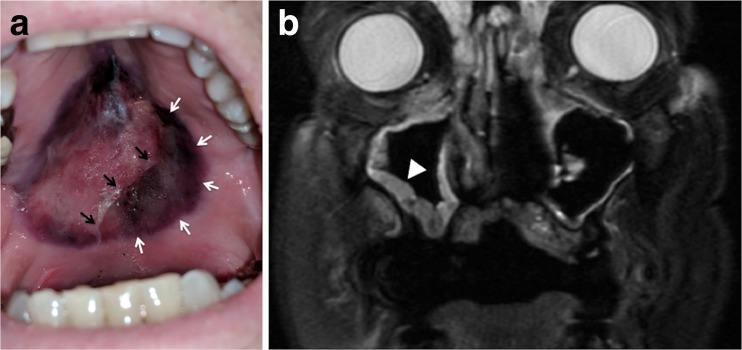
A 75-year-old man with refractory acute lymphoblastic leukemia was hospitalized for dyspnea, hypotension, and neutropenia (< 0.10 × 10⁹/L). Four weeks earlier, he initiated treatment with inotuzumab ozogamicin (an anti-CD22 antibody-drug conjugate). Home medications included prophylactic voriconazole, acyclovir, and trimethoprim-sulfamethoxazole. Computed tomography of the chest showed scattered pulmonary masses up to 3 cm in diameter. Bacterial and fungal cultures from blood and bronchoalveolar lavage were sterile. His condition improved with vancomycin, cefepime, and isavuconazole. On hospital day 16, he reported mouth pain. Examination revealed bilateral maxillary sinus tenderness and an ulcerative hard palate lesion with necrosis (Fig. 1 panel a). Absolute neutrophil count was 0.02 × 10⁹/L. Magnetic resonance imaging showed diffuse paranasal sinus mucosal thickening (Fig. 1 panel b). Palatal biopsy and culture confirmed angioinvasive mucormycosis. Rhino-orbital-cerebral mucormycosis develops following the inhalation of fungal spores by an immunocompromised host. Palatal mucormycosis reflects tissue invasion from paranasal sinus infection and is a sign of aggressive disease portending a poor prognosis.1,2 Palatal ulceration, necrosis, and perforation can result.2 Treatment for rhino-orbital-cerebral mucormycosis (with or without palatal invasion) is intravenous antifungal therapy and surgical debridement.3 Resection was too morbid given the poor prognosis of his refractory leukemia. Amphotericin B and caspofungin were administered without improvement. The patient died 1 week later.
Fig. 1.
a Photograph of the patient’s hard palate showing an ulcerative lesion with necrotic eschar (white arrows) and sloughing necrotic mucosa (black arrows). b Magnetic resonance imaging showing diffuse paranasal sinus mucosal thickening, most prominent on the right (arrowhead).
Compliance with Ethical Standards
Conflict of Interest
Dr. Dhaliwal reports receiving honoraria from ISMIE Mutual Insurance Company and Physicians’ Reciprocal Insurers. All remaining authors declare that they do not have a conflict of interest.
References
- 1.Akhrass FA, Debiane L, Abdallah L, et al. Palatal mucormycosis in patients with hematologic malignancy and stem cell transplantation. Med Mycol. 2011;49:400–5. doi: 10.3109/13693786.2010.533391. [DOI] [PubMed] [Google Scholar]
- 2.Barrak HA. Hard palate perforation due to mucormycosis: report of four cases. J Laryngol Otol. 2007;121:1099–102. doi: 10.1017/S0022215107006354. [DOI] [PubMed] [Google Scholar]
- 3.Gamaletsou MN, Sipsas NM, Roilides E, Walsh TJ. Rhino-orbital-cerebral mucormycosis. Curr Infect Dis Rep. 2012;14:423–34. doi: 10.1007/s11908-012-0272-6. [DOI] [PubMed] [Google Scholar]


