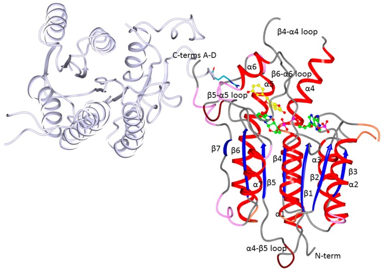
Figure 2.
Ribbon diagram of the LmPTR1 subunit A showing the tertiary and secondary structures as well as the location of the cofactor (green ball and sticks) and an inhibitor (Compound 1; yellow ball and stick) in the active site. The subunit D (pale lilac cartoon) contributing to the active site of the A subunit with Arg287 (cyan sticks) is also shown. The helices of subunit A are colored red, the β-strands blue, and the secondary structure elements are numbered following the sequence. TbPTR1 has the same fold and topology as LmPTR1.

